Anatomy Lesson 4: “Jamie’s Chest – 8th Wonder of the World!”

Greetings friends of Outlander Anatomy and welcome to today’s lesson! Do you recognize the classic seven wonders of the world?

  • Colossus of Rhodes.
  • Great Pyramid of Giza.
  • Hanging Gardens of Babylon.
  • Lighthouse of Alexandria.
  • Mausoleum at Halicarnassus.
  • Statue of Zeus at Olympia.
  • Temple of Artemis at Ephesus.

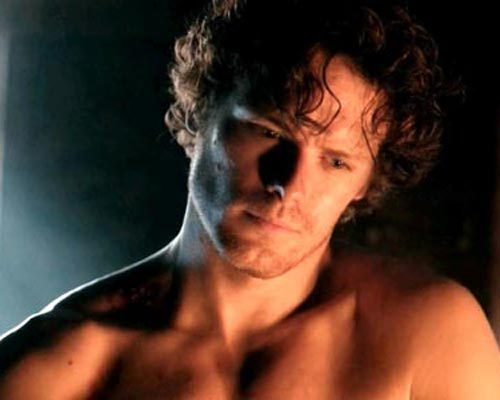
The 8th wonder is, of course, James Fraser’s chest!

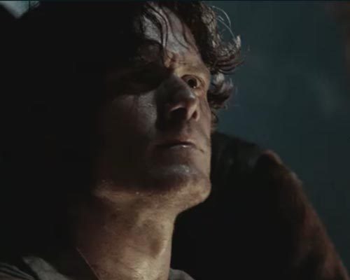
Getting in a mood, let’s take a wee keek at the breadth (Uncle Dougal, ye are a crud for exposing your nephew like this!)…

image

And the depth (BJR, man, ye are darkness itself!):

image

Of this very timely topic!

Now, we all ken that Claire gets her first TV looksee at Jamie’s blest-chest in Starz episode 102, Castle Leoch. But, Herself wrote in Outlander that Claire made touchdown with his chest when he fainted from blood loss after Cocknammon Rock:

“Stop! Help!” I yelled. “He’s going over!” … Jamie slid off headfirst like a sack of stones, luckily landing in someone’s arms. The rest of the men were off their horses and had him laid in a field by the time I had scrambled down. “He’s breathin’,” said one.

“Well, how very helpful,” I snapped, groping frantically for a pulse in the blackness… Putting a hand on his chest and an ear to his mouth, I could feel a regular rise and fall, with less of that gasping note. I straightened up. “I think he’s just fainted,” I said.

He groaned and opened his eyes… “I’m all right,” he said, trying to sit up. “Just a bit dizzy is all.” I put a hand on his chest and pushed him flat.

The instant the bandages were tied, the patient tried to sit up. I pushed him flat and put a knee on his chest to keep him there. “You are not to move,” I said fiercely.

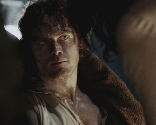
So, leading up to Nurse Claire actually seeing his chest, let’s start with the castle courtyard. I have to begin the lesson here because I LOVE the courtyard scene with all its swirling undercurrents!

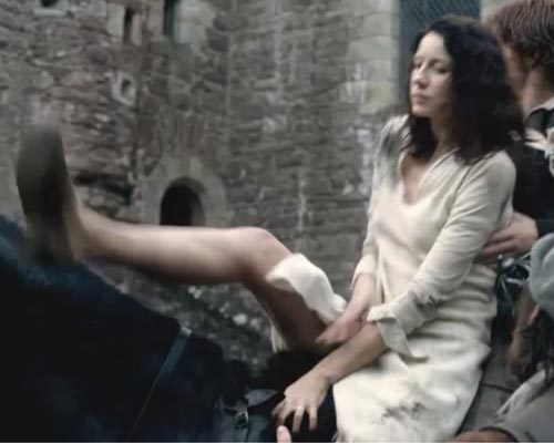
After dismounting, our bedraggled Claire stands there saying nothing but watching everything.

image

Puir lass, the front of her slip and dress have gone bye-bye, having given those up for Jamie’s field dressing. Left in her ruined, oxford walking shoes and hair in straggles, she stands out like a wee sore thumb! She is scairt and confused but tough as nails.

image

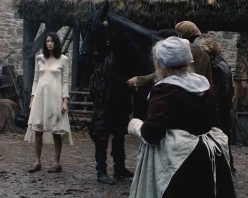
Our stalwart heroine stands completely IMMERSED in a maelstrom of male testosterone and ribald jokes. And, Jamie hovering…
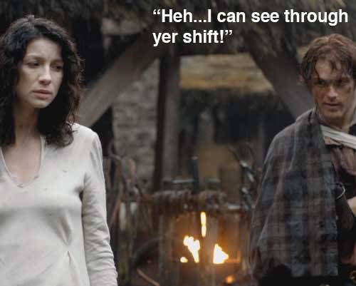
image

…hovering I say!

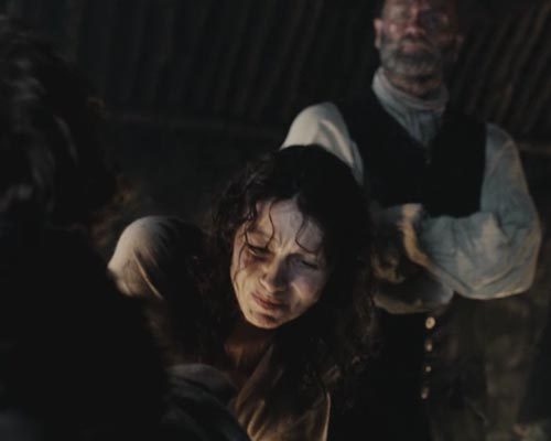
Next, we witness a fabulous tete-a-tete between Claire, Mrs. Fitz, and Jamie where a lot is said, but a whole lot more remains unsaid!

image

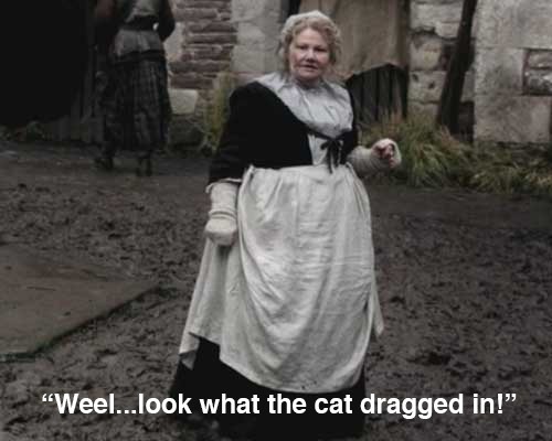
Jamie says Murtagh found her and Dougal said to bring her along. But, it’s NO HIS FAULT that Ms. Fitz has another mouth to feed, bed to make, and body to clothe. Tcha! Just like a man!

image

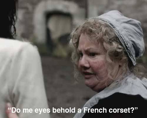
Mrs. F is shocked at Claire’s scandalous appearance and doesn’t mind saying so!

image

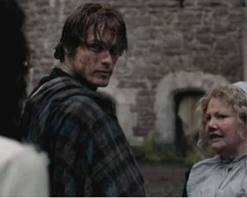
And, Jamie still hovering in the background finding any inane thing to do with his horsey, even though there are several hostlers to do the work, all the while listening and watching the two lasses size each other up.

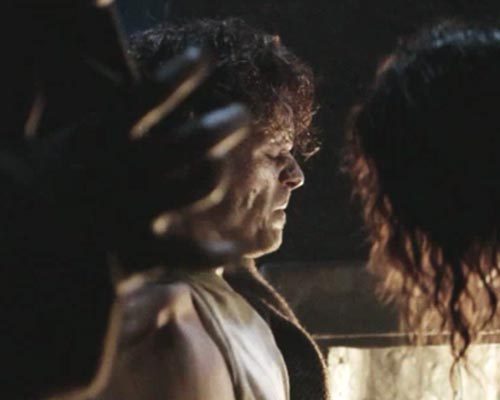
As Grand Dame, readies to drag Claire away for cleaning up, Claire is adamant that she must properly care for Jamie’s wound as Jamie brawly boasts “I can fend fer meself!”

image

Mrs. Fitz upon hearing Claire’s credentials, declares “Jamie, ye need fixin’, git yerself indoors!”  Ye heard the leddy, Jamie! 

And just like that, Jamie  finds himself seated on a stool covered with a blanket and ALONE with Mistress Beauchamp. (Being a wee bit nosey here, who washed Jamie’s face? Whoever it was, thanks – he cleans up nicely!). Claire gently removes the blanket to cleanse the wound and hears the awful truth that her hubby’s sixth, great grandfather gleefully produced the horrific scars on Jamie’s back.

image

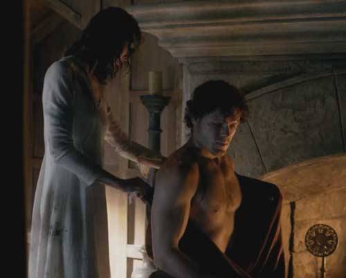
Claire takes time to properly clean Jamie’s gunshot and apply an ingenious dressing involving a pressure bandage and strips of linen crossed under both oxters, an excellent technique for anchoring a shoulder dressing. Jamie tries moving the injured shoulder and winces, so Claire promptly straps his arm to his chest by the golden glow of firelight. Why? Claire knows the arm must immobilized for at least a week to promote wound healing, a truly complicated process! By next day, someone has replaced the strap with a sling, which is not the same at all!

image

Sadly, we must leave this tender scene to start Anatomy Lesson #4. Sniff!

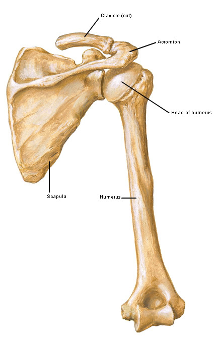
Terms: Let’s come to terms with it, anatomists do not use the terms upper and lower arms.  Instead, the entire upper appendage is called the upper limb.  The region between shoulder and elbow joints is the arm and the region between elbow and wrist joints is the forearm (Image A). This is important to know for this lesson.
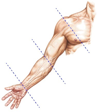
image

Image A

Rib Cage: The bony foundation of the chest is the thoracic (rib) cage, including 12 pairs of ribs, their costal cartilages (blue structures), sternum (breast bone) and 12 thoracic vertebrae (Image B).  This spring,  bony enclosure not only protects heart, lungs, airways, esophagus, and great blood vessels, it also provides attachments for important chest muscles! Above the thoracic cage lies the clavicle (collar bone) which articulates (forms a joint) with it.
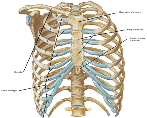
image

Image B

Pectoralis Major: The chest muscles or “pecs” as trainers call them, actually include two pairs of muscles on each side of the chest. The word pectoralis derives from the Latin pectus meaning “breast.”

Pectoralis major muscles are the largest, most superficial, and most powerful of the two pairs. Each fan-shaped pec major covers half of the chest and is divided into three heads. The clavicular head (Image C) arises from the clavicle.
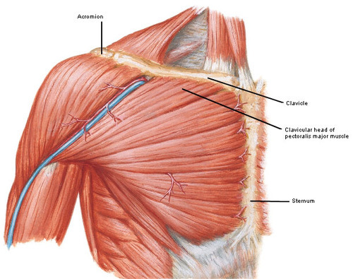
image

Image C

The larger sternocostal head arises from 1 through 6 costal cartilages and sternum (Image D).
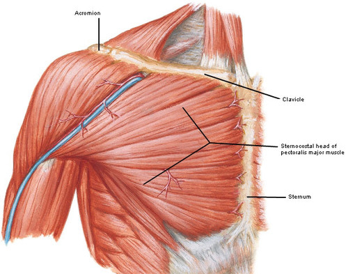
image

Image D

A smallish, third part (Image E) arises from an abdominal muscle. This is a fairly insignificant part unless torn in which case, the pain surpasses all conscious thought. Just kidding, except it does hurt!
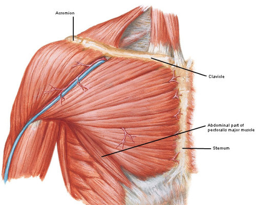
image

Image E

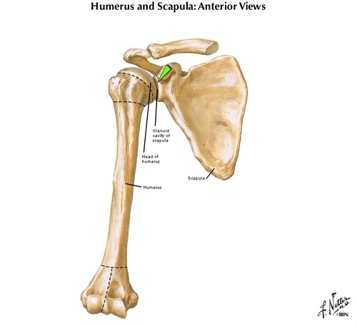
Pectoralis major muscle fibers converge toward the arm inserting into the humerus or arm bone (Image F).
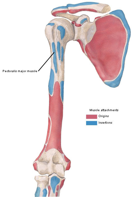
image

Image F

Heh! Wake up, fledgling anatomists!

image

Yep, we can do pretty much anything for the Great Scot!

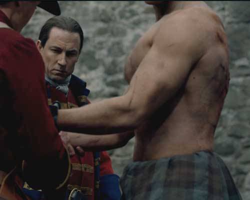
On their way to the humerus, pec muscle fibers create  the anterior axillary fold (Image G). This fold forms the front border of the arm pit, oxter, or axilla! (I still want to see Claire stick her foot in Jamie’s oxter!) Grab this fold on a pal or sibling and give it a wee pinch. Gives them a jolt, so not a fab idea to try someone without a sense of humor!
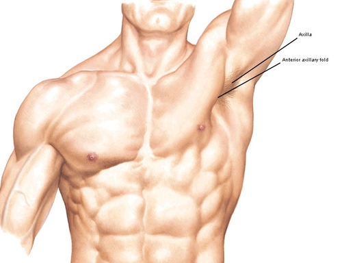
image

Image G

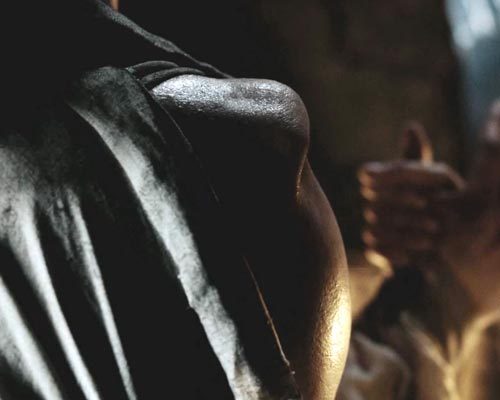
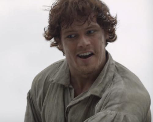
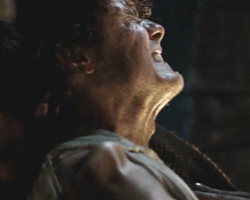
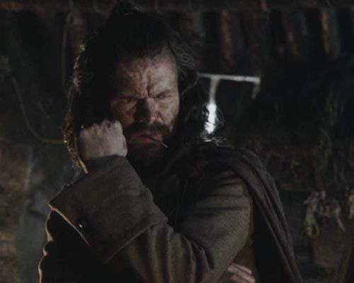
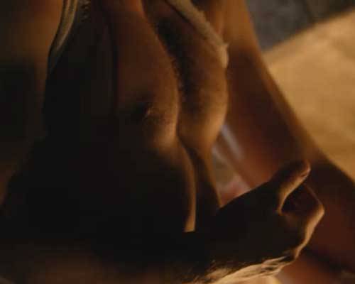
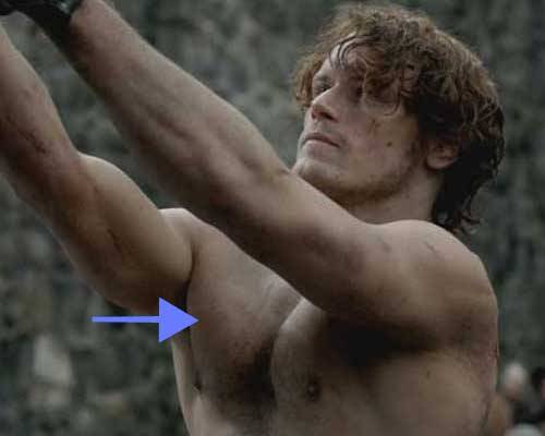
In this horrifying image from Starz episode 6, The Garrison Commander, Jamie’s massive right anterior axillary fold is very easy to spot. Our darling hero  near freezes to death as BJR parades, preens, and prepares for his fav outdoor sport!

image

Function: Each pec major is a very hard working muscle. And, because it has three heads, they do quite a bit of work!

  • raises arm forward (as in lifting a child)
  • returns arm against the torso (as in setting down the child)
  • pulls arm from spread eagle to the sides of torso (as in standing in mountain pose)
  • rotates arm (internally) toward the chest

And, for those who wonder, pec major is best developed by standard pushups (not triceps type!), bench presses and weighted flyes.

Try This: You can see the tendons of your own pec majors this way: place palms together about 6” in front of your chest as in prayer mode. Now, press the palms firmly together. Your own pec majors should stand out strongly as the anterior axillary folds. Just don’t get them pinched!

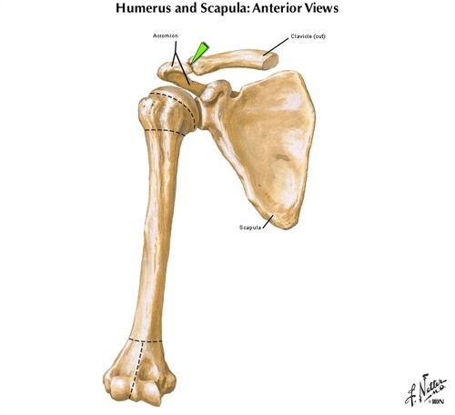
Pectoralis Minor: In anatomy (like baseball), if there is a major – there is a minor. So, deep to each pectoralis major is a pectoralis minor muscle (Image H). This smaller fan-shaped muscle is also very important.  It arises from the ribs (2, 3 & 4) and inserts on a small bony knob of the should blade known as the coracoid process (Greek for “like a raven’s beak”). Its contraction pulls forward on the corcoid process aiding in shoulder mobility and stability.
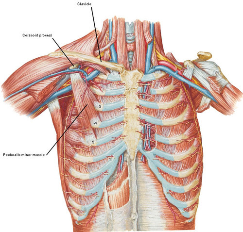
image

Image H

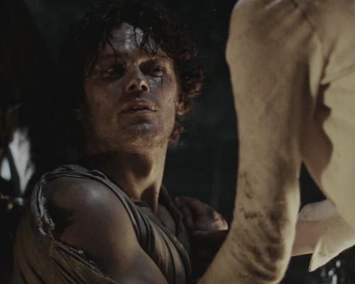
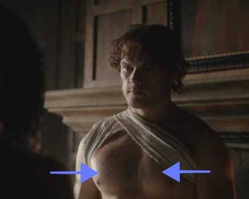
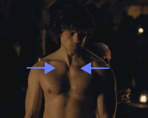
Applied Anatomy: OK, with the thoracic cage and two pairs of pecs  done, let’s find them on our wonderful warrior! After Claire trusses Jamie up, the only parts of his chest still showing are the verra fine sternocostal heads of his pec major muscles, covered with skin, of course (blue arrows). Ye can see them fair keeking out from under the dressing. Awesome sauce! And, good reason to consider them the 8th wonder of the world.

image

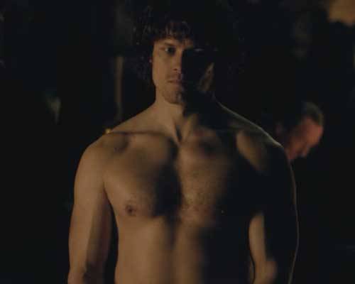
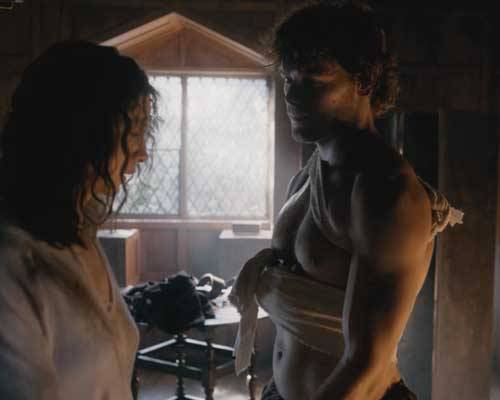
But, wait, here’s more! The clavicular heads of Jamie’s pec major muscles are unusually well developed! Check out this image (Starz episode 5, Rent) for prominent bulges just under those bonny clavicles (blue arrows). These are the clavicular heads of each pec major, an oft neglected part in body building – but not here! Props to the trainer! Gasp! They’re bloody awesome!

image

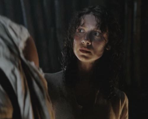
Finally, Claire has done all she can legally do for Jamie – he stands there with his glorious chest peeking through the bandages and his nipped waist and.. GAH! Lord, gimme a dram! 

image

Now, returning to anatomy (I am a professional after all)… about 1 in about 50,000 people are born without some or all of pec major and minor muscles. Known as Poland Syndrome, it may include hand and finger anomalies and shortened forearm bones. Interestingly, people with the syndrome compensate quite well using other arm and shoulder muscles. In fact, there’s a well-known PGA player, an Olympic boxer, and a Formula 1 World Champion (car racing) that have Poland syndrome and, clearly, it didn’t hinder them! The lady shown below (Image I), from a theatrical production, has it; she is missing the sternocostal head of pec major (blue arrow).
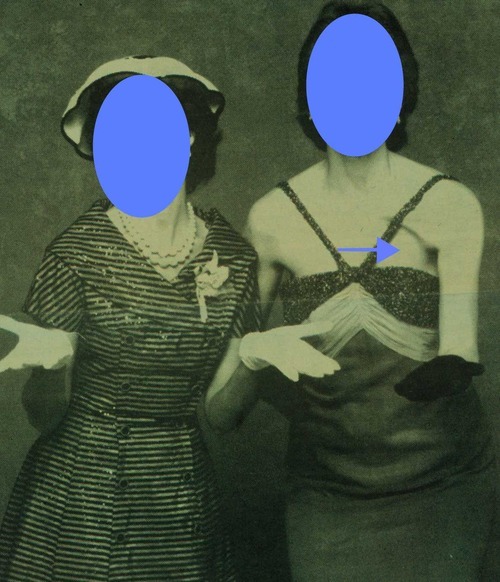
image

Image I

That’s it! Let’s close this lesson with a lovely poem nicely expressing the growing attraction between Nurse and Highlander!

Place your hand upon my chest.
It reminds me how it feels when it’s mended.
Then use it to cradle your head while you rest.
The worst of it, like the day, has ended.


I hope this lesson helps you more fully appreciate the chest muscles and their bony attachments.  Fare thee well for now.  Am thinking that Claire might be the subject of my next posting! We have ignored our amazing heroine for far too long!

How many days left before Starz episode 9? 156 days or so but who’s counting?! Sigh.

The deeply grateful,


Outlander Anatomist

Photo credits:

All photos are credited to Starz or Frank Netter’s Atlas of Human Anatomy, 4th edition. The lady with Poland’s syndrome is an archival photo from my lectures and I do not know the photographer.

Anatomy Lesson #2: “Claire and Jamie Meet at a Joint!”

Haha. Just joshing. Claire doesn’t meets Jamie at a joint. Claire meets Jamie because of a joint! Welcome to Anatomy Lesson #2, the shoulder joint. Now, hold on just a sec, please don’t leave this lesson! This is great stuff – truly – and I promise to keep it interesting. I’ll even throw in a gratuitous shot of our favorite #JAMMF just to hold yer interest! Hee, hee, here ye go…

image

Better? Great!  So, in Starz episode 101, Sassenach, Claire first sees Jamie seated on a low stool in a dimly-lit cottage. He is in pain. Herself enlightens us in Outlander book:

a dreadful hump rose on that side, and the arm hung at an impossible angle.

Now, what is the dreadful hump, what might have caused it, and how might it be resolved?

Anatomy to the rescue! Yep, it explains everything!

image

Hump: The hump is an bony part of the shoulder joint. The should joint is an essential part of each upper limb. Let me assure you, its anatomy is absolutely awesome! First, it is the MOST moveable joint of the entire human body (think of the shoulder joint during slow pitch or with a butterfly stroke). Second, the shoulder joint is a slightly misleading term because it is not one joint, but two – the glenohumeral joint and the acromioclavicular joint:

  • glenohumeral joint: between humerus (arm bone) and scapula (shoulder blade) –  note the spelling: humerus not humorous!
  • acromioclavicular joint: between clavicle (collar bone) and scapula – this one is oft ignored but is crucial for normal function.

Together both joints form the “shoulder joint!”

Glenohumeral Joint: At the glenohumeral joint, the head of humerus moves in the glenoid cavity (a.k.a. glenoid fossa), a small and shallow socket on the side of scapula.

The good news: because the glenoid cavity is shallow, the humeral head is highly moveable allowing for at least nine different defined motions.

The bad news: unfortunately, the shallow socket also allows the head of humerus to dislocate with relative ease! Get it? Grand!

Turns out that puir Jamie dislocated his glenohumeral joint (Image A – green arrow, right front view). Och! 

image

Image A

Acromioclavicular Joint: The acromioclavicular joint (green arrow in Image B – right front view) is a tight joint between acromion (also part of scapula) and clavicle. The clavicle acts as a strut holding the acromion away from the torso. Associated muscles contract to adjust positions of both clavicle and scapula (e.g. up, down, back, forward). And, amazingly, the acromioclavicular joint also guarantees greater range of motion at the glenohumeral joint.

Try this: Place fingers of one hand on the contralateral (opposite) clavicle. Now, move clavicle forward, backward, up, down. Now, roll it forward (as in doing the hoochie koo) and then backward. See how mobile the clavicle is? Amazing!

Now, move fingers to the scapula of the same side and repeat the same clavicular (adj.) movements. See how the scapula is carried along with the clavicle? This occurs because they are united at the acromioclavicular joint! And, increased scapular mobility means increased mobility of humerus as it is concurrently moved at the glenohumeral joint. Normally, the whole apparatus works together like a finely-oiled machine.

So, for example, each time you lift the clavicle and scapula, you can also raise your arm higher. Prove this to yourself: hold your scapula still and raise the arm of the same side. Find that you cannot raise the arm very high, but once you release the scapula to lift, the humerus can be raised to the full vertical position. Hoping this makes sense!

image

Image B

Acromion: The acromion (Image C – right back view) overreaches and protects the underlying glenohumeral joint. Acromion shares the same Greek word root with the Acropolis of Athens, meaning “peak” because both structures overlook what lies below. In lay terms, the acromion is better known as the “point of the shoulder.”

The “dreadful hump” on Jamie’s right shoulder is his right acromion, left high and dry because the humeral head is no longer in its socket (glenoid cavity). Gah! Deprived of the rounded contour normally provided by the humeral head, the acromion is revealed as a huge bulge of the shoulder area.

image

Image C

Try this: Palpate the point of one shoulder and feel the big bony chunk; this is the acromion of scapula. Now, check this area out on your wee dog or perhaps on a horse (‘cuz Jamie does like the horsies)! The acromion faces forward, coming to lie under the neck of these animals. Why? Because dogs and horses have no clavicles to hold the shoulder joint away from the torso, so, the entire scapula and its acromion are located on the sides of their chests – ours are on our backs.

The Bottom Line: Glenohumeral joints of dogs and horses are much more stable than ours but their range of motion is also greatly limited. We don’t see dogs running with front legs whirling like windmills. The photo is of a Vizsla, BTW.

Rotator Cuff:  Now, if the glenohumeral joint is relatively unstable, what holds it in place? This is accomplished by some very strong ligaments and four muscles forming the rotator cuff. Also, the powerful deltoid muscle (Image D – green arrow denotes head of humerus) covers the joint, ligaments and part of the rotator cuff with a thick, protective muscular padding (especially in a lad like Jamie – <G>!).

image

Image D

Ok, I can tell I am losing you. Wake up! Here, have another dose of our gorgeous hero!

image

Now, back to Starz episode 101, Sassenach: We hear the ever “tender” uncle Dougal mutter ”it’s out-o-joint, poor bugger.” Well, duh!

In  Outlander book, Jamie explains:

I fell wi’ my hand out, when the musket ball knocked me off my saddle. I landed with all my weight on the hand, and crunch!, there it went.

Now, folks, falling on an outstretched arm/hand is the classic cause of an anterior dislocation of the glenohumeral joint, wherein the humeral head is thrust forward (anterior) and out of the glenoid cavity. This stretches and tears ligaments and muscles and, sometimes, cartilage.

Back to episode 101, Sassenach Our favorite goof, Angus, delicately raises one brow and declares in his best practitioner’s voice:

I’ll have to force the joint back!

Yeah, right, Angus! He and his brawny pals start forward only to be stopped in their shoon by awesome Claire using her best Hospital Matron voice:

Don’t you dare…stand aside at once!”

She then enlightens Dougal, who, no doubt, is one of the brighter bulbs in that crofter’s cottage:

You have to get the bone of the upper arm into the proper position before it slips back into joint.

Now, at this point, our sweet hero is looking a wee bit dubious about this whole “girlie” thing. Well, it’s either Claire or Angus. Who would you choose?  I’d go with the shift, too, sweet lad.

image

After obtaining Jamie’s nod of assent, Claire grips his right wrist in one hand and his bent elbow in the other. Lifting with considerable effort, she rotates his forearm to his right side (external rotation), a maneuver that aligns the humeral head with glenoid cavity.

The considerable resistance Claire encounters (just look at our strong, winsome heroine!) is due to swollen muscles, torn ligaments, and the weight of his “heavy as lead” arm!

image

Then, warning Jamie that the worst is yet to come, Claire holds his bent elbow against his torso, and gripping his right hand, rotates his forearm to his left (internal rotation), a maneuver designed to reseat the humeral head in the glenoid cavity!

image

Suddenly, Jamie’s shoulder gives a soft, crunching pop as the humeral head slides home into the glenoid cavity and the dislocated joint is reduced.  And, folks, it hurts, especially several hours post-injury – truly! Kudos to the special effects crew for the CGI/prosthesis!

image

Just ask Angus…Och!

image

Next and this is very sweet, Jamie says he’s

takin’ a guess she’s done this afore.

Trust me, Claire assures him:

I’m a nurse!

Har, har! Jamie eyes immediately drop to her bosom and he responds:

Aye.

Ah, well, he may be injured, but his other brain seems intact! What was he thinking’? Do me eyes behold a sweet avenging angel? A wee demon? A biting vixen? But, whatever she is, thank the stones she’s NOT A WET NURSE!

image

Now, just in case ye missed it, take a look at Claire’s eyes when she sees Jamie’s muckle size as he rises off the stool. She even has the good sense to blink a few times. Yup, he got her attention alright and she got his. There’s a sizzle on the griddle, now!

image

Fun Fact: Did you know that Claire could have employed a much older method of reducing Jamie’s dislocation? Hippocrates (460-370 B.C.), father of western medicine, devised the Hippocratic maneuver, wherein the practitioner places the heel of his/her foot into the armpit of the dislocated side and pulls on the corresponding wrist to reduce the dislocation. I would love to see Claire pull that one off! Without a doubt, she is flexible enough to stick her foot in Jamie’s oxter. Check out her graceful dismount from Jamie’s steed in the castle courtyard, Starz episode 102, Castle Leoch! Watch out, Rockettes!

image

Realism: OK…question: was the scene from episode 101 realistic? Is it likely that a WWII combat-trained nurse such as Claire might know how to reduce the dislocated humeral head using the method shown? Oh, aye, because the procedure she employs looks verra much like the Kocher maneuver, developed in the 19th century by a Swiss surgeon of the same name. Although other reduction methods are now available, Kocher was widely used for over a century and Claire could very well have learned it in training or as a combat nurse.

Another realistic feature, check out Jamie’s eyes in episode 101. Throughout the entire scene, his pupils are VERY dilated. This is also highly realistic because the pain of dislocation would initiate the fright/flight/fight reflex, dilated pupil being a prime feature of that response! His pupils are soooo dilated that the rims of his irises are barely visible!

image

Overall, this vivid scene was realistic, dramatic, and extraordinary in most every detail. Congratulations to the cast and crew! If you readers are interested, here is a link to a YouTube demo of the Kocher reduction.

My next post may well review Jamie’s other injury sustained in Starz episode 101, Sassenach. Please stay tuned and see what’s next! After all, we have to keep engaged until April of 2015! Geez, Starz…soooo harsh!

Just for fun, consider a few shoulder idioms, use used to convey important emotions:

  • broad shoulders
  • cold shoulder
  • shoulder to cry on (I like this one)
  • have a chip on the shoulder
  • put shoulder to wheel
  • square one’s shoulders
  • on the shoulders of giants
  • a weight off the shoulders
  • rub shoulders with
  • looking over shoulder

Yadda, yadda, yadda. You get the drill.  Hope to see you at Anatomy Lesson #3!

The deeply grateful,

Outlander Anatomist

Photo credits:

Starz; Frank Netter’s Atlas of Human Anatomy, 4th edition, Outlander Anatomist private collection.