Welcome all students to today’s Anatomy Lesson #32, The eye – Part 4! Remarkable sensory organ that it is, this will be our fourth lesson of the eye series. Although I thought this would be the last eye lesson, a 5th and last lesson on eye anatomy is required. But, you’ll see why this fascinating and wonderful organ requires yet another lesson!
We know that the eye is designed to provide us with vision. Understand that the air around us is filled with a sea of electromagnetic energy from the shortest gamma waves to the longest radio waves. Humans are blind to almost all of this energy because our eyes detect only that part of the electromagnetic spectrum known as the visible spectrum or, in more common terms, visible light.
Let’s begin the lesson with a quick review. In Anatomy Lesson #31, we learned parts of the eyeball. Photo A is a horizontal section through a left eyeball (as viewed from above). The sclera is the white outer coat. The choroid is a coat of blood vessels internal to the sclera. The retina with its specialized sites is inside the choroid. The transparent cornea projects from the front surface. Iris and pupil (Anatomy Lesson #31) are behind the cornea. The biconvex lens is behind the iris. The ciliary body with its ciliary muscle forms a ring around the lens. The anterior chamber is a space between cornea and iris that is filled with the watery aqueous humor. A space between iris and lens is the poorly named posterior chamber which is also filled with aqueous humor. Behind the lens is the largest space of the eyeball, the vitreous chamber containing vitreous humor or vitreous body.
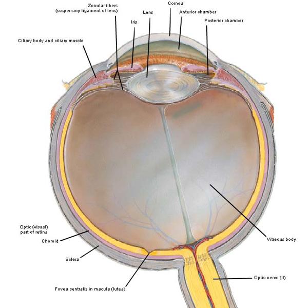
Photo A
NOTE: this lesson shows images of the eyeball in horizontal section oriented with the cornea directed upward (Photo A) or rotated 90° so the cornea faces left or right (Photo C). Don’t be distressed when you see this flip flop; it just reflects preference by the creator of the image.
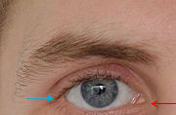
This lesson will cover sclera, cornea and lens. The sclera is the tough, white outer fibrous coat of the eyeball (Photo B – red arrow); it provides shape and resistance as well as attachment for extraocular muscles (Anatomy Lesson #30) and some intraocular muscles (Anatomy Lesson #31). The tiny red blood vessels seen in Photo B are actually located in the bulbar conjunctiva (Anatomy Lesson #30) which adheres to the anterior sclera.
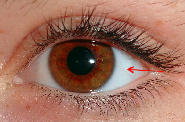
Photo B
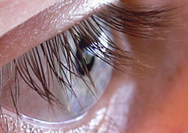
The cornea (Latin meaning horn) is a transparent dome covering the anterior 1/6 of the globe. It bulges because its radius is smaller than that of the remaining eyeball. This creates a small sulcus (groove) at the corneoscleral junction known as the limbus (Latin meaning edge or border; Photo C).
Try this: Ask a partner to sit while you look at one of their eyes from a side view. Identify the white sclera. Find the transparent cornea bulging from the front of the eyeball. Find the limbus, the groove where cornea and sclera meet.
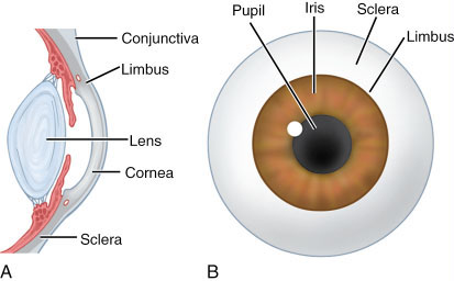
Photo C
The cornea is a truly fascinating part of the eye being composed of cells and extracellular molecules arranged in five microscopic layers (Photo D). With the exception of the stroma or middle layer, the names of the other layers don’t concern us today. A normal cornea is transparent because the stroma is made of regularly arranged layers of collagen molecules and because it lacks blood vessels.
Question: if it has no blood supply, how does it get oxygen and nutrients?
Answer: these are delivered to the superficial cornea by the tear film and to the deep cornea by aqueous humor (or humour). The cornea is well-equipped with pain fibers as anyone knows who has injured it. And, as you realize, if the cornea is touched an automatic reflex is evoked that closes the eyelid for protection.
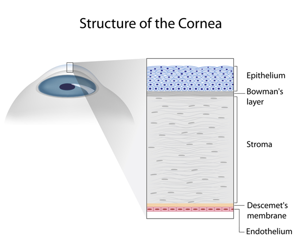
Photo D
The cornea is not of the same thickness throughout: it is thinnest at its middle (~0.5 mm) and thickest near its rim (~0.8 mm). This change in thickness is clearly evident in this magnificent image from Lesson #31 (Photo E – yellow arrow at middle of lens; red arrow at rim).
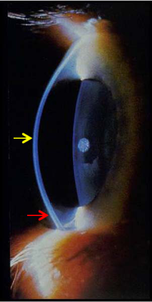
Photo E
The normal cornea is smooth and clear like glass, but it is also strong and durable so it helps the eye in three ways:
- Refracts (bends) light rays to help focus them on the retina (Photo F – black arrow).
- Shields the eyeball from germs, dust, and other harmful matter.
- Filters out some of the damaging ultraviolet (UV) wavelengths in sunlight.
Light rays entering the eyeball must be bent to focus them on the retina. The cornea is powerful providing roughly 2/3 of the overall refractive power of the globe. But, understand that its curvature is fixed and normally does not change.
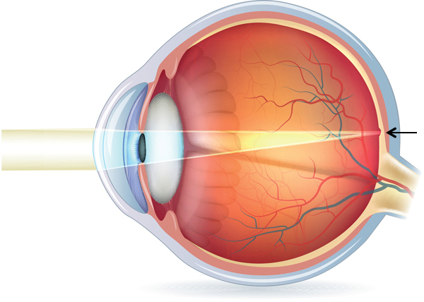
Photo F
Corneas are subject to a variety of diseases and conditions including allergies, conjunctivitis (pink eye), infection, dryness and dystrophies (cloudiness) to name a few. A seriously damaged cornea may be injured such that light cannot penetrate the eyeball to reach the light-sensitive retina. Sometimes a corneal transplant is required wherein a surgeon removes the central portion of the cloudy cornea and replaces it with a clear cornea, usually donated through an eye bank.
Here are two pairs of clear corneas going at it eye-to-eye (Starz episode 109, The Reckoning). Look me in the corneas lass, when I’m talking to ye! I am your 18th century hubby and ye will obey me! This Sassynach looks Jamie in the eye and while she’s at it, she’ll spit in it too! She doesn’t have to do what Jamie says. Aye ye do; If ye dinna do what I say then there’s a price ye’ll have to pay! Ouch!

And, just when we think that we have seen it all, well, we haven’t! Have you ever heard of corneal jewelry? Yep, small crystals and other gewgaws hang via a thread from a contact-type lens! Not sure that swinging counterweight would feel very good dropping from the cornea but can’t say as I’ve never tried one.
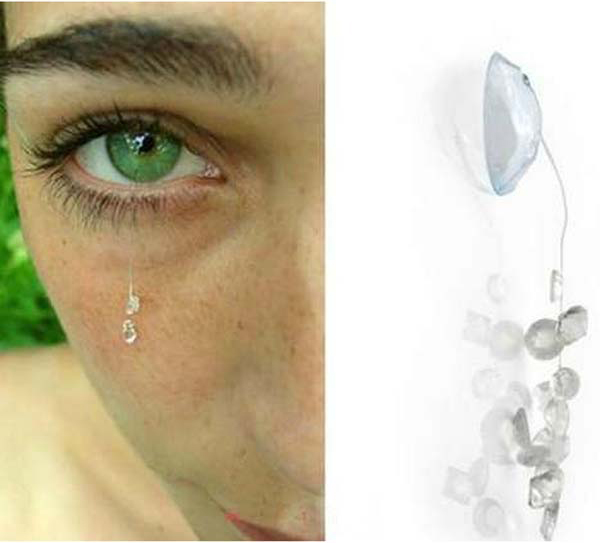
Photo G
Moving along, the lens (Latin meaning lentil) is next. The lens is a transparent, bi-convex structure suspended between iris and vitreous humor that continues to grow throughout life. The lens is shaped like a flying saucer although its front surface is flatter than its back surface (Photo H). Near the pupillary rim, the iris is in contact with the front surface of the lens but the two diverge at the sides. The back surface of the lens sits in a divot of the vitreous humor. Like the cornea, its purpose is to refract light rays to focus on the retina and is responsible for about 1/3 of the overall refractive power of the eyeball.
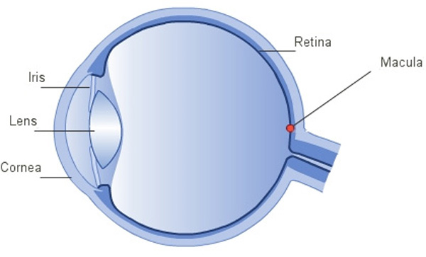
Photo H
Lens structure is amazing and complex but simplified for this lesson. Like the cornea, the lens is avascular to assist with transparency. It is made of very long (12 mm) and very thin cells known as lens fibers (Photo I). These cells are arranged in layers around a central core much like the rings of an onion. The lens has also a capsule, some cube-shaped cells in front and a few other features but these do not concern us for this lesson.
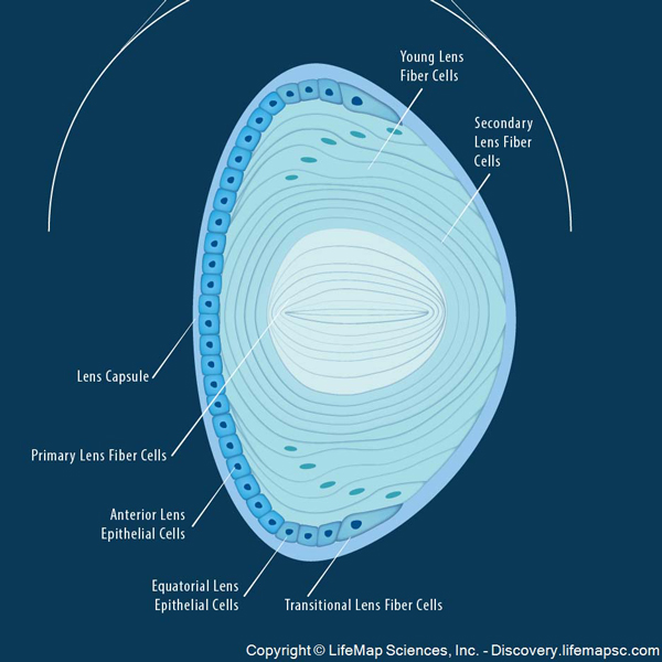
Photo I
Photo J is an image of lens fibers taken with a scanning electron microscope (Anatomy Lesson #6), an instrument that takes 3-D images of microscopic structures. Very cool! With this type of microscopy, lens fibers look like tightly-packed lasagna noodles. Nuclei and other cell structures (organelles) are absent from mature lens fibers to boost transparency. Lastly, the fibers are filled with crystalin protein and their water content is very low.
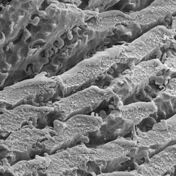
Photo J
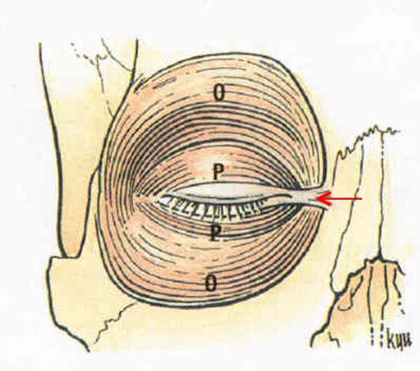
The lens is suspended from the ciliary body by a ring of fibrous strands known as zonular fibers. Although difficult to appreciate in a two dimensional image, zonular fibers attach all the way around the equator of the lens (Photo K – red arrow) much like a round trampoline pad that is suspended from a frame by its coils.
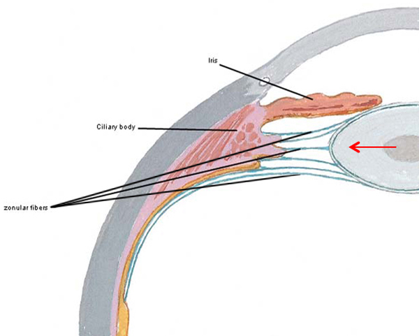
Photo K
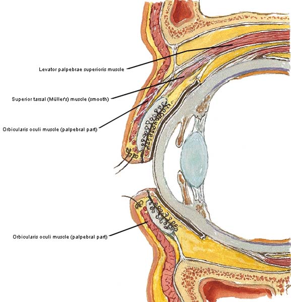
A closer look at the suspensory mechanism reveals that several layers of zonular fibers attach to the lens at various points at or near its equator (Photo L – red arrow). Collectively zonular fibers form the suspensory ligament of the lens. Another important feature, the ciliary body contains bundles of (involuntary) smooth muscle cells (Photo L – black arrows).
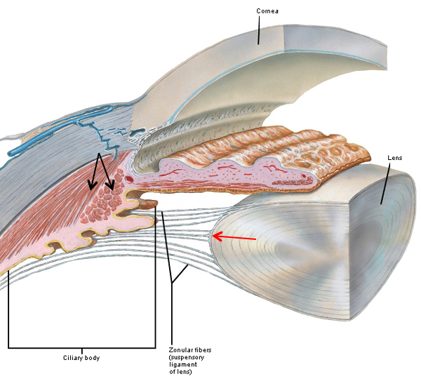
Photo L
Like the cornea, the lens refracts (bends) light rays to help focus them on the retina. Unlike the cornea with its fixed curvature, the lens is flexible and its curvature can be adjusted to fine tune focus, an ability which decreases with age. Based on the distance from a viewed object, the lens flattens or fattens.
Simply put #1: light rays from far objects enter the eyeball roughly parallel (Photo M – top image) and not much lens refraction is required to focus them on the retina. With this scenario, the ciliary muscle is relaxed and the lens is flat because it is under tension by zonular fibers of the suspensory ligament.
Simply put #2: light rays from near objects (Photo M – bottom image) diverge before reaching the eye so the lens thickens to refract these rays and bring them into focus at the retina (if the lens doesn’t thicken, near light rays would come to focus behind the retina and the image would be blurry). With this scenario, ciliary muscles contract releasing tension on zonular fibers (i.e. the suspensory ligament) so the lens fattens, an action known as accommodation of the lens. Accommodation is accomplished by, yes, the parasympathetic division (PSNS) of the autonomic nervous system causing ciliary muscles to contract!
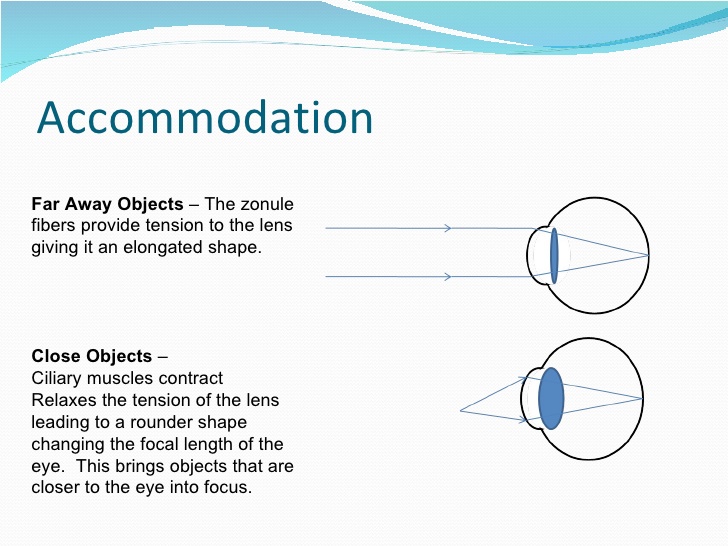
Photo M
A clarification is important here – accommodation for near vision actually involves three important changes, all of which occur automatically:
- Eyeballs converge because both medial rectus muscles contract (Anatomy Lesson #30)
- Pupils shrink in size because the sphincter pupillae muscles contract (Anatomy Lesson #31)
- Lens thickens because ciliary muscles contract, releasing tension on zonular fibers and the lens assumes its natural fat state (This description may sound counterintuitive but it is correct)
Although scientists know that lens curvature increases with near vision to ensure that the light entering the eye focuses on the retina, the way this happens is still unresolved. Photo N, left panel shows the flattened shape of the lens with far vision. The middle and right panels show two theories about the shape change of the lens during near vision/accommodation. The Helmholtz theory (1855) proposes that the ciliary muscle contracts, all zonular fibers relax and the lens thickens. The Scharchar theory (1992) suggests that the ciliary muscle contracts, selective zonular fibers relax and only the central part of the lens thickens. There is yet a third theory but too technical for today’s lesson. It has been claimed that most modern eye specialists hold with the Helmholtz theory.
Try This: Have a pal sit at one end of a room and focus on something at the far end. Now, step in front of the pal, bend down and ask him/her to look at your nose. If you do this carefully, you can see the eyeballs draw together and the pupils shrink with accommodation. You will not see the lens change shape because it lies too deeply. Try it a couple of more times with the pal looking at something over your shoulder and then at your nose to be sure you got it. This is accommodation.
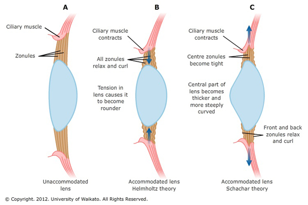
Photo N
Claire presents us with a wonderful example of accommodation as she looks into the bonny orbs of her handsome Highland Hero (Starz episode 110, By the Pricking of My Thumbs)! Her pupils are constricted and her eyes converge to focus on the object of her desire. Thickening of her lens is not visible. Come back to her, James Fraser!

Not to be outdone, Jamie’s eyes can accommodate too (Starz episode 111, The Devil’s Mark). Ahhhh, where did ye say ye were from, Sassylass??? Pupils constricted and eyes convergent…great job, laddie!

With age, the lens and ciliary muscle undergo changes. Known as presbyopia (Greek meaning old eye), the lens gradually loses its flexibility and the ciliary muscle weakens so focusing on near objects is impaired (Photo O – top image) and the image comes to focus behind the retina. Thankfully, distance vision usually remains unaffected. This condition is easily treated with eyeglasses or contact lenses (Photo O – bottom image) which refract the light rays to bring them to focus at the retina. Please note that at present, presbyopia cannot be treated effectively with surgery. Presbyopia usually becomes apparent in our early- to mid-40s, but this is actually a gradual process that often begins earlier.
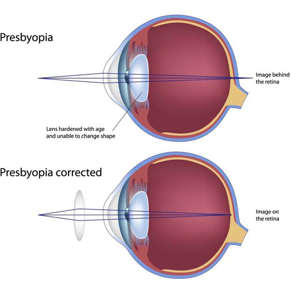
Photo O
Next, let’s consider cataracts: not a torrent of water but opacities of the lens that can result in impaired vision or blindness (Photo P). Cataracts have been around for a very long time with the earliest documented case being depicted in a small wooden Egyptian statue from about 2400 B.C.E.
A cataract can occur in either or both eyes but does not spread from one eye to the other. Eye cataracts are common in older people as the aging lens and its fibers become more and more opaque, but cataracts can also form congenitally, after injury, following treatment with some medications, or from disorders such as glaucoma. A startling statistic: half of the US population 80 years old and older have cataracts or have had cataract surgery. Now days, there are several different surgical approaches to cataract treatment but they typically involve removal of the natural lens followed by implanting an artificial intraocular lens.
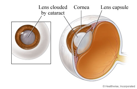
Photo P
Let’s take a look at some interesting history about the cataract. It may be difficult to believe, but the earliest recorded surgical treatment of cataracts was 800 B.C.E., in India. Using a procedure known as couching, a curved needle was inserted through the coats of the eyeball. The lens was pushed downwards and backwards into the vitreous humor and out of the field of vision. Later, couching spread from India to China, Babylonia and Egypt. Centuries later, couching was practiced in Europe (Photo Q) by traveling barber-surgeons with little or no training and the procedure often left patients blind or at best with only partially restored vision. Other strange and startling treatments were also popular. Nicholas Culpeper records a cataract treatment in his School of Physick (1659):
“The head of a cat that is all black burned in a new pot or crucible, and made into fine ashes, and a little of it blown with a quill into an eye that hath a web or pearl growing in it, three times a day is a sovereign remedy”
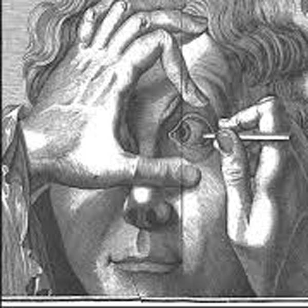
Photo Q
During Outlander’s time, John Taylor (1703-1772) was the first in a long line of British oculists. Dubbing himself “Chevalier” and “Ophthalmiater Royal,” Taylor became the personal eye surgeon to King George II (Yep; same sovereign that Claire mentions to Colum in Starz episode 102, Castle Leoch), the pope, and a number of European royal families (Photo R). A colorful character who many consider a charlatan, he traveled throughout Europe in a coach painted with images of eyes to advertise his expertise. Taylor removed cataracts either by couching or breaking them up and extracting the bits and pieces. Prior to performing each surgery, he delivered to the audience (yes, groups of people often observed such surgeries) a long, grandiose self-promoting speech.
Purportedly, Taylor once confessed that he had blinded hundreds of patients. Two of Taylor’s botched cataract surgeries included famous composers George Frederic Handel (1685–1759) and Johann Sebastian Bach (1685-1750). Taylor performed couching on Handel but it was unsuccessful and the composer suffered blindness throughout his remaining years. Taylor also used couching to treat Bach’s bilateral cataracts. A week later, Bach was re-operated on but he remained blind and, weakened by the surgeries, he died four months later.

Photo R
Many of us are indeed fortunate to be able to choose good eye surgeons with superior training and expertise than those of Jamie’s time. But, I would be amiss not to note that today couching is still practiced in remote areas of third world countries. Let’s give a shout out (and a donation if possible) to those surgeons who travel to dangerous regions of the globe and often at their own expense to bring current therapies to those who are less fortunate.
Let’s close the lesson with a parting keek at Jamie as he keeks at Claire (Starz episode 102, Castle Leoch)! Herself reveals in her 8th book, Written in My Own Heart’s Blood, that Jamie often surreptitiously appraised Claire with his bonny orbs before they wed. He declares that anyone would have thought them well-suited for each other had they noticed his thorough perusal of her:
“If they’d seen the way I looked at ye, Sassenach, when ye didna see me lookin’—then, aye, they would.”
OR, to quote Rick’s famous line from Casablanca:
“Here’s looking at you kid!”

Yum! Yum! Gah!
Next lesson will be the last one on the eye: we’ll cover the retina and its parts, nearsightedness, farsightedness and some grand experiments with our own vision. For now, let’s end with a fun poem about eyes by Mr. R! The last Anatomy Lesson to feature one of his delightful science poems was in Anatomy Lesson #24.
Eyes Poem: sense of sight
I need my eyes,
So I can see,
I don’t have 5,

Or 1,

Or 3…

Just 2 eyes,
On my face,

It seems to be,
A common place,
For eyes to sit,
On animals,
From frogs, to fish,
From bears, to gulls…

2 eyes,
1 face,
That’s a match,
But really here’s,
A little catch…
Spiders see,
With 8 bug-eyes,
And here’s another,
Eye surprise,
Scallops have,
Most of all,
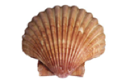
More than 50,
Must see all!
I’ll stick with 2,
They let me see,
My 2 eyes,
Are good for me…
A deeply grateful
Outlander Anatomist
photo creds: Starz, Netter’s Atlas of Human Anatomy, 4th ed. (Photos A, K, L), Wheater’s Functional Histology, 4th edition (images of structure of eye and of retina), Outlander Anatomy collection (Photo E), www.archopht.jamanetwork.com (Photo Q – couching lenses), www.discovery.lifemapsc.com (Photo I – lens anatomy), www.en.wikipedia.org (Photos R – John Taylor), www.lasik-center.com (Photo H – lens), www.medicine.academic.ru (Photo C – limbus), www.optics.rochester.edu (Photo J – SEM of lens fibers), www.rosineyecare.com (Photo O – presbyopia), www.sciencelearn.comm.nz (Photo N – theories of accommodation), www.sciencepoems.net (-Eyes poem, Sense of Sight; Mr. R’s World of Math and Science), www.slideshare.net (Photo M – accommodation), www.timelessnaturalhealth.com (Photo B – sclera), www.toovia.com (Photo G – corneal jewelry), www.visioncarespecialists.com (Photo D – corneal structure), www.webmd.com (Photo P – cataracts), www.wynneeeyeassociates.com (Photo F – refraction of light rays by cornea)