Let’s Mull the Skull tomorrow in Anatomy Lesson #60!
A deeply grateful,
Outlander Anatomist

Human Anatomy taught through the lens of the Outlander books by Diana Gabaldon and the Starz television series
Let’s Mull the Skull tomorrow in Anatomy Lesson #60!
A deeply grateful,
Outlander Anatomist
Greetings, anatomy students! Several of you have asked for an exploration of the “skeleton scene” shown in Outlander episode 305, Whiskey and Freedom. Although Anatomy Lesson #39, Dem Bones – Human Skeleton, examined the entire human skeleton, this lesson focuses on skeletal analysis performed by Dr. Abernathy with input from Dr. Claire.
The “skeleton scene” is a great adaptation of Chapter 20, “Diagnosis,” from Voyager book, by Diana Gabaldon. So, put on your forensic cap and let’s begin with a summary of that scene.
Here, the anthropology office at Harvard seeks Dr. Abernathy’s expertise to determine cause of death using skeletal remains. In the book, the skeleton arrives at his office in a box labelled “PICT-SWEET CORN.” I remember that brand!
In the episode, bones are laid out on a desk top (Image A). From Voyager:
Horace Thompson was probably someone from the coroner’s office, I thought. Sometimes they brought bodies to Joe that had been found in the countryside, badly deteriorated, for an expert opinion as to the cause of death. This one looked considerably deteriorated.“
…from the anthropology department at Harvard,” he said… “asked me would I have a look at this skeleton, to tell them what I could about it.”
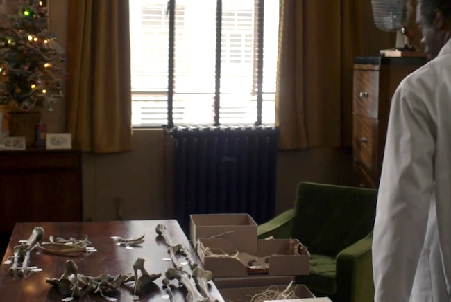
Image A
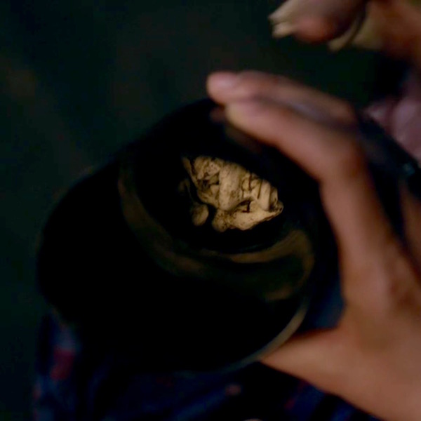
Dr. Abernathy reaches into the box and removes the skull (Image B).
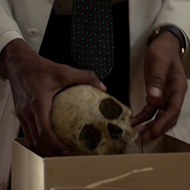
Image B
Examining the skull, the good doctor concludes it belonged to a pretty lady who was mature and middle-aged (Image C). And, from Voyager:
As the saying goes, “Pretty is as pretty does.” The owner of this skull definitely did not do pretty things! 😳
“Oh, pretty,” he said in delight, turning the object gently to and fro. “Pretty” was not the first adjective that struck me; the skull was stained and greatly discolored, the bone gone a deep streaky brown. Joe carried it to the window and held it in the light, his thumbs gently stroking the small bony ridges over the eye sockets. “Pretty lady,” he said softly, … “Full-grown, mature. Maybe late forties, middle fifties.”
Image C
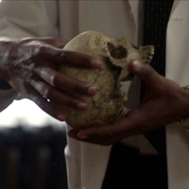
Clairvoyant Claire picks up the skull, which speaks to her (Image D). Well, not really – that would be weird – though Hallowe’en does draw nigh! Another quote from Voyager:
Then I held it close against my stomach, eyes closed, and felt the shifting sadness, filling the cavity of the skull like running water. And an odd faint sense—of surprise?
“Someone killed her,” I said. “She didn’t want to die.”
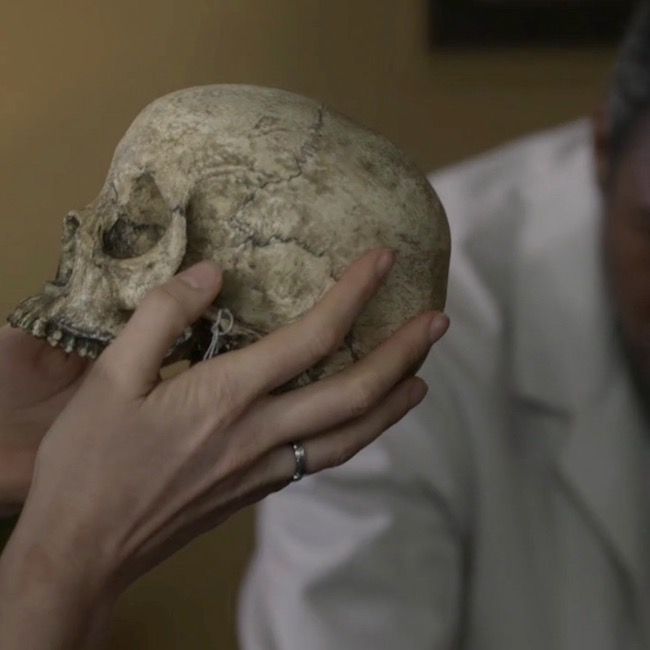
Image D
Dr. Abernathy fixes Dr. Claire with a gimlet eye. Lady Jane, have you lost your scalpels??? After all, how could she possibly conjure such details using a touchy-feely method of scientific inquiry? Well, the lass does harbor some awesome powers that seem to grow with time (Image E)? Again, from Voyager:
… “Where did you find her?” I asked….
“She’s from a cave in the Caribbean,” he said. “There were a lot of artifacts with her. We think she’s maybe between a hundred-fifty and two hundred years old.”
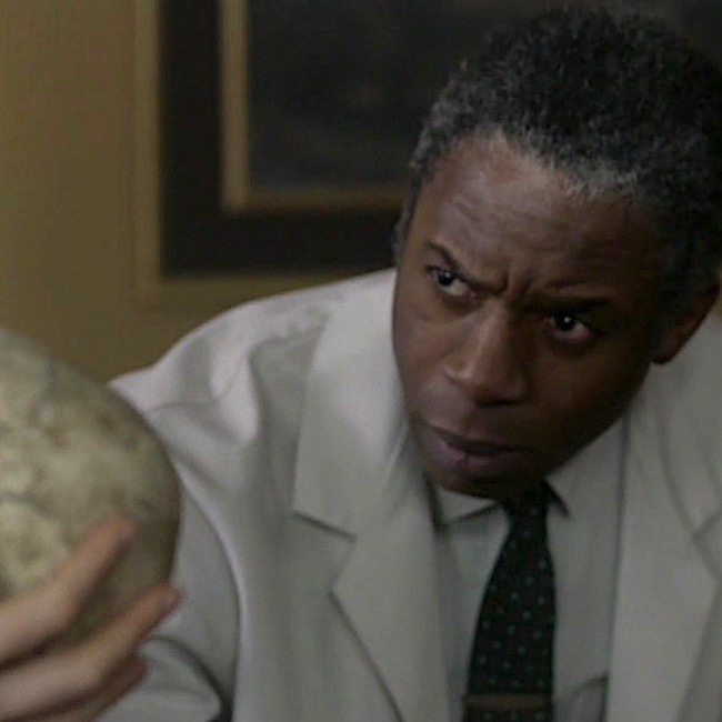
Image E
Next, Dr. Abernathy plucks two pieces of bone from the corny box (Image F). “You were right,” he says. Looking at them, he observes that the fracture plane runs through the centrum, although he doesn’t identify which bone has been fractured.
Voyager book reveals the fractured bone as a vertebra (bone of spine) – more specifically, it is the axis, aka the second cervical vertebra (C2).
The wide body of the axis had a deep gouge; the posterior zygapophysis had broken clean off, and the fracture plane went completely through the centrum of the bone.
Joe’s finger moved over the line of the fracture plane. “See here? The bone’s not just cracked, it’s gone right there. Somebody tried to cut this lady’s head clean off. With a dull blade,” he concluded with relish.
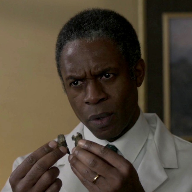
Image F
Then, Dr. Abernathy notes that although the burial site was a cave filled with slave artifacts, this lady was not a slave! He points to two leg bones (Image G), the tibia (Anatomy Lesson #9, The Gathering or Boar Gore) and the femur (Anatomy Lesson #7, Jamie’s Thighs or Ode to Joy!). Ahhh, Claire sagely nods, “the crural index.” Back to Voyager, again:
“Not a slave,” he said… “No,” Joe said flatly. He tapped the long femur, where it rested on his desk. His fingernail clicked on the dry bone. “She wasn’t black.”
“Take a look at this,” Joe invited. “You can see the differences in a lot of bones, but especially in the leg bones. Blacks have a completely different femur-to-tibia ratio than whites do. And that lady”—he pointed to the skeleton on his desk—“ was white. Caucasian. No question about it.”
…“If you want to think blacks and whites are equal under the skin, be my guest, but it ain’t scientifically so.” He turned and pulled a book from the shelf behind him. Tables of Skeletal Variance, the title read.
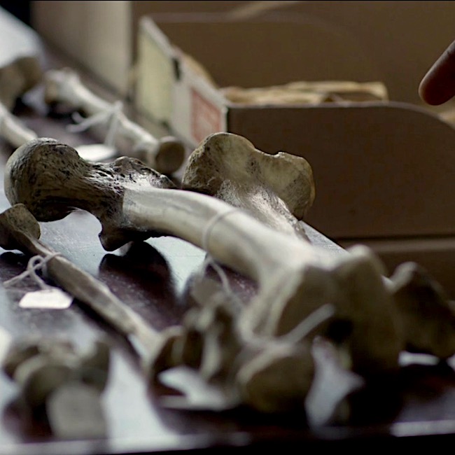
Image G
OK, that pretty much summarizes the salient scientific points of this scene, although I see three issues that warrant comment:
Issue #1: Can bones reveal sex, beauty and/or age of the owner?
The answer is a qualified yes.
When Claire arrives at the office, Dr. Abernathy has already laid out most of the skeleton and presumably, has already examined each of these bones. It’s pretty iffy to hazard a reliable guess with one measurement. But, if a series of measurements tend to fall within a given range, a forensic scientist can venture an educated guess. So, assuming he examined the skull and pelvis, then sex can be surmised with a resonable degree of confidence. Here’s why.
Sex & Skull: “Typical” female and male skulls exhibit differences (Image H). A female skull is usually smaller, with rounder orbits (red dot), prominent frontal eminences (yellow dot), smaller mandible (pink dot), less prominent temporal lines (green dot), smaller brow ridges (dark blue dot), and less pronounced glabella (turquoise dot), and the brow ridges are sharper. There are also other, more subtle differences, but understand that not all skulls show all structural differences, regardless of genetic sex.
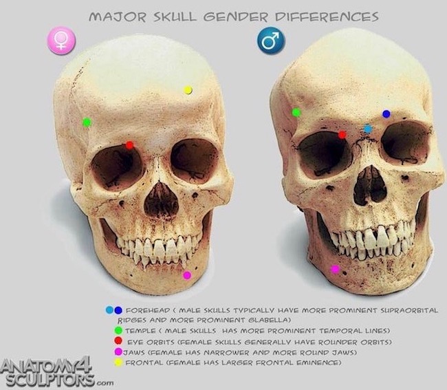
Image H
Sex & Pelvis: The bony pelvis also supplies important clues to sex. “Pretty lady’s” bony pelvis lies in three pieces (two hip bones plus sacrum). The pelvic width, shape of sacrum, sub-pubic angle and shape of obturator foramen (two front holes) are consistent with a female pelvis (Image I – obturator foramen not labeled). Pelvic inlet and outlet are difficult to demonstrate in a dissembled bony pelvis but, assembled they would be similar to those shown in Image I, lower figure. Ergo, pretty lady’s pelvic features are consistent with those of a female.
Age: Age can be estimated if cranial sutures (sites where cranial bones meet) are thin, bony ends are fused to bone shafts (growth plates are ossified), teeth are mature and bones are hard, along with the presence of wear-and-tear diseases, etc. So, yes, with careful analysis, general age can be estmated. Presumably, Dr. Abernathy considered these before Claire’s arrival. So far, so good!
Beauty: As we all ken, beauty is in the eye of the beholder. Dr. Abernathy expresses an opinion when he dubs the owner a “pretty lady.” Of course, he cannot know how flesh draped those bones, but he considers the skull to be delicate and beautiful to his practiced eye. This is a subjective response on his part, but it is arguably consistent with the appearance of the skull which is delicate with good teeth. Nowadays, forensic scientists can reconstruct a face using computer programs, or the older clay sculpting technique.
Thus, sex and age can be assessed with a fair degree of confidence if and only if multiple measurements and observations are considered, collectively. But, as beauty remains in the eye of the beholder, this issue receives a qualified yes.
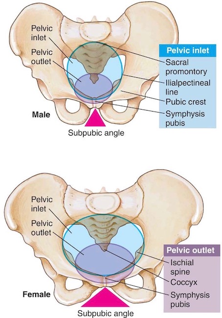
Image I
Issue #2: Does the fractured bone match with “pretty lady’s” death?
Because I have some issues with this issue, the answer is mostly yes for the book, but no for the episode. Here’s why. 🤓
Dr. Abernathy holds up a wee bone, which is broken vertically into two nearly symmetrical pieces. Voyager identifies these fragments as belonging to the axis. The axis is one of seven neck bones (numbered 1-7 from skull downwards); it is also designated as C2, meaning it is second of the seven cervical vertebrae. The purpose of cervical vertebrae is to support the head and supply flexibility to the neck, augmenting movements of the head and shoulders.
Voyager book states:
The wide body of the axis had a deep gouge; the posterior zygapophysis had broken clean off, and the fracture plane went completely through the centrum of the bone.
Although the bone fragments of shown in episode 305 are supposed to represent a vertebra, the fragments do not appear to be an axis. The axis is an atypical, weird-looking bone (Image J – gif):
Image J
However, please appreciate that the axis is a splendid bone, allowing us to rotate our head from side-to-side in a “no-no” gesture (Anatomy Lesson #12 Claire’s Neck – the Ivory Tower!).
Try this: Cervical vertebrae are buried rather deeply in the neck and difficult to demonstrate. But, if you able, sit up straight with chin level, place a finger in the groove of your neck just below the skull. You may feel a small bulge under your fingers. This is the spine of the axis.
Let’s examine the parts of the axis (Image K – front view) to help us understand the quote from Voyager.
To reiterate, Voyager book states:
The wide body of the axis had a deep gouge; the posterior zygapophysis had broken clean off, and the fracture plane went completely through the centrum of the bone.
Analysis: The book statement is a wee bit awkward because if the fracture plane passed through the centrum, it must necessarily cleave the body, so a deep gouge would not have been left to discern.
Also our vertebrae don’t have posterior zygapophyses – I suspect Diana intended inferior rather than posterior. Assuming this is correct, then: The dull blade cut horizontally (as in cutting off a head), breaking off the inferior zygapophyses (forming zygapophyseal joints with C3), and passing through the axial body and its centrum. Otherwise, the description makes sense.
One may also conclude, the blade must have passed through the lower part of the axis. Had the blade passed through its upper part, the stroke would have sheared off dens and superior zygapophyses but completely missed the centrum! Make sense? Yay! 👏🏻👏🏻👏🏻
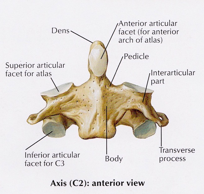
Image K
Conclusion: Dr. Abernathy holds two bony fragments of the fractured bone and I think, hum… that ring of bone appears to have been cleaved in halves by a vertical blow (Image L)! The only way a vertical fracture could occur is if the blade sliced downward, vertically cleaving the skull and its supporting cervical vertebrae. Gah! I think you will agree, this is pretty unlikely plus, the skull remains intact. So, no vertical swipe of the blade!
Ergo, although dramatic and interesting, the bone fragments do not reflect the axis damage as described in Voyager or by episodic Dr. Abernathy. Now, does all this keep me up at night? Hardly – I loved this scene despite its wee anatomical issues!
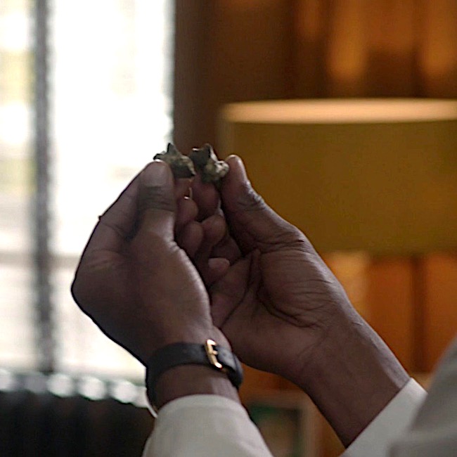
Image L
Issue #3: Can crural index determine race?
The answer is a qualified no.
This topic is a super sticky-wicket but very important to consider, so bear with me.
Best start with a definition: the crural index, established in 1933, is the ratio of tibia-to-femur (not femur-to-tibia, as this yields different results). The formula to determine crural index is:
(length of tibia x 100) / length of femur OR
(length of tibia/length of femur) x 100
Using this ratio, many studies showed that individuals of African descent had higher crural indices than those of European descent. Image M is a simplified, summary version of such findings.
In 1968, Dr. Abernathy was positive that a low crural index meant “pretty lady” was white….“No doubt about it.”
(BTW, I am pretty sure Claire pronounces this “cruel index;” but, it should be crural <kru-ral>, from the Latin crus meaning “leg”)
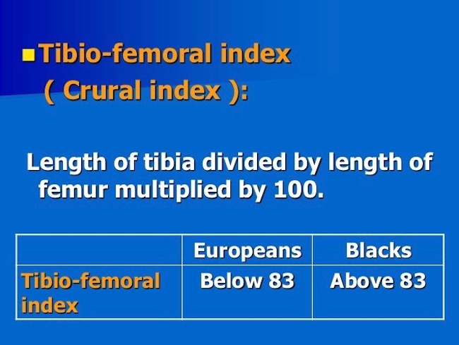
Image M
So, can race be determined from the crural index?
When I started graduate school in 1965, we were taught there were three human races: negroid, caucasoid, mongoloid. Fast forward 52 years and much has changed! Today, many biologists say race cannot be determined from bones because there is no such thing as race. These scientists posit that all living humans belong to one species (Image N): Homo sapiens sapiens (the second sapiens denotes the subspecies – that would be us).
Many designate the term ancestry, because race and even ethnicity have confusing connotations and definitions. Furthermore, they point out, more genetic variations can occur within “racial” groups than between them, meaning findings are limited by the sample studied. What a conundrum!
Just to clarify, some bony physical traits are characteristic of ancestry and can be traced to a particular global location. But, bear in mind, people of mixed ancestry may present features which do not fall neatly into any category. Also, humans are so similar that all bone morphologies are present in all groups, just at varying rates. Despite such variations, skeletal analysis remains part and parcel of human identification especially when numerous skeletal measurements are obtained. Today, using calipers, x-rays, microscopy, DNA, and a mess of other tools, some of which were unavailable in 1968, forensic researchers can make reasoned guesses as to a person’s ancestry based on skeletal remains.
Summary: Nowadays, before a scientist suggests ancestry based on skeletal remains, (s)he makes multiple measurements, never relying on just one. And, prudent scientists avoid stating “we are sure” (even if they are). Instead, they posit, the data suggests or indicates or is consistent with or is likely. Verra prudent!
Hence the qualified “no” regarding the crural index; it is only one skeletal measurement and insufficient to make a judgement if a person was white or a not. However, Voyager accurately expresses views prevalent in the 1960’s. Make sense? Ta da!
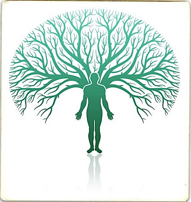
Image N
Bottom line:

Image O
So, this concludes a brief analysis of the “skeleton scene.” Much more could be added, but would likely be too technical for most students. Hopefully, this summary was enlightening and will generate some thoughtful discussion and consideration in our ever expanding Outlander world. Buh-bye, pretty lady!
Ode to Pretty Lady
Your bones tell a tale.
Who are you?
Were you well?
Were you pretty?
Were you witty?
Were you sweet?
Did you cheat?
Were you bad?
Were you sad?
Or, were you mad?
Your bones tell a tale.
No Spoilers! Who are you “pretty lady?” Mayhap, we will find out during Outlander S.3!
The deeply grateful,
Outlander Anatomist
Follow me on:
Photo Credits: Sony/Starz (Images A-G, L, O), Clinically Oriented Anatomy by Moore and Dalley, 5th edition (Image K), www.pinterest.com (Image H), www.medical-dictionary.thefreedictionary.com (Image I), www.premiersourceshopping.com (Image O), www.slideshare.net (Image M), www.wikipedia.com (Image J – gif)
Hello, Outlander anatomy students, and welcome to today’s Anatomy Lesson #39, the Human Skeleton. This is a whopping subject so it will take two lesson to cover the bones!
Dem bones or dem dry bones refer to a spiritual song inspired by a vision recorded in the biblical Book of Ezekiel, 37:1-14. Ezekiel stands in a valley filled with dry human bones. Before his eyes and with the promise of hope, the bones join into human skeletons which become enshrouded with flesh. This wonderful spiritual has been rewritten for children:
Skeleton Lesson over! Naw, just kidding. The skeleton is a wee bit more complicated than the song lets on.
As if on cue, Starz Outlander team offers up a S.2 treasure trove of bone images just in time for our skeleton lesson.
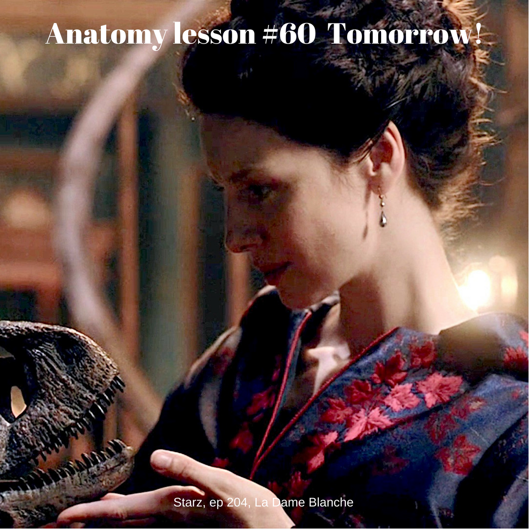
Hum, look again, Claire, that cup isn’t half empty – it’s half full (Starz episode 204, La Madame Blanche)! Thank you, Outlander!
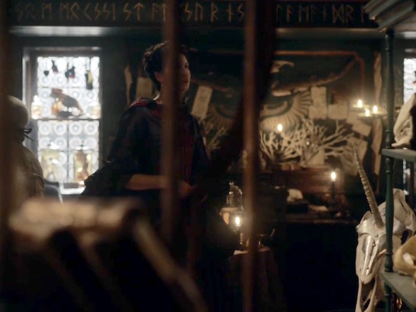
Claire glides into Master Raymond’s secret “little shop of horrors” overflowing with marvelous skulls from real and imagined beasties (Starz episode 204, La Madame Blanche). His wonderful ossuary includes a unicorn skull (lower right) embellished with head armor (chanfron for horses) complete with horn hole! A whimsical nod to the national animal of Scotland, no doubt. Love it!
Diana elaborates in Dragonfly in Amber.
Two walls of the hidden room were taken up by a honeycomb of shelves, each cell dustless and immaculate, each displaying the skull of a beast. …Tiny skulls, of bat, mouse and shrew, the bones transparent, little teeth spiked in pinpoints of carnivorous ferocity. …They had a certain appeal, so still and so beautiful, as though each object held still the essence of its owner, as if the lines of bone held the ghost of the flesh and fur that once they had borne.
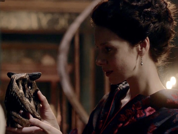
Claire examines an unusual skull, perhaps the remains of a horned carnivorous dinosaur such as carnotaurus (Latin meaning flesh bull). There are loads of horny creatures on Outlander. Ha, ha. Alas, such animals are no more declares Master Raymond. True, unless one takes a Spielberg detour to Isla Nublar!
Goddess of the Pen continues in DIA:
I reached out and touched one of the skulls, the bone not cold as I would have expected, but strangely inert, as though the vanished warmth, long gone, hovered not far off. ..“You see the teeth? An eater of fish, of meat”—a small finger traced the long, wicked curve of the canine… “Such beasts are no more, madonna.”
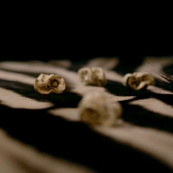
Interested in old stuff, Master Raymond might like this bony specimen for his awesome collection (Image A), a 130,000 y.o. Neanderthal skull. Found in a limestone cavern near Altamura, Italy, the bones are shrouded with cave popcorn, limestone formations caused by splashes of mineral-rich water. Wondrous!
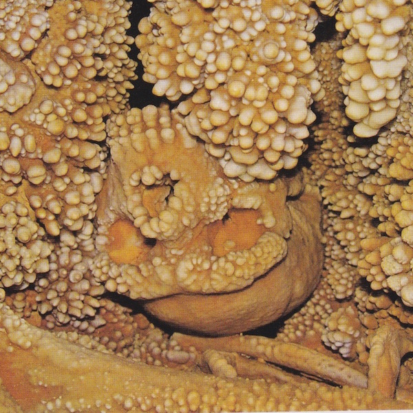
Image A
Master Raymond and his coy toys are endlessly fascinating, but it’s time to study the skeleton.
Gross anatomy (Anatomy Lesson #34, “History of Anatomy”) teaches bones and their relationships as they appear in the visible skeleton. We haven’t been exactly idle as prior anatomy lessons have discussed many individual bones. Some of these will be referenced in this lesson. Microscopic anatomy (Anatomy Lesson #34) studies bones as organs and tissues. Slightly different approaches and today’s lesson considers both.
Adult Human Skeleton: The word skeleton derives from the Greek skeletós, meaning “dried up”, because long after flesh has withered, the skeleton steadfastly remains (remains, get it? Hee hee). However, despite the definition, our skeletons are very much alive!
The skeleton forms the supporting structure of an organism. Some creatures, such as the Japanese beetle (Image B), have an exoskeleton (Greek exo- meaning outside), a stable outer shell to protect delicate innards, a type of organic armor. But exoskeletons present a couple of major disadvantages as grown and movement are limited.
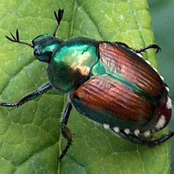
Image B
We humans don’t walk around with our skeletons exposed – unless we suffered a Randall-scandal with BJ! Rather, humans enjoy an endoskeleton (Greek endo- meaning within); our skeleton lies inside the body, wrapped in flesh. The endoskeleton is a genuine boon because it gifts its owner with freer movement and growth potential.
The skeleton (Image C) is a composite of all bones in a human body. It is also heavy, accounting for 20% of our total body weight. The adult human skeleton includes 206 individually named bones whereas, the infant human skeleton contains about 300. The overall count drops during maturation as many bones fuse (e.g. skull bones), usually completing the process after three decades of life.
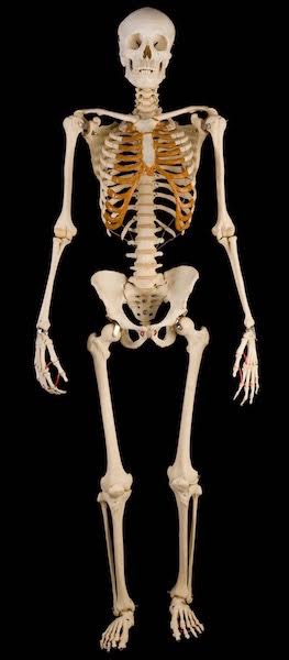
Image C
Interestingly, the number of bones comprising the adult skeleton is always higher than 206, but, because some bones are not present in all people, are small, or are variable in number, they are excluded from the overall count. A couple of good examples follow.
Humans have small sesamoid bones (Latin meaning like a seed) housed within tendons of thumbs and great toes (Image D) where they influence the pull of muscles. These are not counted. The paired patellae (pl., knee caps) are the largest and best known sesamoid bones but, because of size and constancy, these are routinely included in the 206 count.
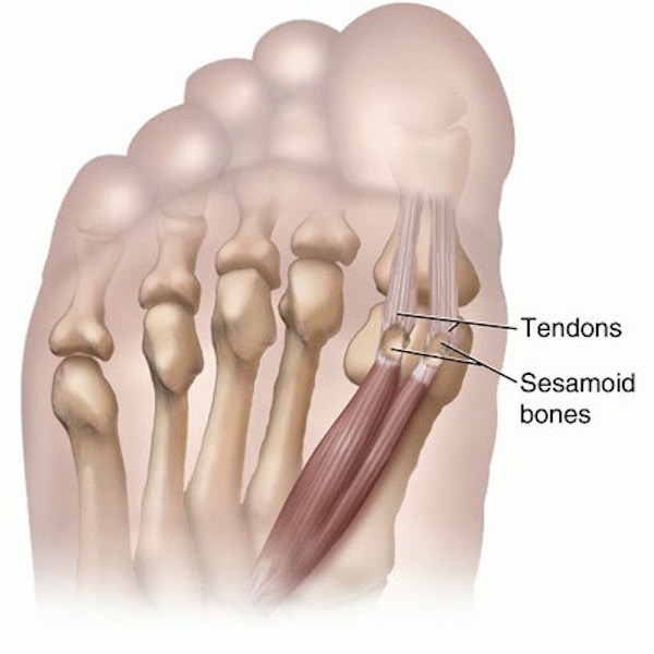
Image D
Another example of bones eliminated from the count are small, irregular wormian bones, which develop in sutures of the skull (Image E – red arrows). Although not rare, most skulls do not have them. Wormian bones can be markers of diseases such as brittle bone, but in normal individuals, their significance is unknown.
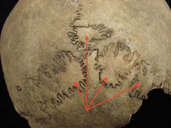
Image E
Back to the adult skeleton. The skeleton is divided into axial (Image F – yellow) and appendicular (Image F – green) parts. The axial skeleton includes bones of skull, vertebrae, sacrum, coccyx, ribs, and sternum, for a total of 80 bones. The appendicular skeleton houses bones of upper and lower limbs, including clavicle, scapula, and hip bones, for a total of 126.
Notice this, all bones of the adult appendicular skeleton are duplicated on each side of the body: two femurs, two humeri, (pl.) etc. However, the adult adult axial skeleton is variable: ribs and some facial bones are duplicated but all remaining axial bones are singular, lacking a counterpart. Three pairs of ear ossicles are good examples of duplicated skull bones (Anatomy Lesson #25, “If a Tree Falls – The Ear”). Whereas, the frontal bone of the skull is unpaired (Anatomy Lesson #11, “Jamie’s Face” or “Ye do it Face to Face?”).
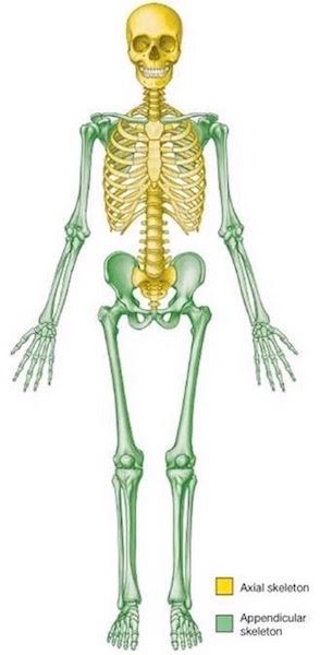
Image F
Bones are Secure: Bones of the skeleton don’t swing in the breeze; they are joined by connective tissue elements. Unmoveable joints (think skull sutures) are united by strong layers of collagen. Moveable joints (think elbows – Anatomy Lesson #20, “Arms! Arms! Arms! – Redux”) are sites where adjacent bones move on each other. To be more precise, the articular cartilages of such bones move on each other.
I cringe when my yoga teachers mention the bad “bone-on-bone” plank position. They really don’t mean this because bone-on-bone means the articular cartilages are worn away ensuring arthritis as one’s constant companion!
Image G shows the moveable knee joint, one of the largest and most complex of the body. The femur (Anatomy Lesson #7, “Jamie’s Thighs” or “Ode to Joy!”) and tibia (ditto) are stabilized by four major ligaments, muscle tendons, joint capsule (collagen again), and shock-absorbing menisci (pl.). Similar anatomical elements (sans menisci) compliment all moveable joints.
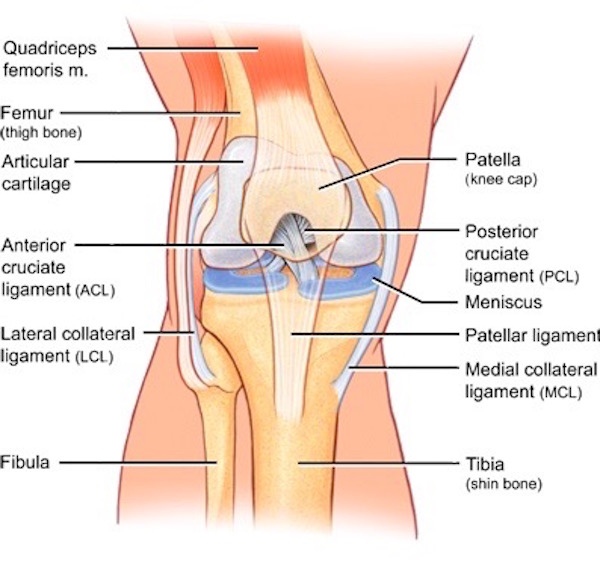
Image G
Bone Shape: Bones are classified into four categories based on shape: long, short, flat, and irregular (Image H). Most anatomists eliminate sesamoid and suture (wormian) bones from shape categories but all agree on the following four.
Long bones are longer than they are wide and are mostly confined to the appendicular skeleton where they engage in weight bearing and movement. Examples are femur, humerus, and phalanges. The femur (Anatomy Lesson #7, “Jamie’s Thighs” or “Ode to Joy!”) is the skeleton’s longest bone.
Short bones are as wide as they are long (cube-like), providing support and stability. Good examples are carpals of wrist (Anatomy Lesson #22, ”Jamie’s Hand – Symbol of Sacrifice”). The stapes (Anatomy Lesson #25, ”If a Tree Falls – The Ear”) of the middle ear is the skeleton’s shortest bone.
Flat bones are expanded into broad, flat planes. They protect underlying elements and/or provide wide muscle attachments. Good examples are bones of cranium, scapula, and sternum (Anatomy Lesson #15, “Crouching Grants – Hidden Dagger”).
Irregular bones have peculiar shapes; they provide protection and muscle attachment. Vertebrae and facial bones are good examples.
Now, the shape classification scheme is not without controversy as some bones cross boundaries, sharing elements of more than one category…. think of the flat and oddly-shaped scapula (Anatomy Lesson #2, “When Claire Meets Jamie” or “How to Fall in Love While Reducing a Dislocated Shoulder Joint!”).
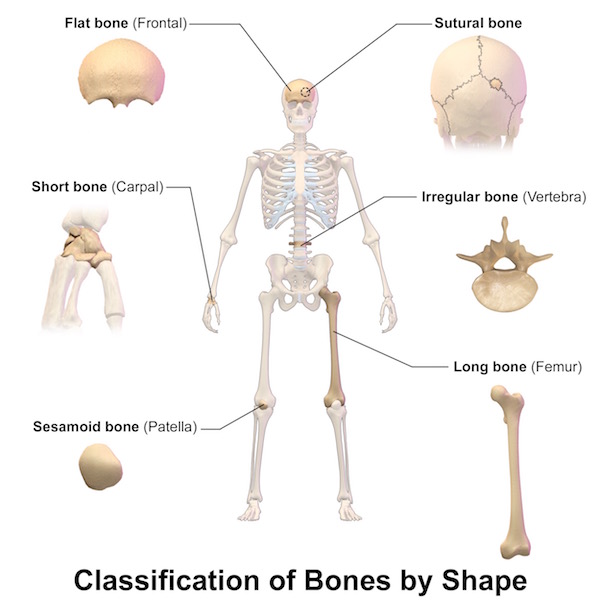
Image H
Bones as Organs: Now for some microscopic anatomy… an organ is a group of tissues that perform a function or group of functions. Like heart, lungs, liver, and brain, our bones are also living organs.
Bones are characterized by hard outer shells and “spongy interiors.” The outer shell is compact (cortical) bone; the interior is spongy (cancellous) bone, a distribution best illustrated using the femur (Image I). BTW, spongy bone is so named because it is riddled with holes, not because it is soft and pliable like the animal known as a sponge.
Parts of a Long Bone: Now for a crash course in bone anatomy. A long bone displays shaft (diaphysis), marrow cavity, and articular (epiphyses) ends (Image I). A connective tissue periosteum envelops the shaft and is richly endowed with pain fibers; anyone who has broken a bone kens this very well! Depending on the bone and age of its owner, the marrow cavity is filled with either fat (yellow marrow) or blood-forming tissue (red marrow). Both epiphyses are covered with smooth, firm articular cartilage (blue in Image I), which augments movement at the joints. Cancellous bone is abundant deep to the articular cartilage caps. Epiphyseal (growth) plates separate epiphyses (pl.) from shaft.
Growth plates are sites where long bones grow in length. Long bones continue lengthen until growth plates ossify, about two years after the onset of menstruation in girls and late teens for boys (there are variations). Whew, a packed mini-lesson!
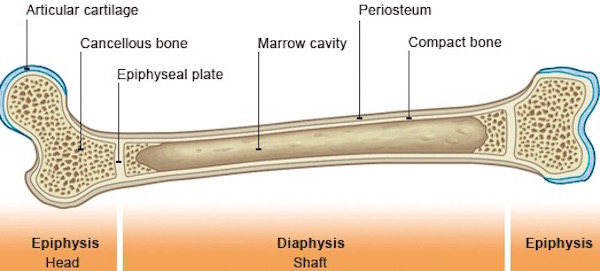
Image I
Bone as Tissue: Named bones are organs, but bone is also classified as a type of connective tissue. Tissue is an aggregation of similar cells and extracellular material acting together to perform specific functions. Connective tissues range from fluids, such as lymph and blood, to semi-solids or solids such as cartilage and bone.
As a tissue, bone is a composite of organic proteins and inorganic bone mineral. The organic protein is collagen, the most ubiquitous structural protein of the human body. Collagen is abundant in bone where it acts as a scaffold for the deposition of minerals. Inorganic bone mineral is made of hydroxyapatite, tiny crystals of calcium, phosphate, and magnesium which endow bone with rigidity. Understand that normal deposition of bone minerals is a complex process involving several hormones (e.g. calcitonin), dietary calcium, phosphorus, and magnesium,, and sufficient Vitamin D (via sunlight and/or supplements)!
Tidy Test: This bitty bone test demonstrates the relative roles of organic and inorganic components of bone. Bake a raw chicken bone at low temperature for a few hours. Immerse another raw chicken bone in acid (vinegar works OK but something stronger is better) for many days. Baking destroys collagen (organic protein) leaving the bone brittle and friable; it readily snaps in two. Soaking removes inorganic minerals leaving a rubbery bone that can be easily bent. I don’t expect you to try this demonstration, but it has been done for many years in school science labs.
A dry femur (Image J), offers a superb example of how bony tissue is organized. Covering the surface is a rind of hard, cortical bone of varying thickness. The cartilage covering the epiphysis is absent. The interior of the epiphysis is filled with spongy bone, a lattice of thin, hard bony shards. Spaces in the marrow cavity (red arrow) and amid the spongy bone are filled with either blood-forming tissue or fat, depending on the bone and one’s age.
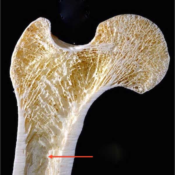
Image J
Time for another outlandish image (Starz episode 204, La Madame Blanche). No, lass, dinna ask Master Raymond about future Frank! Dem bones, dem bones, goin’ talk about… you don’t want to know the answer!
Which brings us to the very entertaining topic of sexual dimorphism. Don’t ask how I made that leap, it just seems to fit here! <G>
Sexual Dimorphism: Like many primates, the adult human skeleton exhibits sexual dimorphism; differences in form based on sex. Male skeletons are typically larger, heavier, and more massive than those of the female. There are also gender differences between some skull bones, canine teeth, and long bones (e.g. femur). But, the most reliable difference in discerning gender is via the bony pelvis. In 95% of cases, a skilled observer can assign an adult bony pelvis to the correct gender, although such differences are not evident before puberty.
To understand these sex-based differences, we must first consider anatomy of the bony pelvis (Image K), a ring formed by sacrum (yellow) and two hip bones (peachy-red). These three bones are held together by some of the body’s strongest ligaments. Why? Because, they bear the entire weight of torso, head and upper limbs, more than half our body weight!
BTW, I can immediately discern (with ~ 95% accuracy) that image F illustrates a female bony pelvis. You’ll understand how in a moment.
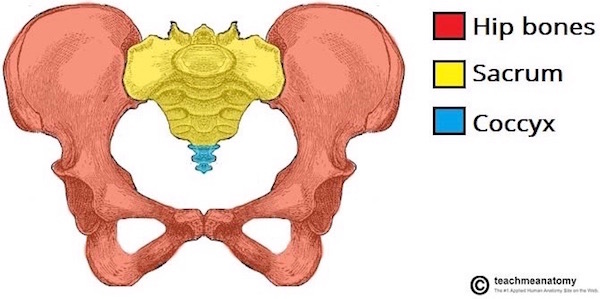
Image K
Another consideration about the bony pelvis: at birth, each hip bone consists of three separate bones, ilium, ischium, and pubis, joined together by cartilage – one reason why there is righteous concern over young children doing rigorous gymnastics. By age 25, the three bones of each side, fuse and ossify into a single hip bone (Image L – right hip bone). From a lateral (side) view, the three bones meet at the acetabulum, the socket for the femoral head.
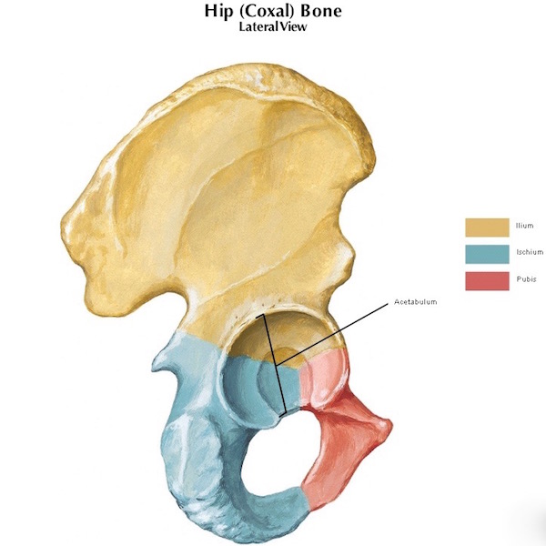
Image L
Pubic bones join in the midline at the pubic symphysis (Image M). The ilia (pl.) form relatively immovable joints with the sacrum at the infamous sacroiliac (SI) joints (Image M – black arrows). The top opening of the bony pelvis is the pelvic inlet (Image M – dashed black oval). Flip the bony ring upside down and the bottom opening is the pelvic outlet (not shown, so use your imagination). The space beneath the pubic symphysis is the sub-pubic angle (Image M – red arrow).
Interestingly, if x-ray reveals a break in the bony pelvis, there will always be at least one or more additional breaks. Try breaking a round pretzel… one cannot break just one side… same with the bony pelvis.
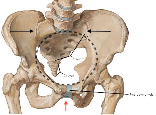
Image M
Now that we understand the bony pelvis, back to sexual dimorphism… Table A summarizes typical differences between adult male and female bony pelves (pl.). Although there are always outliers, female pelves generally express traits that augment pregnancy and childbirth.
TABLE A
| Males | Female |
| Thick and heavy | Thin and light |
| Narrower, taller pelvis | Wider, shallower pelvis |
| Sub-pubic angle < 90° | Sub-pubic angle > 90° |
| Pelvic inlet heart-shaped | Pelvic inlet oval and rounded |
| Sacrum tilted forward | Sacrum tilted backward |
| Small pelvic outlet | Large pelvic outlet |
Following puberty, the female bony pelvis grows becoming wider and shallower than the male pelvis (compare Table A with Image N). Let’s consider these differences.
The female sub-pubic angle is typically 90° or greater (Image N – green inverted V). The male sub-pubic angle is less than 90° (Image N – red inverted V). If one opens the space between thumb and index finger to a right or 90°angle; this is the typical extent of the female sub-pubic angle. If one spreads index and middle fingers as far apart as possible; this angle is less than 90° and is typical of the male sub-pubic angle. This is how anthropologists, forensic experts, and anatomists quickly identify the gender (historically, anatomists use the terms sex and gender interchangeably) of a bony pelvis.
Next, the female pelvic inlet is usually large and oval-shaped (Image N – top, right); the male pelvic inlet is small and heart-shaped (Image N – top, left).
Lastly, the female sacrum typically tilts backward such that the pelvic inlet of a standing woman faces mostly forward. Because the male sacrum tilts forward, the pelvic inlet faces mostly upwards.
Pop quiz! Return to Image F and determine if the yellow and green skeleton belongs to a female or a male. Answer follows Image N.
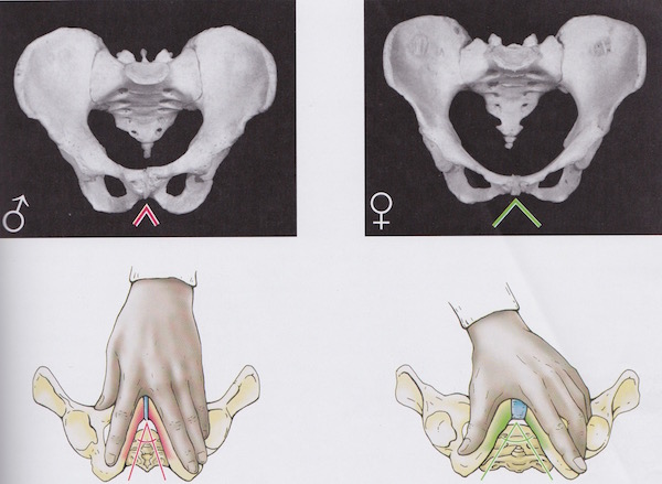
Image N
If you answered female, you are ready for a guest spot on one of those forensic TV shows. Congrats!
We now understand anatomy of the skeleton but what purpose does it serve? Well, it actually serves at whopping six purposes:
Support: Our flesh in the form of muscles, ligaments, tendons, blood and lymphatic vessels, nerves, and so forth, enshrouds the skeleton. If we did not have an endoskeleton for support and movement, we might look something like Mr. Blobfish (Image O), a deep sea fellow living off the coasts of Australia, Tasmania and New Zealand!
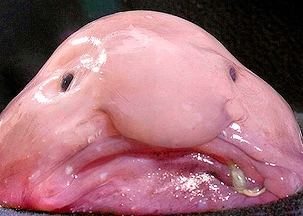
Image O
Movement: The body contains three different types of muscle: smooth, cardiac and skeletal. Skeletal muscles attach to bones via origins and insertions. As a skeletal muscle contracts, it moves the bone(s) to which it attaches. It goes without saying that movement allows us to negotiate our environment for survival.
We have over 700 named skeletal muscles accounting for over half our body weight! The foot of a running man from a 2008 Body World’s exhibit (Image P) shows 12 muscles (there are more that are not shown) of the right foot and leg engaged in lower limb locomotion… add the left leg and foot and the number doubles! Add muscles of thigh and buttocks and, the numbers continue to climb. What a wonder!
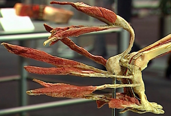
Image P: Running man KLS edited
Protection: The skeleton offers sanctuary for vital organs such as brain and heart (Image Q). Seated in its bony thoracic cage (Anatomy Lesson #15, “Crouching Grants – Hidden Dagger”), all sides of the heart except its diaphragmatic surface are surrounded by bone. Ditto for trachea (most of it), lungs, bronchi, aorta, kidneys, brain, pituitary gland, eyes, tongue, etc., etc., etc. Our well-being has a vested interest in preserving vital organs from injury so surrounding them with bone is an ingenious devise.
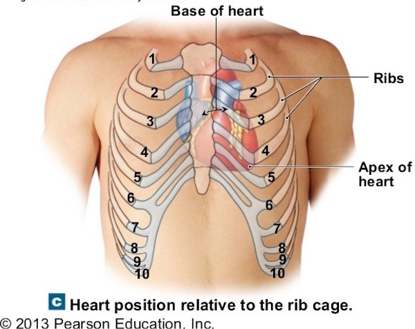
Image Q
Hematopoiesis: Aaaah… What does this term mean? Hematopoiesis means the production of blood cells (Image R). Greatly simplifying a very complex process, circulating blood contains six classes of blood cells plus platelets (Anatomy Lesson #37, “Outlander Owies! – Part 3, Mars and Scars”), all of which arise in bone marrow. Side note: one class of blood cells (lymphocytes) also develop during immune responses in sites outside bone marrow.
Adding another layer of complexity, hematopoiesis is a tortuous process which varies throughout life (Image R). In utero, blood cells arise in the yolk sac (human embryos have one), liver, spleen, lymph nodes, and by the third trimester, in bone marrow. In children, the entire skeleton is engaged in hematopoiesis (it stops in the earlier organs). By adulthood, hematopoiesis confines itself to the ends of long bones and the axial skeleton. Sadly, in the aged, hematopoiesis declines even more, leaving elderly people challenged to produce enough blood cells for good health (exercise helps thwart this decline). Interestingly, blood-forming potential is retained such that under rare conditions, adult liver and spleen can resume hematopoiesis.
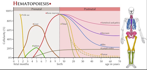
Image R
Storage of Minerals: Remember calcium, phosphate, and magnesium that crystalize into bony hydroxyapatite deposits? As we know, these microscopic crystals form either compact bone or spongy bone (Image S – spongy bone). Either way, given the proper signals, the stored minerals can be mobilized from bone and released into the blood stream for other needs in the body. Thus, bones play an important role as storage depots for minerals.
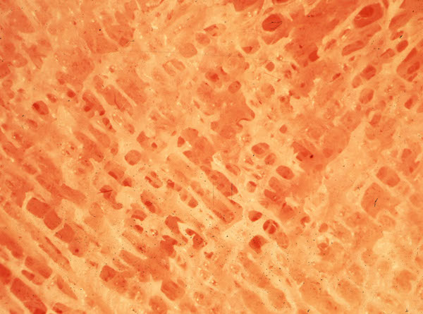
Image S
Hormone Regulation: Last but never least, bone plays a hormonal role. Yes! Bone cells mostly in the medullary (marrow) cavity (Image T) produce two hormones. One, phosphatonin, targets the kidneys causing them to increase phosphate loss in urine thus regulating levels in the blood stream. A second, osteocalcin, stimulates pancreatic cells to release insulin and testicular (Leydig) cells to release testosterone. Ergo, bones are awesome, low-paid multi-taskers!
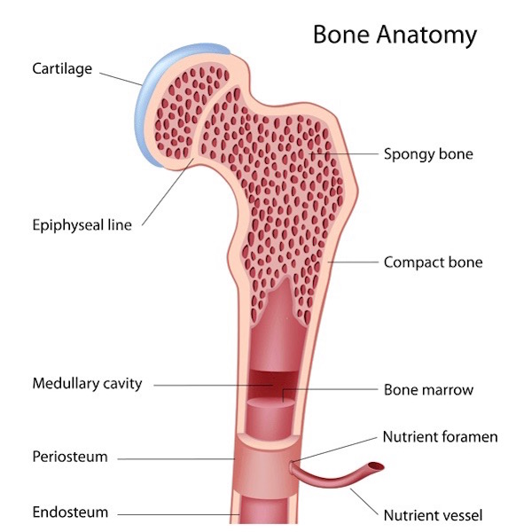
Image T
One last point before this lesson goes bye-bye. What about teeth? Where do they fit into the bony scheme? Most anatomists group teeth with the skeleton, in part because like the skeleton, they remain after all other tissues succumb. And, like bone, tooth enamel is made of specialized hydroxyapatite crystals, although harder, as enamel is the hardest substance in the body.
But, the real reason teeth are saved for the end of this lesson is because the devil made me do it! Yep, BJR is my “go to” guy (Starz episode 206, Best Laid Schemes)!
Moralizing Moment – S.2… Dueling with Jamie, the Snap-Dragon eschews codes duello as he sinks a Munch-Crunch into Jamie’s right arm (puir Jamie, he gets bitten a bunch in S.2)!
A duel bite? Shocking ! What “officer and gentleman” would disarm an opponent’s arm via a carnassal-chomp?
Where’s Murtagh, Jamie’s second, to demand the “field of honour” remains honorable? Sadly, Godfather is long gone – off to Portugal selling hijacked wine.
This is a perfect spot for Moralizing Moment – S.1. Something has been bugging me for months!
Some high level Outlander folks once opined that BJ has a “code of honor” because he kept his word, allowing Claire to escape Wentworth in exchange for Jamie’s surrender. Och! I beg to differ. He is as despicable about that “promise” as about Battle-Bites.
I posit that Claire was “dishonorably discharged” from that hell-hole. Does shoving an unsuspecting lass down a 3-4 meter shaft seem honorable to you (Starz episode 115, Wentworth Prison)? Snort!
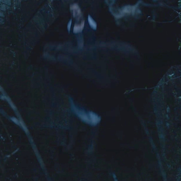
We have come to expect mind-boggling acts from Jack-the-Nipper – that fall could have broken Claire’s back. And, ugh, he pushes her into Wentworth’s garbage/dead body dump, where she finds Taran. We miss you big guy!
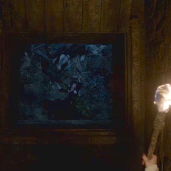
Holding a torch aloft, Jack-Jaw surveys his handiwork. Seeing Claire move, he can now “honorably” inform Jamie that his beloved has “left the building” (Starz episode 115, Wentworth Prison)! Ruadh, being a man of honor, honors his “end” of the bargain. Moral to the story: never dance with that dishonorable devil!
But, take comfort, budding anatomists……. Diana reminds us in a quote from Dragonfly in Amber that love redeems all, even dem bones (and teeth) – ! Yay, the skeleton scores!
” ’Blood of my blood, bone of my bone …’” “I give ye my body, that we may be one,” he finished. “Aye, and I have kept that vow, Sassenach, and so have you.” He turned me slightly, and one hand cupped itself gently over the tiny swell of my stomach.
Whose your daddy (Starz, episode 206, Best Laid Schemes)? Hee hee!
“Let us rise up and be thankful. For if we didn’t learn a lot today, at least we learned a little… ”
-Buddha
Next lesson: how bones heal.
See you later, alligator….. that creature hanging in the apothecary shop. Oops! It’s an “after a while crocodile” dangling from Raymond’s rafters! Ta-ta!
A deeply grateful,
Outlander Anatomist
Photo creds: , Starz, Sony Pictures, Archival image from Outlander Anatomy collection (Image S), National Geographic Magazine, March 2016 (Image A), Body Worlds specimen (Image P), Frank H. Netter, 4th ed., Atlas of Human Anatomy (Image L; Image M), Kieth L. Moore and Arthur F. Dalley, 5th ed.,Clinically Oriented Anatomy (Image N), YouTube (dem bones), www.anthropology.si.edu (Image C), www.arbordoctor.net (Image B), www.archive.museumoflondon.org.uk (Image E), www.bbc.co.uk (Image I), www.en.wikipedia.org (Image H; Image R), www.highlands.edu (Image J), www.nim.nih.gov (Image T), www.skeletalsystemdev.weebly.com (Image F), www.slideshare.net (Image Q), www.teachmescience.info (Image K), www.theinertia.com (Image O), www.theknee.com (Image G), www.wikiradiography.net (Image D), Walt Disney’s Fantasia.