Anatomy Lesson: Mandy’s Malady

Welcome all anatomy students! It has been a while since I posted a lesson because, frankly, I have been dealing with my own malady – a shattered left ankle! Six months out and beginning to feel and function better. 🥳

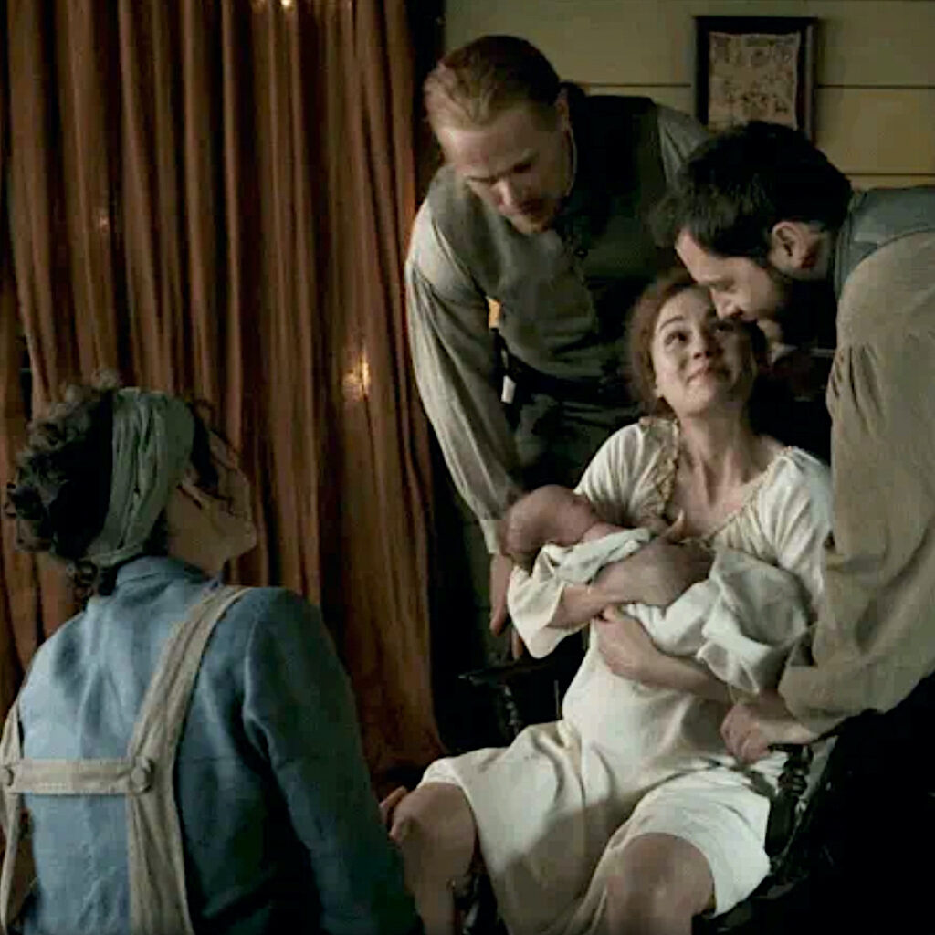
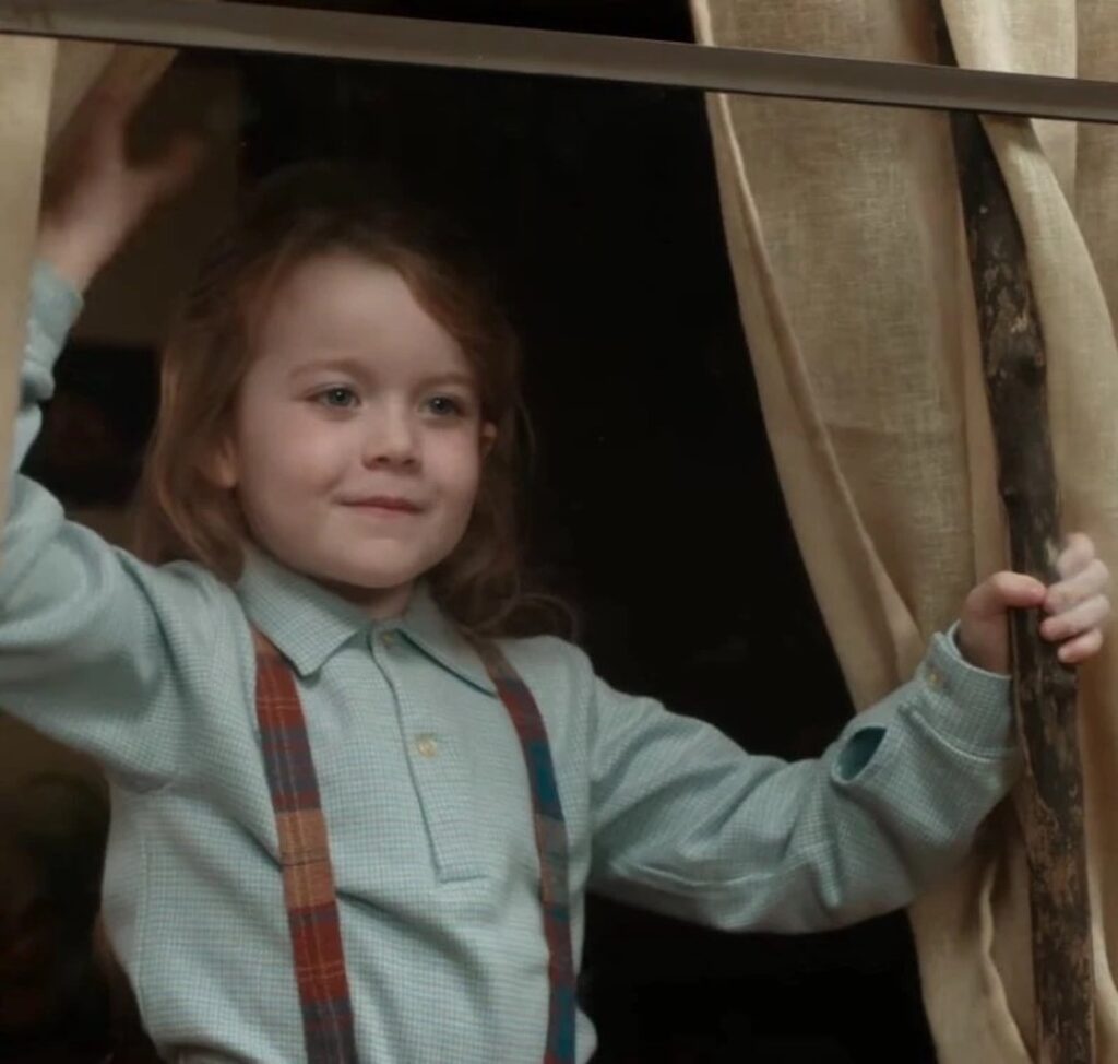
Outlander fans recall that in episode Episode 702, “The Happiest Place on Earth,” Brianna gives birth to her second child, Amanda Hope Claire MacKenzie-Fraser. Dr. Claire is there to reassure Brianna and ease the process. The wee one is adored by all and affectionately nicknamed, Mandy.  Fraser’s Ridge is the happiest place on earth and all is well! 🥰

Young Mandy is quickly introduced to the Ridge’s denizens as Granda’ Jamie takes her on a wee stroll to meet a new foal at the stable. Do you see it, Mandy? It is a cutie-beauty like you! 😍 

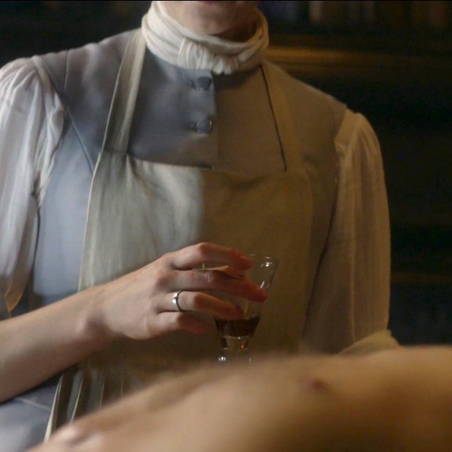
Mandy’s doting Granny takes her on a guided tour of Claire’s fav room – the surgery! Mayhap she will follow in Granny’s footsteps? 👩🏻‍⚕️

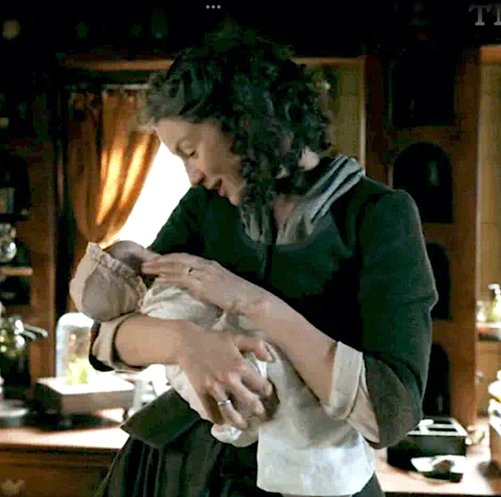
Claire coos to Mandy, admiring the beautiful, wee lass. Then…. she sees Mandy’s fingernails. A closer look and a startled Claire exclaims: “Bloody hell!”  (Couldn’t have said it better myself)  

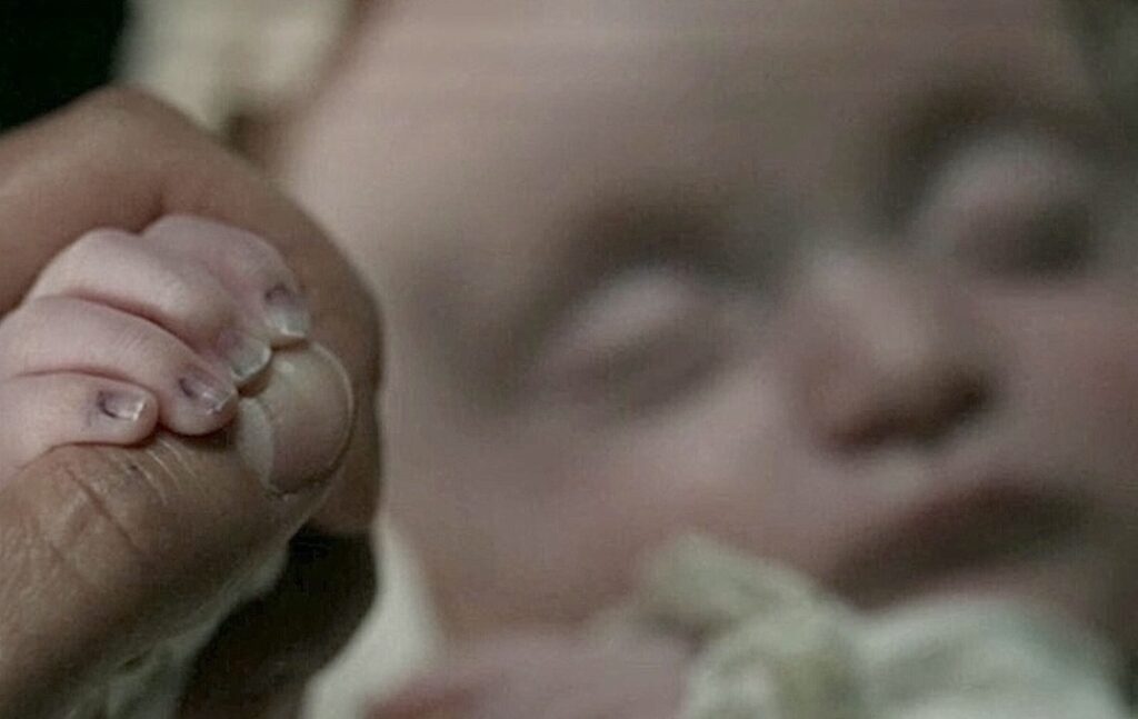
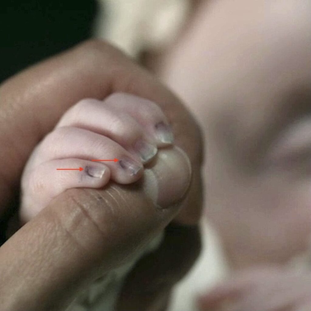
Ever observant Claire spies a bluish tinge at the base of Mandy’s wee fingernails (below, red arrows)!

Fun Fact: This is not hyperbole.  Fingernails and toenails give clues to at least a dozen possible medical conditions that require evaluation and followup. And, Mandy is no exception.

A quote from Diana’s 6th big book, “A Breath of Snow and Ashes,” documents the moment Claire observes Mandy’s nail beds: ”

“The minute nails were faintly tinged with blue.” 

Bree senses Claire’s concern and asks what is wrong.  She kens that look on her mother’s face. After Claire explains, Bree reports her own maternal observations: Mandy does not nurse well nor is she gaining weight like Jem. What is wrong? 😯

Claire determines that the wee lass has a heart defect requiring more advanced care than Claire is able to provide in the 1700s. She is clear that Mandy’s Malady is life-threatening and she likely will not survive for long without corrective cardiac surgery. Brianna and Roger decide to return with their children to the 20th century to obtain the necessary care to save Mandy’s life.

Arrangements are made for the MacKenzie family to travel through time at the standing stones on Ocracoke Island. Everyone agrees this is the best course of action, but this time and place is no longer the “happiest place on earth!” Will they ever see Fraser’s Ridge and one another again? 🤷🏻‍♀️

Time to start our lesson….What leads Claire to her startling diagnosis? The following are symptoms and clues that Doctor Claire considered:

    • Lethargy and weakness
    • Fast or labored breathing
    • Tachycardia (a heart rate exceeding the normal resting rate)
    • Cyanosis (blue-ish skin color due to a lack of oxygen), primarily seen in lower extremities
    • Dyspnea (shortness of breath)
    • Poor feeding
    • Failure to thrive
    • Distinctive murmur

Another pithy quote from “A Breath of Snow and Ashes:” 

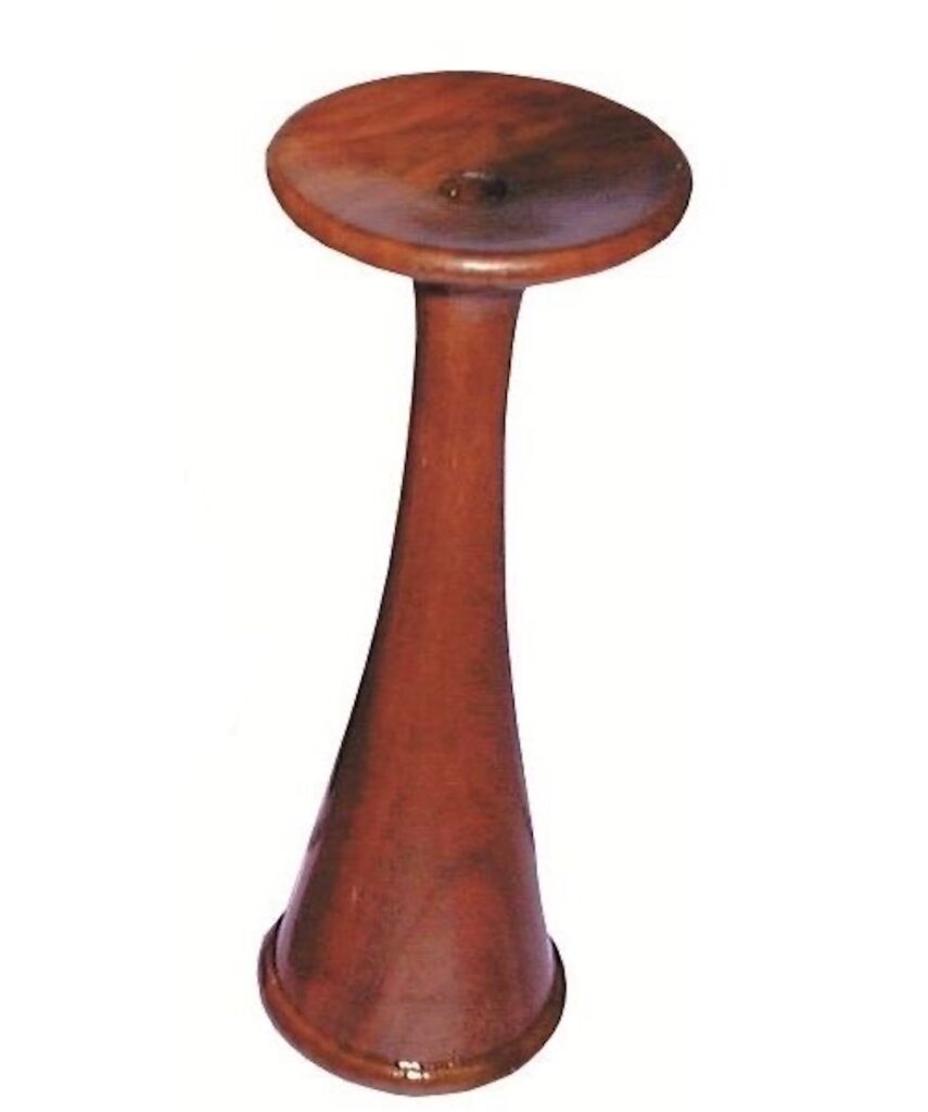
I moved my stethoscope over the tiny chest, ear pressed to it, listening intently. It was my best stethoscope, a model from the nineteenth century called a Pinard—a bell with a flattened disc at one end, to which I pressed my ear. I had one carved of wood; this one was made of pewter; Brianna had sand-cast it for me.

The following image is a wooden Pinard stethoscope, currently for sale on the Internet for about $180 (there are much less expensive versions)! 💰

Claire placed her ear against the cup shaped disc at the top; the bottom of the tube was placed on Mandy’s chest. The stethoscope amplifies the sounds of blood rushing through the heart and striking its valves.  

What did Claire listen for? she was listening for a distinctive murmur: a soft, continuous shushing sound, particularly audible near the base of the neck. It is usually the first diagnostic sign of a problem with a fetal heart vessel.  

Although Mandy exhibits only some of the above symptoms, Claire diagnoses Mandy’s condition as a Patent Ductus Arteriosus (PDA).  

But, before we can understand PDA, let’s look at normal heart anatomy.

Heart Anatomy: To better understand Mandy’s Malady, let’s consider the normal anatomy of the heart and its great vessels. Now, I kid you not, the heart is a very complex organ, both anatomically (structure) and physiologically (function). We will only cover the basics. Understand that there are many more anatomical details that are beyond the scope of this lesson. But, we fearless folk press onward!

Chambers: The human heart has four chambers (next image). Right atrium and left atrium are thin-walled filling chambers. Right ventricle and left ventricle are thick-walled pumping chambers . The walls are composed of cardiac muscle, a specialized tissue found in the heart. (psst….don’t forget, the patient’s left is your right, and vice versa) 🤓 

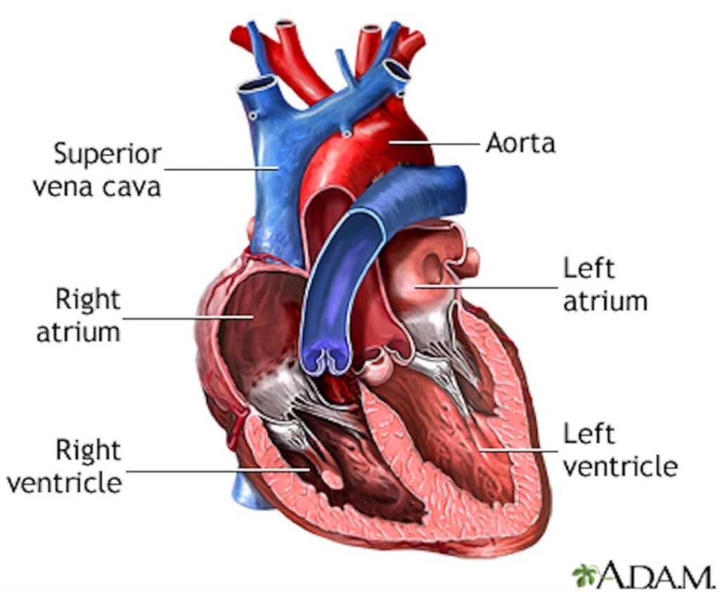
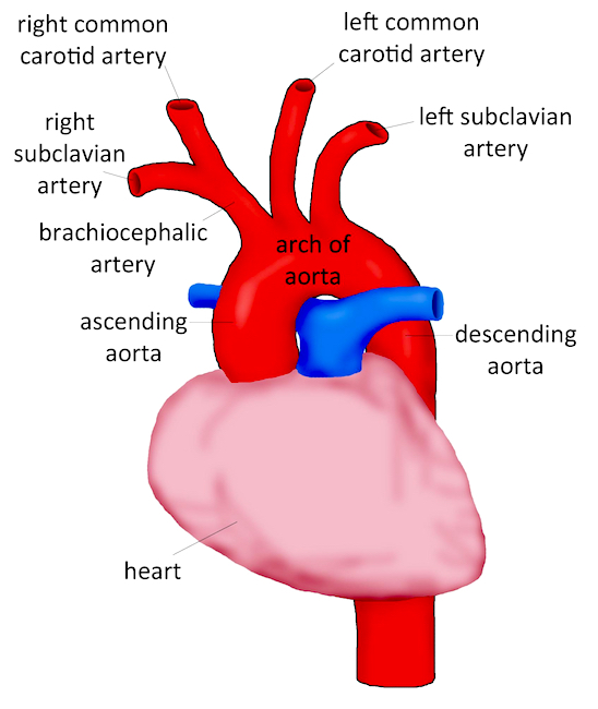
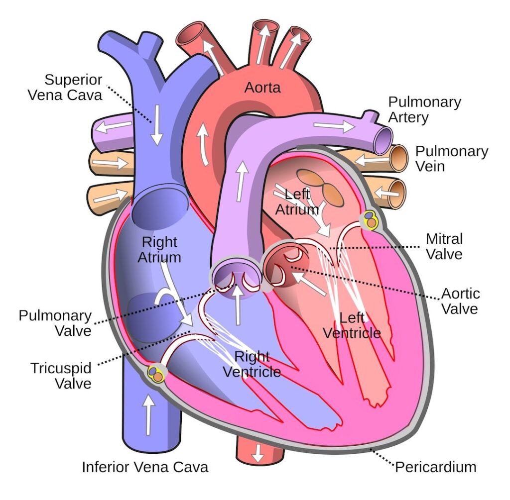
Vessels: The heart has eight (!!!) vessels carrying blood to and from its chambers. These are (next image):

    • Superior vena cava – delivers blood from upper body into right atrium
    • Inferior vena cava – delivers blood from lower body into right atrium
    • Pulmonary artery – deliver blood from right ventricle to lungs
    • Four pulmonary veins – deliver blood from lungs to left atrium
    • Aorta – delivers blood from left ventricle to body 

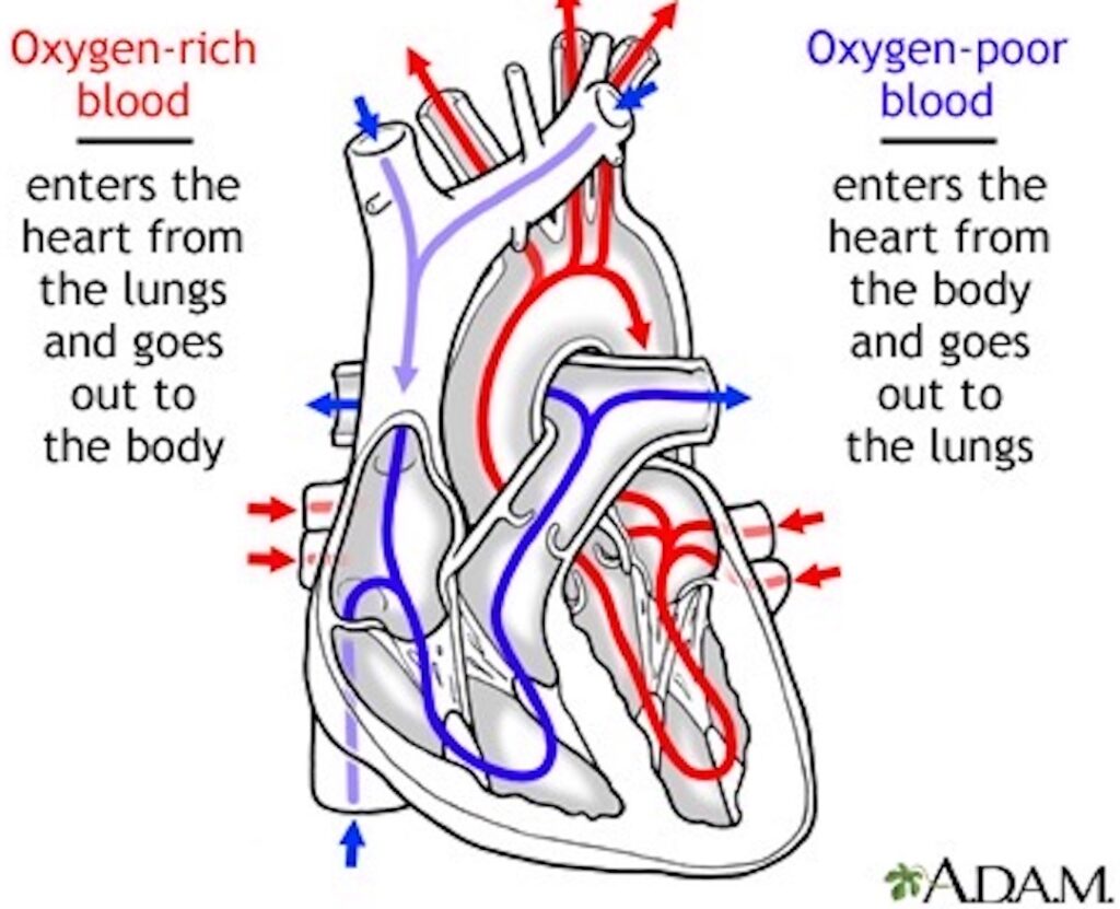
Blood Flow: Next, let’s review the pattern of blood flow through the heart. As you read the details, check with the image below to verify the flow.

    • Deoxygenated blood (low oxygen – O2; high carbon dioxide -CO2) from  superior vena cava and inferior vena cava pours into right atrium and then into right ventricle.
    • Right ventricle contracts and ejects blood into the pulmonary artery which branches to supply left and right lungs. 
    • Blood releases carbon dioxide (CO2) in the lungs which is exhaled and picks up O2 from inhaled air.
    • Oxygenated blood (O2-rich) is carried to left atrium via four pulmonary veins. 
    • Blood pours into the left ventricle which contracts and ejects O2-rich blood into the aorta.
    • Branches of aorta carry blood to all other regions of the body (except lungs).  

OK, students, hang in there! With normal anatomy under our belt, let’s look at the fetal circulation. 

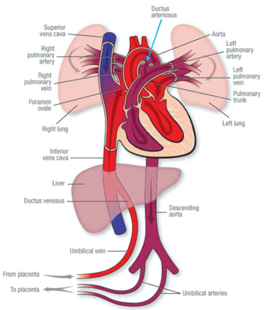
Fetal Circulation: Blood circulation in the fetus is different because the placenta provides the functions of lungs, gastrointestinal tract, and kidneys. Thus, a normal blood supply to these maturing organs is not required.

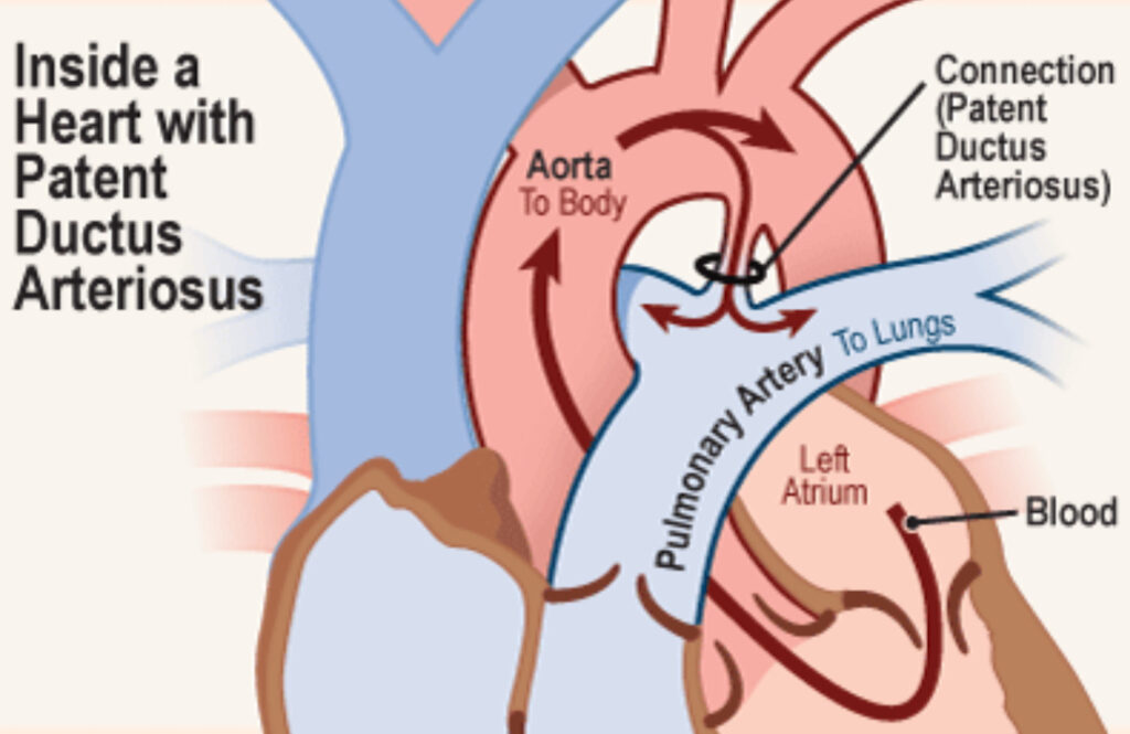
Ductus Arteriosus: The fetal heart has a vascular bridge between pulmonary artery and aorta that shunts blood exiting the right ventricle into the aorta and bypasses the lungs. This vascular bridge is the muscular ductus arteriosus (aqua arrow – below image). Again, because lungs do not process gasses during intrauterine life, most blood is shunted away from them. 

The ductus arteriorsus is patent (open) throughout fetal life and normally closes within 24 hours after birth as blood flow to the lungs is established. Within 2-3 weeks, it turns into a fibrous band, the ligamentum arteriosum. 

All of this is highly regulated by various chemical and physiological substances including oxygen levels. 

Patent Ductus Arteriosus: If the ductus arteriosus does not close soon after birth but remains patent (open), it is diagnosed as PDA, a congenital heart lesion. PDAs are most common in premature babies but can also occur with full term infants. 

Nowdays, if a ductus arteriosus does not spontaneously close after 8 weeks post-birth, it usually is treated with medications, plugged, or surgically closed. Small PDAs may not be a cause for concern and are often not treated.

Adding a bit of perspective – PDA is not new. It was known as early as 129 A.D. to Galen, a Greek anatomist and physician, although he didn’t understand its significance. It wasn’t until 1938, almost two millennia later, that Dr. Robert E. Gross of Harvard Medical School and Children’s Hospital in Boston, Massachusetts, perform the first successful ligation (closure) of a PDA. This was also the first congenital heart lesion to be successful corrected, surgically.

Now, let’s return to Mandy’s Malady. This was her problem. Mandy’s ductus arteriosus did not close after birth and she exhibited some of the concerning symptoms outlined above.  Blood from her aorta flooded her lungs subjecting fragile lung tissues to excessive blood pressure (hypertension). Allowed to go unchecked, the fragile lungs will be permanently damaged and she  eventually will experience right-sided heart failure.

Diana explains all of this with her usual magical writing skills in this excerpt from “A Breath of Snow and Ashes:” 

The ductus arteriosus is a small blood vessel that in the fetus joins the aorta to the pulmonary artery. Babies have lungs, of course, but prior to birth don’t use them; all their oxygen comes from the placenta, via the umbilical cord. Ergo, no need for blood to be circulated to the lungs, save to nourish the developing tissue—and so the ductus arteriosus bypasses the pulmonary circulation. 

At birth, though, the baby takes its first breath, and oxygen sensors in this small vessel cause it to contract—and close permanently. With the ductus arteriosus closed, blood heads out from the heart to the lungs, picks up oxygen, and comes back to be pumped out to the rest of the body. A neat and elegant system—save that it doesn’t always work properly.

The ductus arteriosus doesn’t always close. If it doesn’t, blood still does go to the lungs, of course—but the bypass is still there. Too much blood goes to the lungs, in some cases, and floods them. The lungs swell, become congested, and with diverted blood flow to the body, there are problems with oxygenation—which can become acute.

Lastly, this is a brief video about PDA which you might find useful. It is easy to understand and accurate: https://youtu.be/7DKaCqubuSg. 🤓

Fast forward! It is clear that the MacKenzies arrived safely in the 20th Century and Mandy received the medical intervention needed for her PDA repair. When we meet her next at Lallybroch, she is a healthy, happy, and feisty wee lass, played by Rosa Morris. 🤗

As William Shakespeare once wrote, “All’s Well That Ends Well!”

(Well, it would end better if they were all together, but we shall see) 🤞🏻

The deeply grateful,

Outlander Anatomist

Follow me on:

Photo creds: Sony/Starz; www.commons.wikimedia.org; www.heart.org; www.kidshealth.com; www.medicalsuppliesgh.com; www.medlineplus.gov; www.outlander.fandom.com; www.theoutlandermuse.com; www.twitter.com (now X)

Anatomy Lesson #56: Achy Brachial Artery

Greetings, anatomy students! One might reasonably assume this lesson is about Billy Ray Cyrus, but, one would be wrong. This anatomy lesson discusses the very important, brachial artery, and its parent, the axillary artery

And, what spurs a lesson about these vessels? Why, twisted-sista, Laoghaire, is to blame (Starz Outlander episode 308, First Wife)! 

Who is in her sights – Jamie? Claire? Or, will she fell both with a single shot?

Blamo! A fouling piece discharges a fowling piece!

Dear Laoghaire: 

On the day that you were born,

The angels got together……

And, were severely reprimanded!

Shoot <g>! Laoghaire peppers Jamie with birdshot! Alas-lass, Granny Fitz probably wouldn’t be proud of her progeny. 

Laoghaire, Laoghaire, quite contrary,

How does your vengeance grow?

Jamie fell, amid bloody hell,

And, Claire became your foe!

Dinna mess with the Sassenach. Drop that weapon!  

Now, book readers ken that things didn’t go down quite this way in Voyager book. Herself envisioned Claire fleeing Lallybroch upon finding that Jamie has a harem. Young Ian (bless his wee Scottish heart), catches up to her in the mountains and says:

“But Auntie Claire, it’s not that!” 

“What’s not that?” Caught by his tone of desperation, I glanced up. His long, narrow face was tight with the anguished need to make me understand. 

“Uncle Jamie didna stay to tend Laoghaire!”

“Then why did he send you?” He took a deep breath, renewing his grip on my reins. 

“She shot him. He sent me to find ye, because he’s dying.”

Back to the TV version. Perhaps aiming for his heart, Laoghaire’s birdshot turns Jamie’s left chest, shoulder and arm into mincemeat. But, harboring absolute faith in his beloved he assures us:

 “It’s only birdshot, nothing serious…..It’s nothing Claire canna fix!”

Back to Voyager book, Claire assesses Laoghaire’s assault: 

“Let’s have a look at it.”

The wound itself was a ragged dark hole, scabbed at the edges and faintly blue-tinged. I pressed the flesh on either side of the wound; it was red and angry-looking, and there was a considerable seepage of pus. Jamie stirred uneasily as I drew my fingertips gently but firmly down the length of the muscle. “You have the makings of a very fine little infection there, my lad,” I said. “Young Ian said it went into your side; a second shot, or did it go through your arm?” 

“It went through. Jenny dug the ball out of my side. That wasna so bad, though. Just an inch or so in.”

A bit of difference between the two versions but either way, Jamie needs his Claire! BTW, the special effects are terrific!

Back to TV: No surprise – Claire must remove those pesky pellets! Following the surgeon’s creed: a chance to cut, is a chance to cure, Claire sets to work! 

Several big swigs of Scotch whisky and Jamie lolls unconscious… that stuff is potent! Armed with tools from her medical kit, Claire dives for those wee bits of lead!

One by one, she carefully retrieves the pellets.

Mostly, the bird shot is superficial but one sits in a precarious position. She cut downs to reach the deep-lying pellet explaining to young Ian, she must avoid damaging the artery!  Jamie could bleed to death if the vessel is injured by pellet or nicked by blade. 

The next image exhibits a high yuk factor for some students, but Clair dutifully digs, dives and delves for the perilous pellet! 

Into the brass vessel it goes –  joining its brothers in crime.  

Still royally pissed at Jamie’s deceit, Claire carefully stitches the incision (Anatomy Lesson #35, Outlander Owies!) and our Highland Hero is saved. This lass takes pride in her work!

Anatomy Lesson: Sigh… time to leave Outlander and get to our lesson! Now, which artery does Claire believe is at risk? Let’s tease out the answer. 

Judging from the location of the incision, we may reasonably surmise that Claire’s Concern is directed toward one of these two arteries:

  • Axillary artery
  • Achy brachial artery

Origins: To properly understand how these two arteries might be at risk from bird shot or scalpel blade, let’s study their ancestry.com. We begin at the heart. Image A is a highly simplified view of the heart (pink) and its major arteries (red and blue). 

Aorta: The ascending aorta, the large red vessel, arises from the heart (left ventricle) and curves, becoming the arch of aorta. The arch is then renamed the descending aorta as it dives downward through chest and abdomen.

Anatomic Note #1: Traditionally, arteries appear red to denote they carry oxygenated blood. However, some arteries carry de-oxygenated blood such as the two blue vessels shown in Image A; these are right and left pulmonary arteries (from right ventricle). Their blood becomes oxygenated as it travels through the lungs. Not critical to today’s lesson, so more about these vessels in a later session!

Aortic Branches: Typically, the aortic arch gives rise to three large arteries. In 75% of people, aortic branches follow the pattern shown in Image A. But in 25% of folks, a different branching pattern ensues. My experience in the dissection lab is that blood vessels show the greatest variation of any anatomical structure. None-the-less, the typical branching pattern is:

  • Brachiocephalic artery (dividing into)
    • Right common carotid artery 
    • Right subclavian artery
  • Left common carotid artery
  • Left subclavian artery

Anatomic Note #2: The left side (on your right) typically lacks a brachiocephalic artery, so left common carotid and subclavian arteries branch directly off the aorta. On the right, the short brachiocephalic artery soon branches into right common carotid and subclavian arteries. Embryonic development produces this odd asymmetry.

Image A 

Destination: Once the brachiocephalic artery divides, its branches follow the same pattern as the left side (Image B): 

  • Right and left common carotid arteries ascend to supply head and neck
  • Right and left subclavian arteries arch through base of neck.
  • Name change #1: At outer border of the 1st ribs, subclavian arteries are renamed right and left axillary arteries
  • Name change #2: At lower border of teres major muscle (Anatomy Lesson #10, Jamie’s Back or Aye, Jamie’s Back!), axillary arteries are renamed right and left brachial arteries

Gasp! Pray tell, who thought  so many name changes would prove helpful? Early anatomists are to blame, waaay back in 1578!

Image B 

Axillary Artery: Subclavian arteries give off branches and then, like many city streets, each changes its name to axillary artery

The word, axillary, comes from the Latin axillaris meaning “armpit,” because the axillary artery lies deep in the armpit, skirting outer ribs and angling through the oxter toward the arm (image C). Its important branches supply blood to:

Situated so deep in the armpit, axillary artery is protected and rather difficult to reach except by needle, open dissection, or bird shot! 

Image C 

Brachial Artery: At the lower border of teres major muscle, each axillary artery is renamed the brachial artery, the major artery supplying each arm (Image D). Brachial comes from the Latin brac(c)hium meaning “arm.”  The brachial artery continues into the arm supplying blood to its structures; then, ends near the elbow by dividing into radial and ulnar arteries (Anatomy Lesson #19, To Arms, Too Arms, Two Arms!).

Image D 

Importance: The brachial artery is THE major blood vessel of the upper limb (Anatomy Lesson #19, To Arms, Too Arms, Two Arms!), providing each with almost a liter of blood (.95 qt.) per minute! Understand, this is a huge blood flow given that the entire human body typically contains only 4.7 – 5.5 liters of blood.

Note #1: Blood flow through the subclavian artery, the parent of brachial artery, is difficult to measure because it lies so deep, but it’s blood flow would be slightly higher than the axillary artery.

Try This: This could be fun! The brachial artery carries so much blood, its pulse can be taken to determine heart rate. This is how you can find your brachial pulse. Identify your biceps with the contralateral (opposite) hand. Move middle and ring fingers along its inner border toward the inner elbow and apply moderate pressure (Image E). Feel it? Yay!

Note #2: Today, pulse is often taken using a finger clip device (pulse oximeter) but, a thorough education also includes the manual technique.

Horror: I see many examples on the Internet advising students to take a pulse with their thumb!!! These grieves me, because one should never take a pulse with the thumb. The thumb has its own substantial artery, the princeps pollicis, with its own pulse  – large enough to interfere with determining a patient’s pulse! In some people, the index finger also has a strong pulse, so using the middle and ring fingers are best. 


Image E 

Should you be interested, here is a nice video about locating the brachial artery pulse. The demonstrator uses a slightly different approach than the one described above, but either works well:

Conclusion: Two arteries are candidates for Claire’s Concern: left axillary or left brachial. Each has a huge blood flow and if their wall is torn or cut, a person could easily exsanguinate through the breach. 

But, which one? The location of her incision informs our choice. Back to an earlier image: Claire’s tools are inserted just under the outer border of Jamie’s pectoralis major muscle (mercy!) and above the armpit (oxter hair lies below).  One may reasonable conclude that her tools’ trajectory towards the armpit is most consistent with the pathway of the axillary artery; the brachial artery should be further out towards the arm. Ergo, Claire is concerned that birdshot or tools may harm the axillary artery

But, know this….it is difficult to be absolutely sure sans a proper visual evaluation of the area accompanied with palpation. I volunteer! He he.

So, we have answered our initial query: Likely, Claire was fearful of damaging Jamie’s axillary artery not his achy brachial artery, but either would suffice.  

Cosmic Question: Now, comes the most critical question of all!

How can a surgeon operate on an intoxicated patient, with dim lighting, using fairly cumbersome tools, answering questions from a curious nephew, removing multiple bloody foreign objects, and yet complete her tour of duty with an absolutely pristine apron????  Not a drop of blood in sight! 

Because, she is the Sassynach Surgeon in her bad-ass bat suit and Claire can fix pretty much anything!

Arteries might not be the sexiest of anatomical topics, but we can understand and appreciate their value, especially if they are damaged or dysfunctional. Let’s join Claire with a  toast to our awesome arteries: Slàinte Mhath!

A deeply grateful,

Outlander Anatomist

Photo Creds:

Starz: Outlander episode 308, First Wife; www.biology.stackexchange.com (Image B); www.earthslab.com (Image C); www.humananatomly.com (Image A); www.slideplayer.com (Image D, E)