Welcome anatomy students to another exciting session! Today’s Anatomy Lesson #42 is, The Larynx. Some folks think the larynx only generates “The Voice” (Christina, CeeLo, Blake and Adam, notwithstanding). While this is certainly true, it also serves two other critical functions. Stay tuned for the fascinating saga of laryngeal (adj.) anatomy.
As always, Starz images and Diana’s book quotes are sprinkled throughout to make our studies more endearing. A couple of book spoilers appear, meaning the quotes are from books that have not yet been filmed; these will be preceded by a red flag warning for those who prefer to skip!
First, the sad-bad news. I waited for Starz to depict Dougal’s Demise after a reader asked more than a year ago for a lesson on the voice! I waited patiently because Uncle D’s Death as described in Dragonfly in Amber book is a perfect lead-in for a larynx lesson. Then, horror of all horrors – Starz episode 213, Dragonfly in Amber – didn’t follow the book! Och!
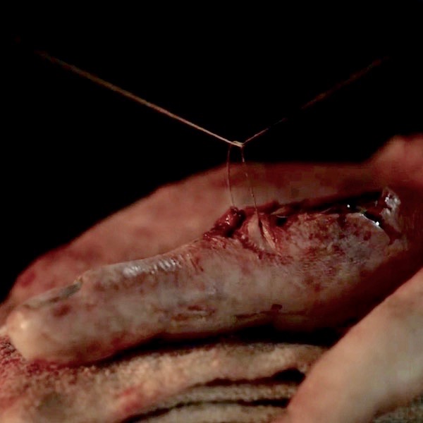
BOOK QUOTE: Dragonfly in Amber describes Jamie’s and Dougal’s shocking Fight with Fate, involving a knife to the throat and a struggle with voice:
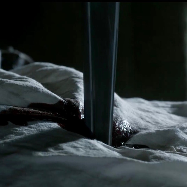
A shift, and a jerk, a sudden grunt of effort, one of pain. Dougal stepped back, staggering, face congested and pouring sweat, the hilt of the dagger socketed at the base of his throat… There was a terrible sound from Dougal, a sound of shock and stifled breath… The big body went limp, then spasmed, sliding out of Jamie’s grasp. Dougal lay crumpled on the floor, muscles jerking with involuntary contractions, struggling like a fish out of water. His head was pillowed on Jamie’s thigh. One heave brought his face into view. It was contorted, and dark red, eyes gone to slits. His mouth moved continuously, saying something, talking with great force—but without sound, save the bubbling rasp from his ruined throat.
Imagine my dismay as the Starz scene unfolds wherein Jamie + Claire stab Dougal in the chest – not in the neck! What? To quote Rabbie Burns: “The best laid schemes o’ mice an’ men – gang aft a-gley.” Ah, well, tha sin mar sin!
Many a time, Jamie casts a wary eye at Dougal (Starz episode 101, Sassenach) and for good reason. Mayhap, the ruadh laddie kens that a battle to the death is the only way a male will prevail? One of these fellows must go!

Now for the lesson: Let’s start at the beginning with how to pronounce the word, larynx. Well, it is pronounced ler-inks, not lar-nix. Emma does a swell job, so check this out:
https://www.youtube.com/watch?v=4mX4-zer2e0
Commonly known as the voice box, the larynx is a splendid structure of startling complexity. A hollow tube with stiff walls, the adult larynx (Image A – orange) lies in the neck where it links pharynx or back of throat (Image A – green) with trachea or windpipe (Image A – trachea).
Just so you are aware, pharynx is pronounced fare-inks, not far-nix. One of my anatomy profs used to declare: “if you say lar-nix one more time, I will rip out your far-nix” (in those days, professors were minor gods)!
The human larynx serves three critical functions:
- Maintains the airway so air enters and leaves the lungs during each breath cycle
- Excludes food, drink and other unwanted stuff from entering trachea
- Creates phonation – “the voice” and other sounds
We will learn how the larynx manages these important functions. But first, the anatomy!
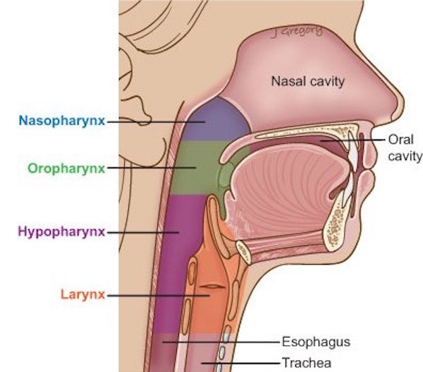
Image A
General Anatomy of Larynx:
A vertical, midline section through the larynx reveals its considerable length, extending from C3 (third cervical) vertebra (Image B – red arrow) through C6 vertebra (Image B – blue arrow). In case you forgot, back in February 2015, we learned about cervical vertebrae in Anatomy Lesson #12, “Claire’s Neck” or “The Ivory Tower.”
The adult larynx is supported by a rigid skeleton of cartilage, ligaments and membranes (Image B – cartilages named). Numerous intrinsic and extrinsic muscles move these cartilages.
Its hollow interior is lined with mucous membrane or wet mucosa (Anatomy Lesson #14 ,“Jamie and Claire” or “Anatomy of a Kiss”), meaning the surface is moist, a necessity because its living cells will die if allowed to desiccate.
The larynx is a dynamic structure that changes throughout life, from the newborn state through old age. It is also a secondary sex organ undergoing marked changes during puberty. Male and female larynges (pl.) respond differently to the internal hormonal milieu, a quality known as sexual dimorphism (Anatomy Lesson #39 “Dem Bones – the Human Skeleton”). We will return to these issues later in this lesson.
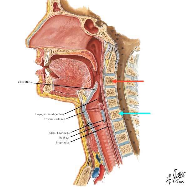
Image B
Laryngeal Skeleton:
The laryngeal (adj.) skeleton features nine cartilages: three large, unpaired and three small, paired. Only unpaired cartilages (epiglottis, thyroid and cricoid) are visible from a front (anterior) view (Image C). The rounded, curved tip of epiglottis peeks above the hyoid bone. The thyroid cartilage is shaped like a helmet visor; two broad plates (laminae) meet anteriorly at a pronounced angle, the laryngeal prominence (Image C – red arrow). The cricoid cartilage is a complete ring, thin in front but expanded in back.
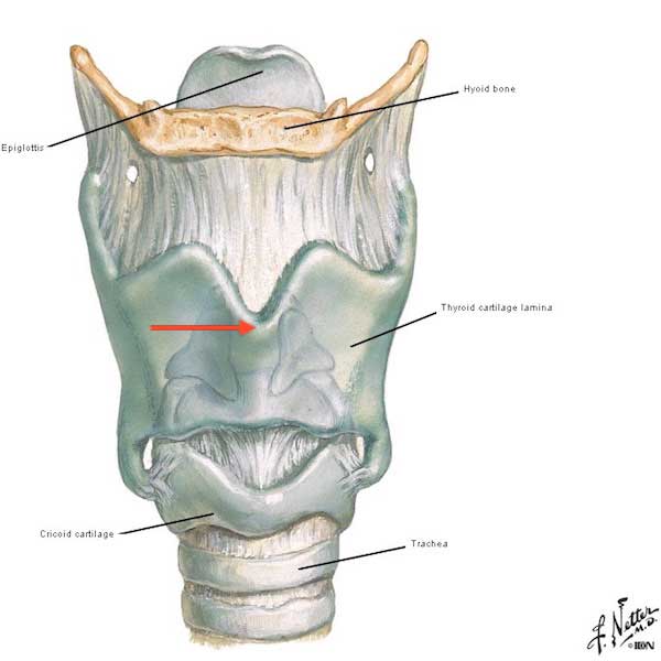
Image C
Laryngeal cartilages do not float, they are anchored by strong connective tissue elements (Image D). The thyroid cartilage hangs from the hyoid bone (Anatomy Lesson #12, “Claire’s Neck” or “The Ivory Tower”) via a tough thyrohyoid membrane. Cricoid and thyroid cartilages are united by the cricothyroid ligament and a cricotracheal ligament (Image D – red arrow) binds cricoid cartilage to trachea. Although not shown in Image D, ligaments also hold the epiglottis in a mostly vertical position. The trachea is not part of the larynx but its wall is also formed of cartilage rings (see below).
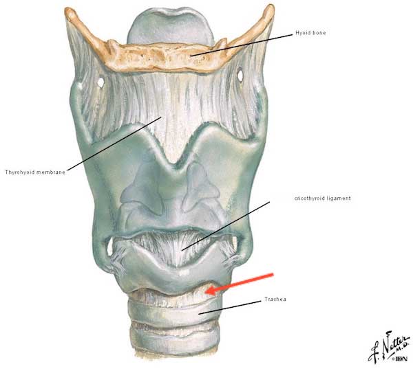
Image D
WARNING! The first RED FLAG means a book spoiler quote is next! Skip, if need be.

BOOK SPOILER: Larynx and hyoid bone appear in Diana’s 9th book, Written in My Own Heart’s Blood. An important character’s name is omitted to “protect the innocent!”
McEwan’s broad fingers were cold on his neck; he felt the icy touch delicate on his skin as it traced the line of the rope scar, then firmer as the healer prodded gently round his damaged larynx… McEwan fitted his hand snugly round _______ neck, just under the chin… “Do you know what a hyoid bone is?” “If I had to guess, it’s something in the throat.” “Why?” … “It’s just there,” the healer said, pressing with his thumb, high up under _______ chin. “And if it had been here”—he moved the thumb down an inch—“ you’d have been dead, sir. It’s a fragile wee bone. Easy to strangle someone by breaking it—with your thumbs or a rope.”
Question #1: Do any of the above structures appear as mounds at the skin surface? Hyoid bone, epiglottis and cricoid cartilages are not visible because these are embedded too deeply or are too small to create a skin mound. But, we often see a bulge that moves during swallowing, talking, singing, etc.; the bulge is created by the laryngeal prominence of the thyroid cartilage, commonly known as the Adam’s apple (Image E). Great tattoo, although most of it lies below the laryngeal prominence. Ha!
Try This: Place two fingers over your laryngeal prominence – don’t press hard as it causes discomfort. If you cannot find it, gently tilt head backwards and feel a firm mound under your fingers. Now, straighten the neck and feel the prominence lift as you prepare to swallow and fall as the act completes. Now cough or sing a few bars and see what happens. I hope you are surprised by its mobility. Remember, this is only a small part of the larynx; much more is happening that you cannot feel.
Try This: Next, tilt head back again and run fingers down the laryngeal prominence until you feel soft tissue; this is the cricothyroid ligament. Below it lies a thin hard line of cricoid cartilage. Below this is another bit of soft tissue, the cricotracheal ligament binding cricoid cartilage to first tracheal ring. From this point, the trachea continues down the throat and into the thorax (Anatomy Lesson #15 “Crouching Grants – Hidden Dagger”). Appreciate that the larynx is solidly built and well-anchored.
Clinical Correlation: If an “owner” desires a smaller laryngeal prominence, then chondrolaryngoplasty is available. The neck is entered via surgical incision and the prominence reduced by shaving. Erroneously called a tracheal shave (trachea isn’t involved), it usually leaves a modest scar.
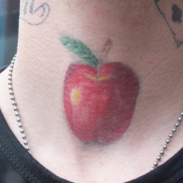
Image E
Time for a Jamie fix! Jamie’s laryngeal prominence is clearly visible (Starz episode 210, Prestonpans – red arrow) as, tilting his head back with immense pleasure, he relieves a full bladder! Hot-shot lad that he is, he attempts to win a bet by peeing into a pot without looking. Splish-splash! Odds?
BOOK QUOTE: Diana describes the delightful scene featuring Jamie’s Adam’s apple (Dragonfly in Amber):
Grinning at the success of his joke, he raised his kilt further, grasped his clearly visible weapon and took careful aim. He squinted his eyes, bent his knees slightly, and his fingers tightened their grip. Nothing happened. “It’s a misfire!” crowed one of the English. “His powder’s wet!… Jamie squinted dubiously at his equipment, bringing on a fresh riot of howls and catcalls. Then his face cleared. “Ha! My chamber’s empty, that’s all!” He snaked an arm toward the array of bottles on the wall, cocked an eyebrow at me, and when I nodded, took one down and upended it over his open mouth. The water splashed over his chin and onto his shirt, and his Adam’s apple bobbed theatrically as he drank. “Ahhh.”

Back to Anatomy: The best way to see remaining laryngeal cartilages is flip the structure into a posterior view (Image F). The odd, leaf-like epiglottis is fully visible from this perspective; it’s curved superior surface peeking above the hyoid bone. Thyroid cartilage, visor-shaped, is incomplete, posteriorly. The cricoid cartilage expands in back, much like the dome of a signet (class) ring. Perched atop the cricoid are the important, paired arytenoid cartilages, each shaped like a three-sided pyramid. Atop each arytenoid is another small curved cartilage (corniculate) which we will ignore along with a couple of other tiny cartilages. Although difficult to visualize from this perspective, a lumen (channel) runs vertically through the center of the laryngeal cartilages; remember, this lumen is lined with mucous membrane.
Posteriorly, the tracheal rings are incomplete, but the C-shaped cartilages are united by strong connective tissue sheets. This brilliant engineering feat allows food blobs and globs to slide and glide down the esophagus (see Image B – esophagus behind trachea) without getting hung up on tracheal rings.
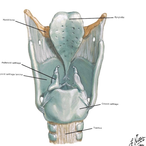
Image F
One last structural component to consider are the paired vocal ligaments. A vertical section of the larynx, through the midline, reveals internal anatomy of the left side of larynx (Image G). Locate the left vocal ligament, a band of elastic tissue reaching from thyroid cartilage in front to arytenoid cartilage, in back. This arrangement is duplicated on the right side. More about the vocal ligaments below.
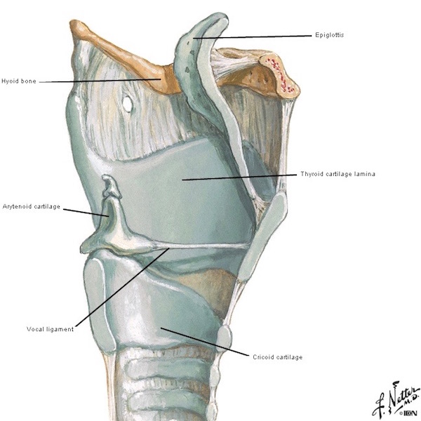
Image G
Intrinsic Laryngeal Muscles: Intrinsic muscles arise and insert within the larynx itself. There are 6 (8 by some counts) pairs of intrinsics; thin, tiny muscles moving various laryngeal cartilages. I can bear witness that these are very difficult to dissect! One of a pair of intrinsic muscles is clearly visible from a side view (Image H – red arrows). Naming intrinsic muscles will bog us down. Suffice it to say, the one in Image H is the cricothyroid, so named because it arises from cricoid cartilage and inserts on thyroid cartilage. Get the idea? Grand!
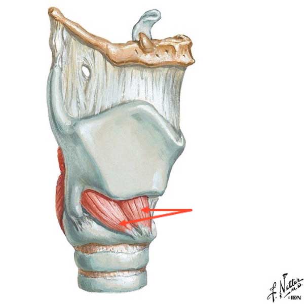
Image H
Remaining intrinsic muscles are visible from a posterior view (Image I). These are strategically oriented to move the various laryngeal cartilages in specific ways and again, named by the cartilages they attach to or the function they perform.
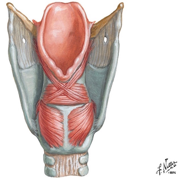
Image I
The following is a short list of how intrinsic muscles act on various laryngeal cartilages.
- Rock thyroid cartilage forward to lengthen vocal ligaments and deepen the voice (Image J – top)
- Rotate arytenoid cartilages together to open airway and allow airflow (Image J – middle, left)
- Rotate arytenoid cartilages apart to close airway and stop airflow (Image J – middle, right)
- Narrow the airway to reduce airflow (Image J – lower, left)
- Tense vocal cords to raise voice pitch (Image J – lower, right)
Ergo, these intricate adjustments, caused by muscle contraction, allow nuanced movements of laryngeal cartilages to manage airflow and to produce and modulate sound. And, these amazing, small muscles contract very rapidly with great capacity for prolonged work, as any singer can verify. Lastly, the lower right image shows muscle fibers stretched from thyroid to arytenoid cartilages; these tiny muscles lie inside the vocal folds (see below).
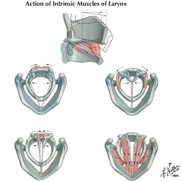
Image J
Extrinsic Laryngeal Muscles: Not only do intrinsic muscles move the larynx, eight pair of strap-like extrinsic muscles also effect laryngeal position. Extrinsic laryngeal muscles are so-named because they take origin from or insert into body parts outside the larynx. Located in the neck or beneath the mandible, extrinsic muscles connect mandible (Anatomy Lesson #26, “Jamie’s Chin – Manly Mentus”), sternum, scapula and skull to thyroid cartilage and hyoid bone. Read more about these muscles in Anatomy Lesson #12 “Claire’s Neck” or “The Ivory Tower.”
Image K identifies extrinsic laryngeal muscles but more importantly, shows upward or downward directional pulls (red arrows); four pair of extrinsic muscles depress (lower) and four pair elevate (lift) hyoid bone and thyroid cartilage. Further, the pulls are not strictly upwards or downwards. Rather, the upper four pair of muscles pull hyoid bone up and forward or up and backward; the lower four pair of muscles pull hyoid bone and thyroid cartilage down and forward or down and backward. The larynx, suspended from that wee hyoid bone, is mostly carried along for the ride!
Thus, these many muscles and their attachments allow for nuanced changes of laryngeal position during talking, laughing, screaming, singing, coughing, clicking, sneezing, snorting, swallowing, etc. Impressive!
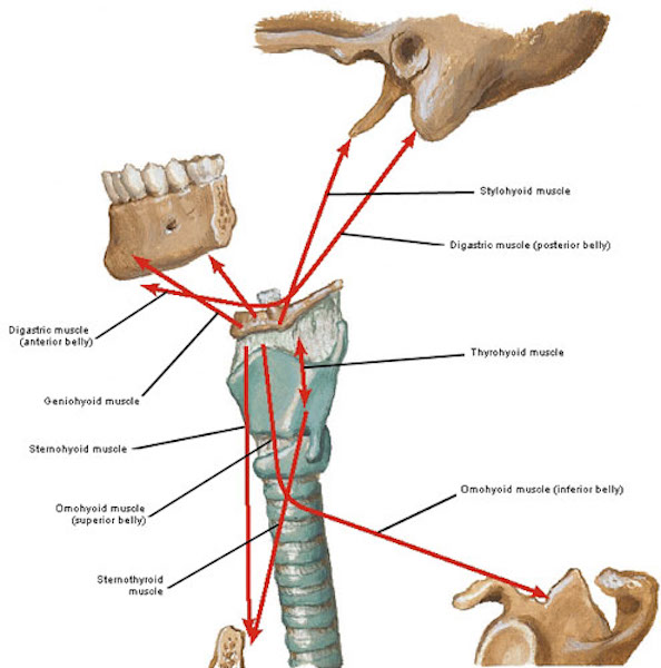
Image K
Thyroid Gland: Remove the extrinsic muscles and we see an important and intimate relationship between larynx and the highly vascular thyroid gland. The bilobed thyroid gland straddles the larynx, its lobes connected by an isthmus (bridge) of thyroid tissue overlying the second and third tracheal rings (Image L).
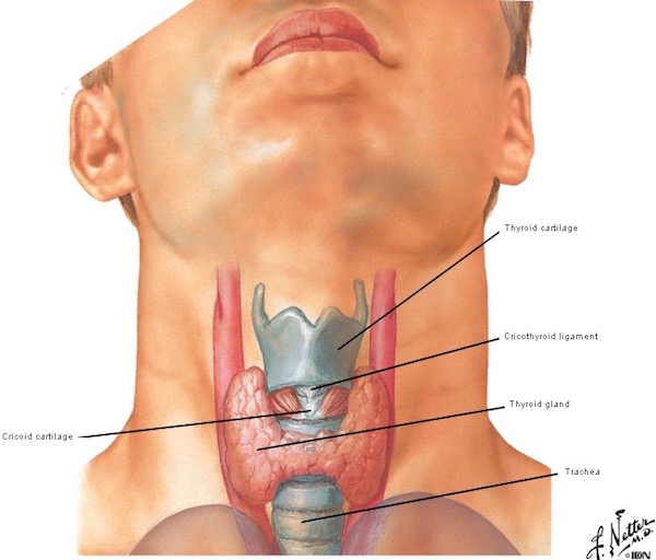
Image L
Vocal Folds: Next, let’s consider vocal folds. Vocal folds, commonly known as vocal cords, are paired folds of mucous membrane, each containing a vocal ligament (see Image G) and the tiny (vocalis) muscle (see image J, lower right).
Looking down the pharynx via laryngoscope (laryngoscopy), true vocal folds appear as pale thin bands (Image M). Below these, are epiglottis and tongue base. Above the folds are bumps created by mucosa overlying arytenoid cartilages. Vocal folds carry out the first and third critical functions listed below:
- provide airway
- exclude unwanted materials
- generate sound
Let’s discuss each of these functions in greater detail.
Provide Airway: As muscles rotate arytenoids together, vocal folds open allowing air to flow through the lumen or glottis (Image M – left side). As other muscles rotate the arytenoids apart, the folds close and air flow slows or stops (Image M – right side). Psst: Just so you know, vocal cords lie horizontally in the neck; their vertical orientation in Image M is how they appear on a screen during laryngoscopy.
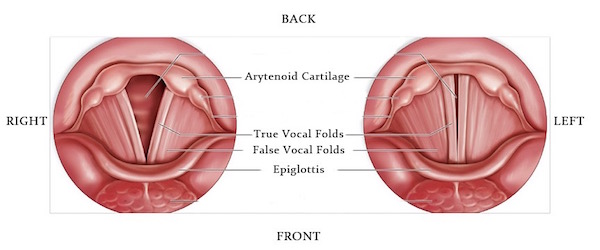
Image M
True vocal folds are so named to distinguish them from false folds. Find the false vocal folds in Image M. False vocal folds do not produce sound but do add resonance to the voice. Two exceptions are Tibetan Chant and Tuvan throat singing, both of these produce an undertone using small spaces (laryngeal ventricles) between true and false vocal folds (ventricles not visible in Image M).
If you find animals endlessly fascinating, get this: Male gorillas and some other primates, such as the gibbon shown in Image N, enlarge their necks by deliberately forcing air into their laryngeal ventricles. The ventricles of some primates are so large, they may extend into the thorax! In most humans, they are small.

Image N
Remember the second crucial function of the larynx? It closes to exclude unwelcome materials. Yep, it discriminates!
Exclude Undesirables: The weird-looking epiglottis helps with the second laryngeal function, to exclude unwanted stuff from the larynx. Here is how it works: During swallowing, hyoid bone lifts and tongue depresses pushing the epiglottis downwards and backwards to cover the laryngeal inlet (Image O – black arrow). This helps prevent food, drink, spit, etc., from entering the larynx as only air is welcome (Image O – white arrow). The epiglottis also has a shape which encourages food and drink to flow around its sides and backwards into the esophagus where it belongs! Does it sometimes, fail? Well, sure, especially if we try to swallow and speak at the same time. In such instances, stuff gets into the larynx , eliciting a paroxysm of coughing! Expel the infidel!
Try this: Wait! Wait! If you have a sensitive gag reflex, don’t try this. Otherwise, do you want to feel the tip of your epiglottis? Here’s how: open mouth wide and relax tongue. Lay one (clean) index finger atop tongue. Slide it gently backward and downward along the tongue surface until you touch the rounded, thin tip of the epiglottis. Don’t gouge it with your fingernail and avoid touching soft palate, sides or back of throat as this initiates the gag reflex! Did you feel it? Good job to those who dared!
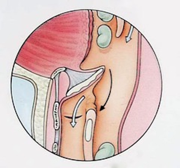
Image O
Generate Sounds: Last but not least, vocal folds produce sounds. During phonation, vocal folds open and close, change shape and vibrate. This brief video shows the fluid movements involved in this process – watch until the end, to see and hear the patient sing. The folds are a pale pink V in the still shot below. The tracheal inlet is the dark hole In middle. Mounds near the top are arytenoid cartilages – these move dramatically during the film clip. The thin mobile curl at the bottom of the video is the tip of epiglottis. Oh, and the sticky material coating the vocal folds is mucus, a viscous fluid that lubricates and protects the folds. Hope you watch all of this fascinating footage!
Clinical Correlation: Please understand that if the larynx is blocked or narrowed by trauma, infection, birth defects, cancer, polyps, scar tissue, etc., the impediment must be removed or bypassed to restore the airway. Two sites commonly used for bypass are cricothyroid membrane and trachea. An opening through the cricothyroid membrane (see Image D) is a cricothyrotomy or cricothyroidotomy. An opening between tracheal rings, is a tracheotomy; if a breathing tube is inserted into the opening, then it is termed a tracheostomy (Image P).
Tracheotomy is not new as it was depicted on Egyptian artifacts dating as far back as 3600 BCE! Here’s an startling tidbit: Alexander the Great purportedly saved a soldier from suffocation by making an incision into the man’s trachea with the point of his sword! Gah! I don’t recall seeing Colin Farrel do that in Alexander.
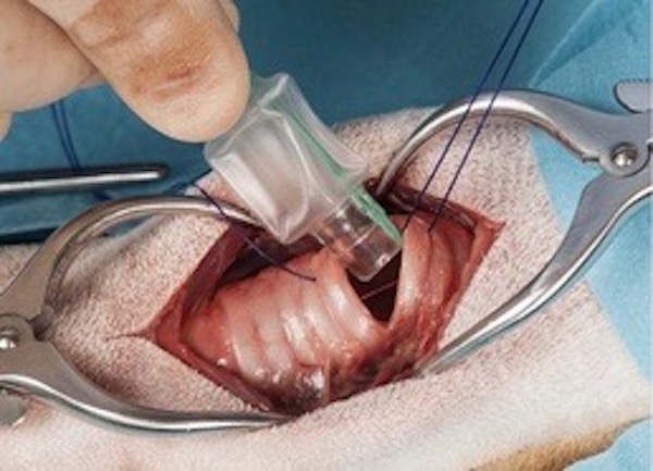
Image P
Red flag and wee black dog are back. Must be another book spoiler! Best skip if you haven’t read ahead of Starz episodes!
BOOK SPOILER: This quote is very pertinent to our lesson, from Herself’s fifth book, The Fiery Cross, as it mentions structures and procedures covered in this lesson. Yay, Diana!
A cricothrotomy? Fast, and requiring no great skill, but difficult to keep open—and it might not be sufficient to relieve the obstruction.
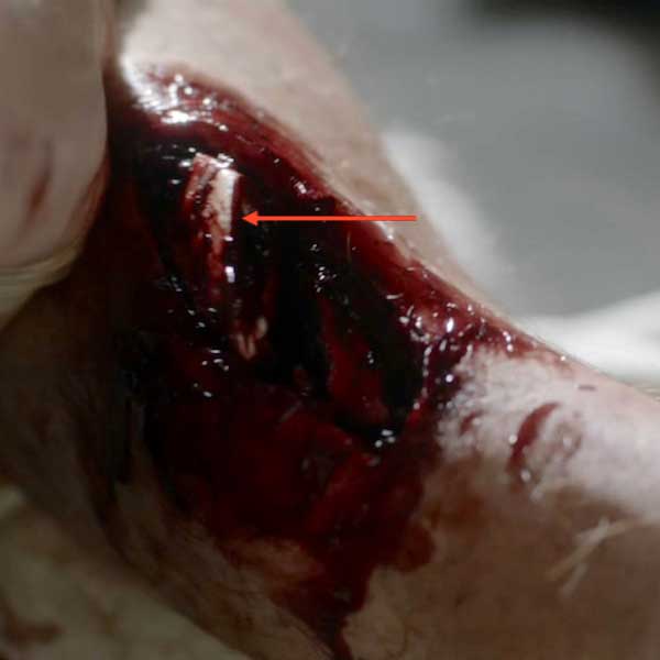
I massaged the isthmus of the thyroid, pushing it out of the way, hard toward his head, and with my other hand, pressed the knife blade down into the fourth tracheal cartilage.
The cartilage here was U-shaped, the esophagus behind it soft and vulnerable; I must not stab too deeply. I felt the fibrous parting of skin and fascia, resistance, then the soft pop as the blade went in. There was a sudden loud gurgle, and a wet kind of whistling noise; the sound of air being sucked through blood.
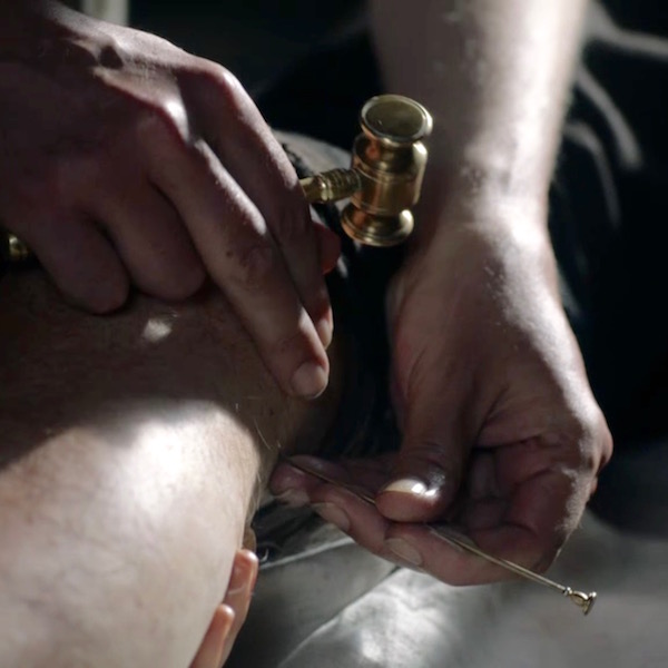
… she could hear the faint whistle of air through the tube in his throat. Claire had commandeered Mr. Caswell’s imported English pipe, ruthlessly breaking off the amber stem. Rinsed hastily with alcohol, it was still stained with tobacco tar, but seemed to be functioning well enough.
Way to Hustle, Claire: Claire assiduously avoids cutting the thyroid gland isthmus, otherwise, blood would quickly flood the surgical field. She also likely pressed her knife through the soft connective tissue between 3rd and 4th tracheal rings. And, she performed a tracheostomy, inserting a pipe stem into the tracheal incision to serve as a breathing tube. Yay, Claire!
Newborn Larynx: At lesson start, I promised to consider age- and sex-related differences of the larynx. Here we go! The larynx is a tiny organ in the newborn which sits much higher in the neck (C2-C3 vertebrae) than in the adult (Image Q – infant left, adult right). The higher position of an infant larynx allows epiglottis and soft palate to touch during suckling (Image Q – left side). This important juxtaposition enables the infant, an obligate nose-breather, to simultaneously suckle and breathe. Otherwise, they would have to swallow, stop, take a breath, stop – repeat. Exhausting for such a tiny body!
Many a nursing mam knows that if her wee bairn bites down (ouch!), she can stop this by gently closing off its nostrils – the babe must let go to breathe!
It should be noted that although infant larynges are much smaller than those of adults, their vocal folds and higher lung pressure enable them to produce very audible howls, yowls and growls!
Childhood Larynx: The larynx continues to grow throughout childhood in proportion to the remaining body with no significant differences between male and female larynges.
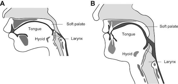
Image Q
Adolescence: Then, puberty raises its provoking head, accompanied by some fascinating changes! Both sexes experience facial development, descent of the larynx in the neck, increased circumference of chest wall and greater lung capacity, all of which typically deepen and strengthen the voice. But, the larynx of a pubescent male is also exposed to a flood of androgens (mainly testosterone) which induces growth of laryngeal cartilages.
Adult Male Larynx: By 20 y.o., the average male larynx is about 40% taller and vocal folds about 60% longer than the female’s (Table A). Plates of the male thyroid cartilage meet at a 90º angle making the thyroid eminence more prominent than that of the 120º female angle (i.e., female angles are more open, so eminence is less pronounced). Male intrinsic muscles become larger and stronger and vocal folds thicker and longer leading to a drop of about one octave in voice pitch. These differences illustrate sexual dimorphism of the larynx; its responsiveness to hormones qualifying it as a secondary sex organ.
Adult Female Larynx: But, did you know that female vocal folds also respond to hormonal changes? Puberty causes the female voice to drop about 1/3 octave. But, female vocal folds also respond to hormone (progesterone and estrogens) fluctuations during each menstrual cycle becoming more edematous in the latter half which often lends the voice a breathy or husky quality. After menopause, the female voice becomes even lower due to increased circulating androgens.
After 20, the larynges of both sexes remain stable until about midlife when cartilages begin to ossify (bone replaces cartilage). Ossification is not usually a problem but one of my young readers (<20 y.o.) has ossified laryngeal cartilages and they are problematic for this person.
An astute student might ask, why would nature make the male voice deeper and stronger? Well, scientists have some suggestions about that: Perhaps, male voices have deepened over the course of evolution to signal dominance and/or to increase the speaker’s attractiveness. Studies do confirm that vocal frequencies correlate with a speaker’s hormonal status – which may or may not be attractive in a potential mate. Gals tend to like Jamie’s baritone voice. Mmphm!
Table A Mean Measurements of Male and Female Larynxes
| MALES | FEMALES | |
| Height | 44 mm | 36 mm |
| Transverse diameter | 43 mm | 41 mm |
| Front-back diameter | 36 mm | 26 mm |
| Circumference | 136 mm | 112 mm |
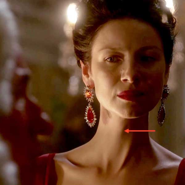
Sexual Dimorphism: Let’s turn to all things Outlander to witness sexual differences of the larynx. Here, in Outlander Starz episode 202, Not in Scotland Anymore, we see a near perfect example of the female Adam’s apple – or is it an Eve’s apple? Bwahahaha!
A small bump of the laryngeal prominence appears on Claire’s lovely ivory tower as she casts stink eye at stinky Duke! Love, love, love those earrings – perfect accessory for her swan-like neck!
Compare and contrast Claire’s gentle laryngeal prominence with that of a typical male. Weil, now, perhaps not entirely typical, but certainly impressive! Jamie obligingly lifts his chin to reveal his prominent Adam’s apple (Starz episode 107, The Wedding). Lots of testosterone plus Claire help him have a vera fine time. Whinny and snort!

The Voice: Let’s end this lesson with a brief discussion of The Voice…No, not the TV show! – just voice in general, the third important function of the vocal folds. Phonation is arguably less critical than maintaining the airway, but very important for communication and quality of life.
Roman physician Galen (130 – c. 200 CE) declared the larynx as the “first and supremely most important instrument of the voice.” Was he correct? Yes, because sounds are normally generated by vibrations of vocal folds and action of laryngeal muscles (side note: people lacking functional vocal folds can be taught esophageal speech and/or use of an electrolarynx). However, sound quality is nuanced by numerous other factors including:
- Lung capacity
- Extrinsic laryngeal muscles
- Tongue movement
- Lip action
- Mouth anatomy
- Pharynx, cheeks and soft palate changes
- Paranasal sinus configuration
The cumulative effect of these factors grants to humans a voice that can be modulated to an amazing degree; never more evident than during singing. Consider the movements of lips, tongue, soft palate, etc. in this wonderful MRI of a man singing opera. The movements are astounding. Try to locate epiglottis and arytenoids. I hope you enjoy watching this internal byplay on the screen!
Question #2: Readers have asked why our taped voices and spoken voices sound differently to our own ears? Answer: Taped voices are recorded via sound waves traveling through air before reaching our ears. The spoken voice creates vibrations transmitted to the inner ear (Anatomy Lesson # 25, “If a Tree Falls – The Ear”) mostly via bone and soft tissues, lending the voice a less tinny quality. Most folks seem to favor the spoken voice over the taped version. Make sense?
Final Outlander Fixes: Happily, Outlander gives us many treacherous throat and larynx scenes to consider!
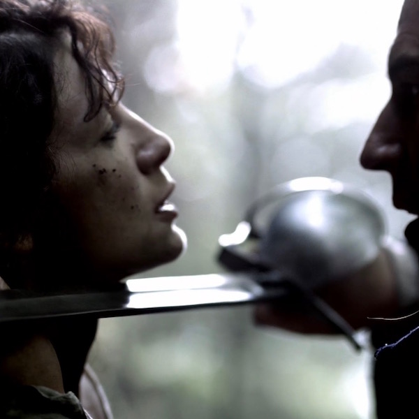
Barely out of the chute, Claire is confronted with Captain of Dragoons’ blade at her neck (Starz episode 101, Sassenach). Good way to lose a Lovely Larynx! Fortunately for our nimble nurse, his threat is only virtual. The man’s got skills, ye ken?
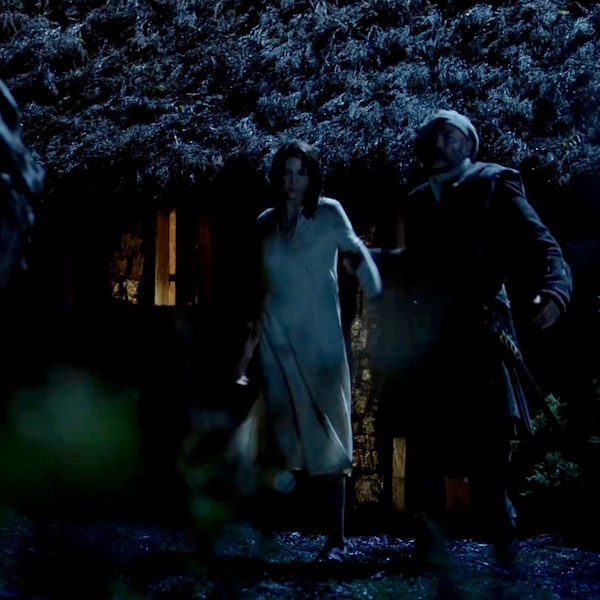
Puir not-a-wet-nurse Claire doesna get a break in episode 101. Next, Dougal jerks her around and tells her to stay with the rest of his troop of hairy-merry lads or he will obligingly slit her throat (Starz episode 101, Sassenach)! Lovely larynx at risk, again!
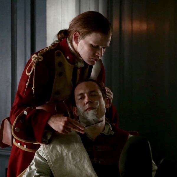
Remember the Sadist’s Shave? Captain Jack-Rat’s larynx is fully exposed to a vera sharp blade (Starz episode 106, The Garrison Commander). Too bad Corporal Milksop’s hand didna slip. Would have saved team Fraser a world of grief!
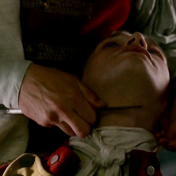
Oops. Flip-flop and the tables are turned. Corporal Hawkins’ hand DID slip – now, he is under the blade, getting the absolute worst: a dry-neck shave (Starz episode 106, The Garrison Commander)! Vera unpleasant, especially at the hands of the foul fearsome fiend!
BOOK QUOTE: Now, a few fabulous apropos lines about a dearly departed larynx from Dragonfly in Amber book:
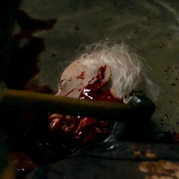
Mary sneezed, and wiped her nose hastily with a fold of her plaid. She stared at Murtagh, eyes wide and baffled. I gazed down at the bulging saddle-bag, feeling a sudden deep chill that owed nothing to the weather outside. But it was Hugh Munro’s widow who sank to her knees, and with steady hands opened the bag and drew out the head of the Duke of Sandringham.
In Starz episode 211, Vengeance is Mine, Jamie, Claire and contrary-Mary witness Sandringham, forever separated from his smooth-speaking larynx. No more mocking jibes from this oily opportunist. Sure and true, Murtagh’s ax seeks its mark!
Diana, author of both Starz episode 211 and Dragonfly in Amber book, offers two different routes to the Duke’s Demise although the outcome is identical…. grand example of “the end justifies the means?”
The Duke, he lost his larynx,
Along with his pharynx.
The ax performed a hi-jinx.
Murtagh’s work is done!
(Let’s hear it for the Godfather!)
Final voice Issue: According to 13 August 2016, New Scientist, and Buzz Feed, Appen, a voice recognition firm working for Google, a call for help has been posted on Reddit’s Edinburgh page for people with Scottish accents to submit recordings of themselves reading certain phrases to help train its software! Users with certain accents – particularly Scottish – have complained that voice recognition systems such as those used by Good Now and Apple’s Siri struggle to understand them. Here is a great comment to illustrate the dilemma. Well, at least Siri got “street” right! Am I right?

I hope you enjoyed learning about the incredible human larynx. Give a brief salute the next time you take a breath or utter a no-no word! Have a wonderful breath-taking day.
A deeply grateful,
Outlander Anatomist
Photo creds: Starz, Netter’s Atlas of Human Anatomy, 4th edition (Images B,C,D,F,G,H,I,J,K,L,M), Grey’s Anatomy, 39th edition (Table A data), www.flickr.com (Image E), www.kit10phish.wordpress.com (Image Q), www.scienceblogs.com (Image N) www.newhealthadvisor.com (Image O), www.quizlet.com (Image P), www.thisiscommonsense.com (red flag and dog), www.vocalclinic.org (Photo A), www.voicedoctorla.com (Image N), Buzzfeed