Hey, anatomy students! How about a brief pilus quiz? No grading, I promise.
What type of pili inhabit your head? Turns out, there are different systems of classifications, but the following one is practical and easy.
Shape of your pili:
Straight?
Wavy?
Curly?
Coily?
None? 😉
Type of pili strand:
Fine?
Medium?
Coarse?
Amount of hair? (based on circumference of “full hair ponytail”). If you don’t have a ponytail, then guess.
Thin (ponytail is 2” or less)
Normal (ponytail is 2”-4”)
Thick (polytail is > 4”)
How did you do? Wouldn’t you know it, all such characteristics have been described and worked out. 🤗 There are even subcategories of hair if you want to read more!
Pili are fascinating for many reason. First and foremost, pili are products of skin, our body’s largest organ! The skin of “average adults” weighs about eight pounds, with a surface area of 22 ft²! 😲 The larger one is, the more weight and more surface area is taken up by skin.
The skin is equipped with various appendages including pili, erector pili muscles , nails, and various glands.
Turns out, hair is far more complex than one might imagine. Read on for more fascinating deets!
Hair growth differs depending on body region (duh 😉):
Glabrous: Regions sans hair – palms, soles, external genitalia, lips, back of ear, and scars.
Vellus: Thin, fine, light-colored hair typical of childhood and adult women. In female adults found on eyelids, face, chest, etc. Vellus hair can convert to terminal hair under the influence of androgens.
Next, anatomy divides the pilus into two parts:
Follicle: Part embedded in the dermis – the only living part of a hair.
Shaft: Thin filamentous part that extends beyond skin surface – non-living part
Follicle: A follicle is the part of a pilus that lies below the skin surface. Pull out a strand of head hair, and observe a pale enlargement on the end that was embedded in skin – this is the bulb or root of the follicle. The shaft is produced by the root.
Follicles are lined with skin stem cells that can re-grow a hair after it is lost. It may also regrow skin after various wounds, such burns. Here, stem cells produce new skin cells that grow out of the follicle and spread across the damaged surface to help cover the injury. This is effective if the wound is relatively small; larger wounds may require skin grafts. Lastly, the new skin is a type of scar tissue which does not regrow appendages.
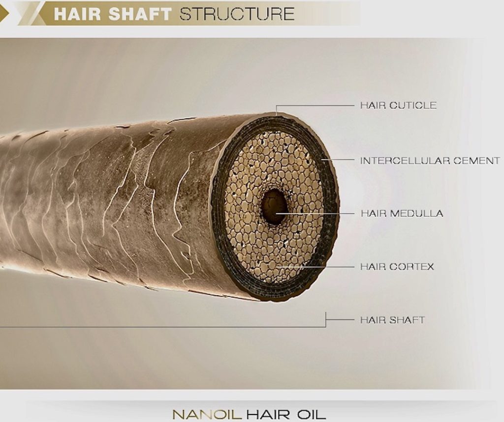
Shaft: The shaft is 2-3 layers of non-living material:
Cuticle: consists of thin, flat cells overlapping like shingles of a roof.
Cortex: Rod-like bundles of alpha keratin, a protein that strengthens the shaft. This layer also gives hair its color.
Medulla: Unstructured area in the center – only present in large pili.
People with straight hair have round shafts. People with wavy, curly or coiled hair have oval or flattened shafts. The follicle itself determines the shaft shape and genetics orchestrates the follicle to do its unique thing🤓!
Growth: Each human hair follows its own cycle, at its own pace, including periods of growth and times of quiescence. Think about it! If all our pili were on the same cycle, we would molt! 😳
Angle: You should also know that the shaft does not grow upright; it emerges at a slant.
Try this: Check the angle of growth of your hair: place your forearm on a flat surface with the palm down. Examine your forearm hairs and see that they are angled toward the little finger side of the forearm. That’s the slant!
Arrector Pili Muscle: Microscopic bundles of smooth muscle (meaning these cannot be voluntarily contracted) are attached to the follicle. If these muscles contract, they pull on the follicles causing shafts to stand upright, creating “goose bumps.” Watch this video about the arrector pili muscle for perspective. Contraction happens when we are cold or creeped out! 🥶😱
Contraction of arrector pili muscles also causes oil glands to release their product (sebum) into their respective follicles following the pilus shaft.
Pili are highly valued in many societies which explains the vast sums of money spent on hair products each year; almost 80 million dollars in US in 2019 –down from 90 million spent two years earlier.
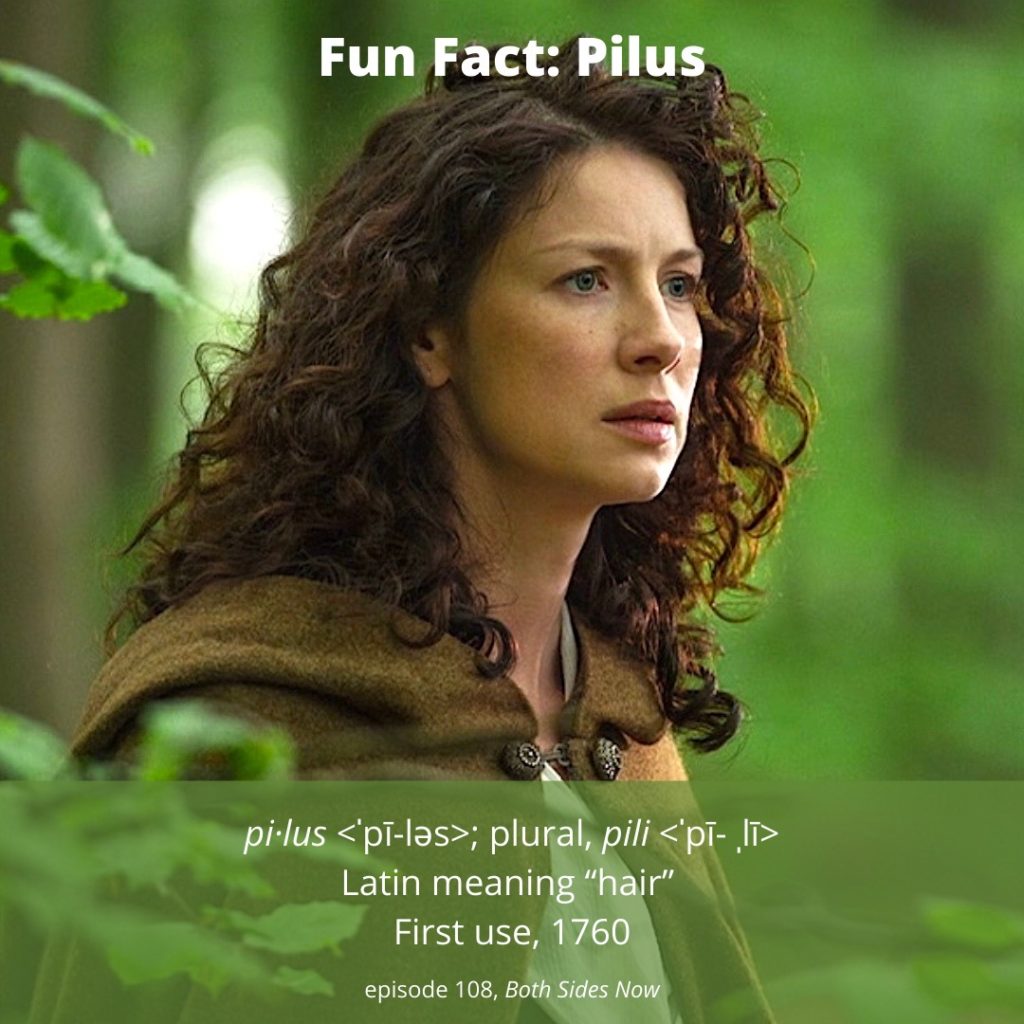
Read about Claire’s-Hair in Outlander book. Diana has provided us with ample descriptions of her follicles and shafts. Here are three iconic descriptions of her amazing pili!!! 😲
The wind was rising and the very air of the bedroom was prickly with electricity. I drew the brush through my hair, making the curls snap with static and spring into knots and furious tangles!
… “Mo duinne?”…“It means ’my brown one.’ ”He raised a lock of hair to his lips and smiled, with a look in his eyes that started all the drops of my own blood chasing each other through my veins. Rather a dull color, brown, I’ve always thought,”….”No, I’d not say that, Sassenach. Not dull at all.” He lifted the mass of my hair with both hands and fanned it out. “It’s like the water in a bern, where it ruffles over the stones. Dark in the wavy spots, with bits of silver on the surface where the sun catches it.”
…”Fretful porpentine, was it?” he asked. He tilted his head, examining me inquisitively. “Mmm,” he said, running a hand over his head to smooth down his own hair. “Fretful, at least. You’re a fuzzy wee thing when ye wake, to be sure.” He rolled over toward me, reaching out a hand. “Come here, my wee milkweed.”🥰
See Claire glorious crown of pili in Outlander, episode 109. Both sides Now!
Grateful for each and every one of my pili! How about you?
Greeting anatomy students, everywhere!Today’s lesson will examine the marvelous, mystical human skull.
Our first lesson about the skeleton was waaay back in Anatomy Lesson #39, Dem Bones – Human Skeleton, but that discussion was pretty general in nature, whereas, the skull alone is quite specific. We do not have sufficient space to cover all details about the skull, so highlights must do.
First, well-earned homage to our fav author, Diana Gabaldon, who wrote this about her life, pre-Outlander:
From the late ’70s to the early ’90s, I wrote anything anybody would pay me for. This ranged from articles on how to clean a longhorn cow’s skull for living-room decoration to manuals on elementary math instruction on the Apple II… to a slew of software reviews and application articles done for the computer press.
Long-horn skulls? Yep. She’s been into head bones for a very long time. 😉
You might not recall, but Outlander has some great scenes involving skulls, so let’s get going!
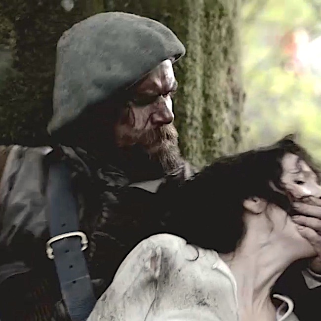
Beginning with Starz ep 101, Sassenach, a mess of skull stuff happens. Murtagh smartly raps Claire’s skull with his dirk hilt to quiet her. Sorry lass, Redcoats everywhere! And, there’s even more skull stuff:
Ep 104, The Gathering, Claire wallops Dougal over the heid with a chair
Ep 104, The Gathering, Rupert whacks Jamie on the skull with his dirk.
Ep 104, The Gathering, Rupert whacks Jamie on the skull with his fists! (Ep 104 was mighty rough!)
Ep 108, Both Sides Now, Frank, related to Black Jack, beats thugs’ skulls with his own blackjack!
Ep 109, The Reckoning, Murtagh thunks a guard on the skull at Fort William.
Ep 204, La Dame Blanche, Murtagh gets a taste of his own medicine from a secret Paris society bent on violating virgins. Skull dunt!
Ep 211, Vengeance is Mine, Murtagh separates Duke S. from his skull. Weil, he was asking for it!
Ep 305, Whisky and Freedom, Dr. Abernathy fondles a pretty lady’s skull in his office. Claire assists. <G>
Ep 307, Creme de Menthe, With help from Claire’s knife, an excise man’s skull strikes stone!
Ep 308, First Wife, young Ian is bonked over the heid by pirates!
Ep 311, Uncharted, Dermestid beetles clean Arabella’s skull!
Ep 312, Eye of the Storm, Claire relieves Mrs. Abernathy’s body of the weight of her skull!
Doubtless, I have missed a few. Help me out here, anatomy students!
Update! A student reminded me of the cave scene wherein Geillis shoots Jamie in the head. The pistol ball travels under his scalp to end up in the back of his head (occipital region). Claire removes it with a blade. The ball failed to penetrate Jamie’s skull because Geillis had failed to load the pistol with sufficient charge. Thank you, Marguerite!
This is the quote from Voyager book. Sadly, it wasn’t filmed so no image to accompany this splendid description:
… I sat Jamie down with a pan of water, to tend the damage to his head. I washed away the blood from face and hair, to find to my surprise that the ball had in fact not plowed a furrow through his scalp as I had thought. Instead, it had pierced the skin just above his hairline and—evidently—vanished into his head. There was no sign of an exit wound. Unnerved by this, I prodded his scalp with increasing agitation, until a sudden cry from the patient announced that I had discovered the bullet. There was a large, tender lump on the back of his head. The pistol ball had traveled under the skin, skimming the curve of his skull, and come to rest just over his occiput.
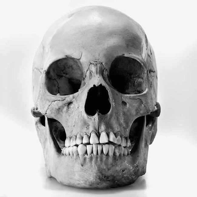
I usually resist descending into the macabre, but let’s introduce the skull with this tidbit. In 2016, 454 human skulls (Image A) were offered for sale on eBay with opening bids ranging from one cent to $5,500!Sources of the skulls were unknown.Since then, eBay has revised its policy to “ban the sale of all human body parts except hair.” Thumbs up! Without informed consent, the sale of body parts is rife with ethical issues.
As a former Director of the body donation program at my medical university, sales of human parts for non-scientific purposes were deemed unethical – a sound policy.
Image A
Definitions: Best to begin our lesson with definitions.
The English word “skull” is likely derived from Old Norse “skulle”, whereas the Latin word cranium comes from the Greek root κρανίον (kranion). What do these words mean?
Skull – all bones of head including mandible (lower jaw). Teeth are not included because they are not bones.
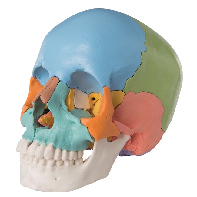
Cranium ( see Image B) – all skull bones (colors) excluding the mandible (white)
Neurocranium – cranial bones that encase the brain
Viscerocranium – facial bones
Function:
Q: Why are skulls so important?
A: Because, skulls are critical elements of the human skeleton which serve to protect the brain and house these major sensory organs:
The skull also fixes the distance between the eyes to allow for stereoscopic vision (depth perception), and positions the ears to enable us to localize direction and distance of sounds.
Image B
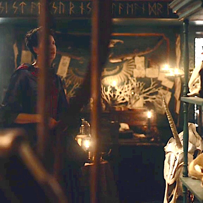
Pause for another deep breath of Outlander! Whisking us into the ‘little shop of horrors” run by conjurer Master Raymond (Starz, ep 204, La Dame Blanche), Claire beholds strange sights.Filled with oddities and ancient bones, curios include the skull of a unicorn! What???? He, he. There it is, with its own shaffron (head armor) complete with a hole for the horn. Only fitting to pay respect to Scotland’s National Animal. Clever!
Back to the anatomy grind…..
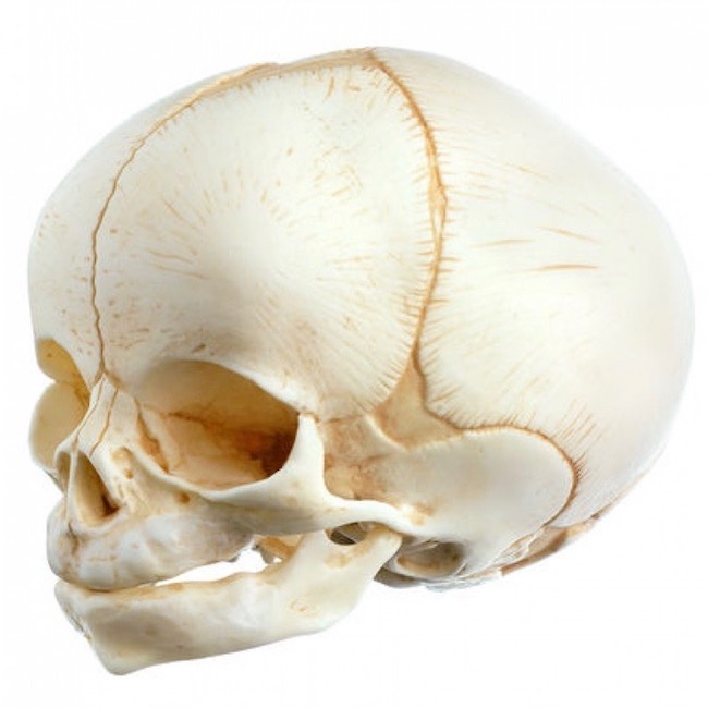
Skull Development: The human skull passes through amazing transformations during development. At birth, the skull is made of 44 different bony elements and the facial skeleton is 1/7 the size of the calvaria (Image C). In other words, big head – small face.
As bony elements fuse, open areas persist; these are the fontanelles (6 of them). With further age, skull bones fuse into unmovable joints known sutures– only the mandible retains a pair of moveable joints throughout life. Some sutures contain little islands of bone. Collectively known as wormian bones, these are inconsistent features of the human skull.
Psssst…No cause to fash about this wee skull; it is a plastic model.
Image C
Adult Skull: Once fusion is complete, the adult skull has 22 or 28 bones depending on how they are counted (anatomists differ on this): 28, if ear ossicles (Anatomy Lesson #25, If a Tree Falls – The Ear) are included in the count or 22, if they are not. And, the adult facial skeleton is 1/2 the size of the calvaria, meaning with age, facial bones grow more than cranial bones. Wormian bones are not included in a skull bone count because they are inconsistent features. Remember? Good!
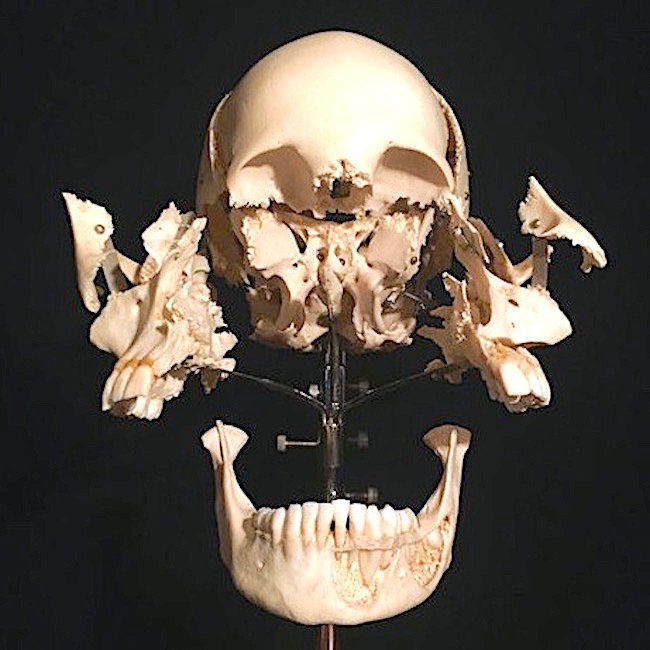
Wow! Image D shows an adult skull which has been “exploded,” exposing its component bones. This view affords an appreciation of the skull and its many varied and complex parts. Such preparations are very expensive but, nonetheless, are rather common exhibits in anatomy labs. Typically, these are encased in glass and unavailable for handling because several bones are paper-thin. Look but do not touch!
Image D
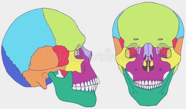
Here are the odd names of the skull bones (Image E):
occipital (1) – royal blue
temporal (2) – orange
parietal (2) –turquoise
sphenoid (1) – red
ethmoid (1) pink
frontal (1) – lime green
nasal (2) – lavender
lacrimal (2) – lavender (guess they ran out of colors?)
zygomatic (2) – yellow
maxillae (2) – purple
mandible (1) – dark green
vomer (1) – peach (part of nasal septum)
inferior turbinate (2)– yellow (sides of nasal cavities)
palatine (2) – not shown (part of roof of mouth)
Image E
Another dram of Outlander!
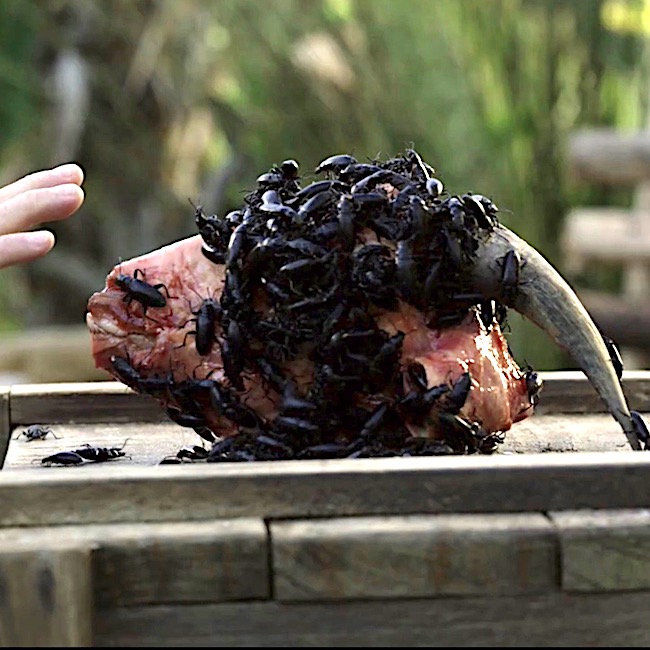
Ever ponder how skulls end up so clean? Anatomical preparers typically utilize insects to do the job. Dermestid beetles are splendid at this icky task and, believe it or not, they are fastidious eaters because they prefer to dine only on carrion! These beasties can even be purchased on line. Or, if beetles don’t suit you, hydrogen peroxide and baking soda are home remedies for an animal skull which demands a thorough cleaning.
Therefore, Outlander accurately depicts Father Fogden using beetles to clean and preserve beloved Arabella’s skull (Starz episode 311, Uncharted). Talk about gross anatomy. Total yuck!!!
If you really wish to see the process in a scientific setting, this is a good YouTube video. But, I advise you to skip, if you are squeamish.
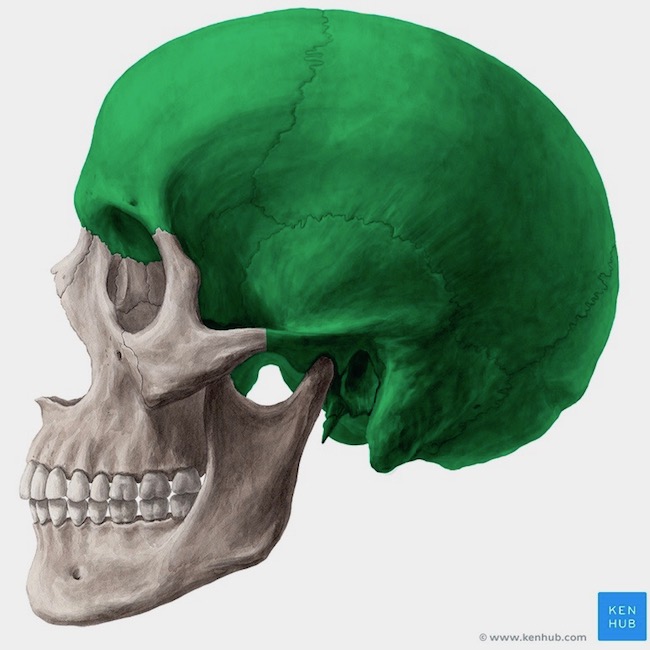
Neurocranium: A few tidbits about the neurocranium. This part of the skull is commonly known as the braincase because it forms a bony hollow housing the brain (Image F). It is composed of all skull bones except mandible and facial bones. It is like a rounded cubical with ceiling, floor, front, back and sides. The shape is a perfect fit providing solid support for soft brain tissue. Got it? Yay!
Image F
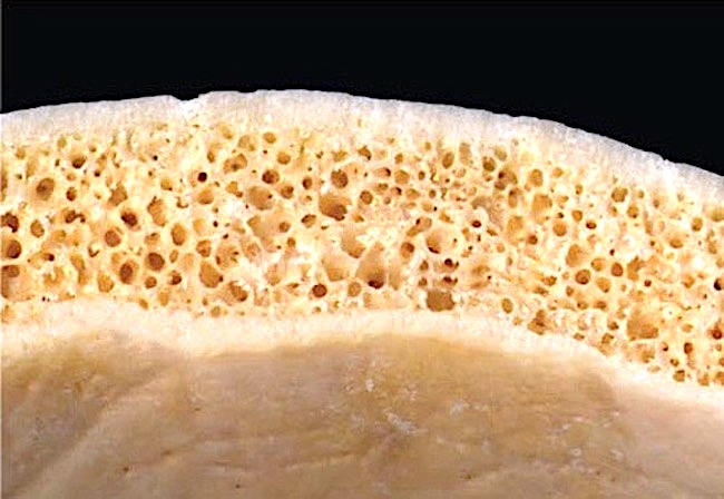
Flat Bones: Bones of the neurocranium come in weird shapes. Some, such as frontal, parietal and part of temporal are thin, flat bones. Flat bones are fascinating because they are curved (go figure, <g>) with outer and inner layers of compact bone sandwiching a core of spongy bone. In image G, the top layer of dense bone is the outer surface, adjacent to the scalp; the bottom layer is closest to the brain. Each compact bony layer is known as a table, so there are outer and inner tables. The spongy core, known as diploe, isn’t spongy at all (go figure, <G>). But, it sure looks spongy. Rather, diploe is a delicate network of bone riddled with holes. In life, the holes aren’t empty; they are filled with blood vessels, developing blood cells and fat cells. Hence, the term “fat heid.” Ha, ha – just kidding!
Image G
Foramina: Another interesting feature – the skull is full of holes (Image H)! Known as foramina (sing. foramen), the holes traverse the skull from outside in or inside out depending on your point of view. Such openings vary from pinpoint size to the largest, the foramen magnum (2.5 – 3.4 cm), at the skull base.
Foramina are ports for the passage of blood vessels and nerves between inside and outside the skull. Foramen magnum is traversed by the spinal cord as it descends to enter the vertebral canal (Anatomy Lesson #10, Jamie’s Back or Aye, Jamie’s Back!).
Try This: Bring palms together with thumbs extended toward the face. Place thumb pads against the eyebrows and move the pads back and forth a bit. They should settle into a pair of divots or depressions. These are the supraorbital notches/foramina through which pass the supraorbital (sensory) nerves. You have just demonstrated the method by which these nerves leave the skull and reach the face. Hurrah!
.
Superior view of the cranial base
Image H
Meningeal Arteries: The brain, nestled inside the neurocranium, is surrounded by three layers of membranes, the meninges. The outermost meninx (sing.), known as dura mater (Latin meaning tough mother), contains several meningeal arteries which supply blood to the dura and adjacent skull. Scroll back to Image H and locate grooves on the inner surface of the bottom table. These imprints are caused by meningeal arteries pressing into the bone.
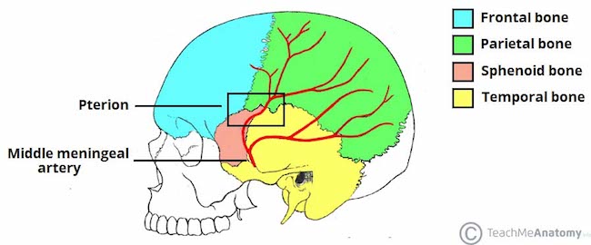
One such vessel is the middle meningeal artery. This important artery is located deep to the temple region where four neurocranial bones meet at the pterion (Image I). Here, the bones are very thin.
Now, because the brain is encased in bone, one might expected it to be impervious to harm, but if so, one would be wrong.A blow, fall or other accident (such as a golf ball to the temple) can burst the middle meningeal artery causing blood to accumulate between the dura and inner bony table, an injury known as an epidural hematoma (a clot between skull and dura mater). The accumulation of blood puts pressure on the brain and interferes with neural function.
This type of brain injury is usually accompanied by loss of consciousness, brief regaining of consciousness, followed by another loss of consciousness. Confusion is typical; bleeding from the ear may occur. Treatment requires immediate surgery, a craniotomy. Without treatment, death typically ensues.
So, can you surmise where this lesson is headed? Of course you can!
Image I
OK, now let’s see how anatomy applies to Outlander!
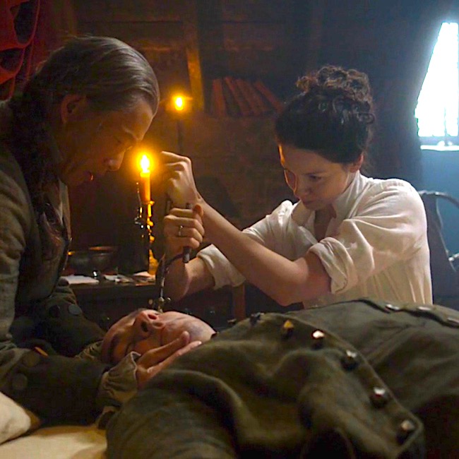
Incise the Excise Man: John Barton, a nasty tax man working for the corrupt Sir Percival, attacks Claire in Jamie’s brothel-nest (Starz ep 307, Creme de Menthe). During his battle with Dr. Dura Mater, she stabs his leg and he falls striking his left temple on the stone hearth. Blood drains from his left ear and Claire (sans modern imaging techniques) quickly diagnosis an epidural hematoma!
Soon, she acquires a trephine (barber surgeons in Edinburgh would likely have these), in essence, a hand drill. She incises the skin over John’s left temple, positions the trephine and proceeds to drill for oil!
Now, drilling through skull bones of the pterion region means the bit must pass through outer table, diploe and inner table of said bones. The good Doctor detects a slight give as the drill completes the traverse. Then, (and, this was thrilling to me!) Claire correctly backs the drill out by reversing direction of the drill handle and voila, a burr hole! What a braw lassie!
Now, blood can drain from the injury giving John a chance at survival. Unfortunately, or fortunately if you belong to team Jamie, he does not. He would surely have died without the surgery, but he died with it, anyway. Warrior Claire fought valiantly for her patient in her own battle joined, but to no avail.
Understand that trephination/trepanation is not a new surgical technique as burr holes been found in prehistoric human remains. In ancient times, holes were drilled into a person’s skull, it is thought, to release evil spirits. BJR surely could have used one! Or how about Geillis?
So, armed with the science of anatomy, we now understand the nitty gritty of what took John Barton’s life! Don’t you feel ever so much wiser?
Today, a craniotomy is performed to release the pressure from an epidural hematoma and other types of brain injuries. Although more sophisticated, it works similarly to a trephination. In an abbreviated explanation, 3-4 burr holes are drilled through the skull and connected by saw. The piece of skull, or bone flap, is freed. The hematoma (blood clot) is usually suctioned out, the bony segment replaced and the scalp secured in place.
If you aren’t squeamish, this video shows an excellent demo of a craniotomy for epidural hematoma:
And, Claire’s version:
Now, lest you depart this lesson thinking the Outlander trepanation/trephination is a total fabrication by the series writers, it isn’t. Diana wrote about trephination in Drums of Autumn. Yes, she did. This woman leaves no stone unturned!
Here is the quote (there is another in An Echo in the bone), but to prevent spoilers, the name of the patient is withheld and another name is blocked out, otherwise the quote is intact:
She was thinner than he remembered, though it was hard to judge of her figure, dressed as she was in a barbaric leather shirt and trouserings. She’d plainly been in the sun and weather for some time; her face and hands had baked a delicate soft brown, that made the big golden eyes that much more startling when they turned full on one—which they now did.
“ ———-says that Dr. Fentiman trephined your skull.” He shifted uncomfortably under the sheets. “I am told that he did. I am afraid I was not aware of it at the time.” Her mouth quirked slightly. “Just as well. Would you mind if I look at it? It’s only curiosity,” she went on, with unaccustomed delicacy. “Not medical necessity. It’s only that I’ve never seen a trepanation.” He closed his eyes, giving up. “Beyond the state of my bowels, I have no secrets from you, madame.” He tilted his head, indicating the location of the hole in his head, and felt her cool fingers slide under the bandage, lifting the gauze and allowing a breath of air to soothe his hot head.
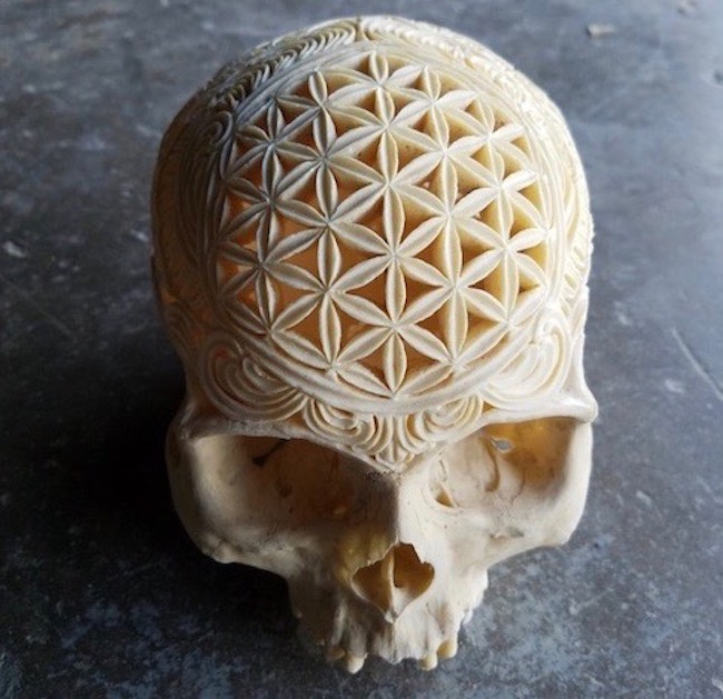
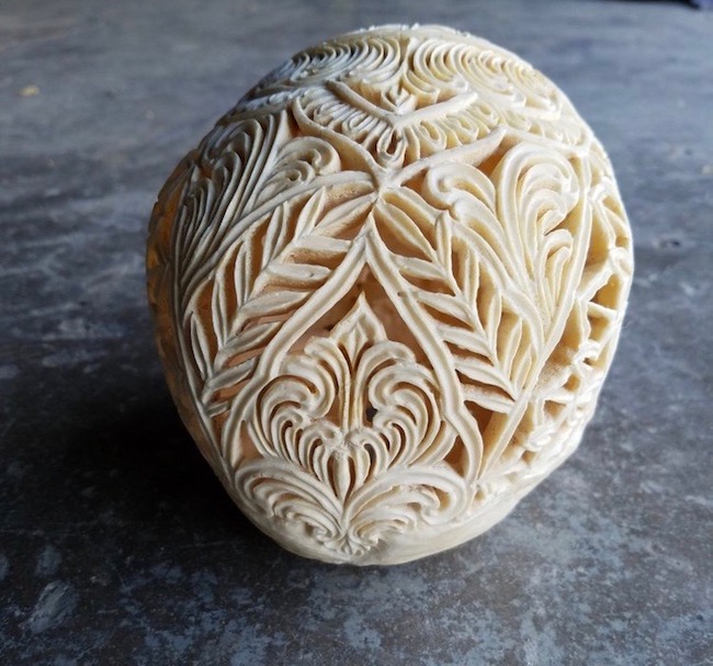
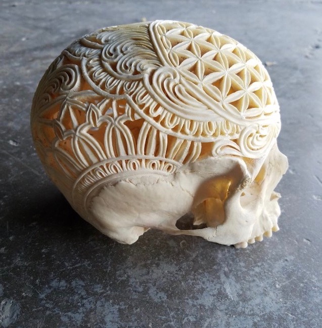
Now, let’s close this lesson with a feeling of satisfaction for knowledge gained and with an appreciation of skull art. The following three images show an intricate and creative carving of a human skull.I do appreciate the skill although I remain ambivalent about using human skulls in this manner.And, it is a human skull. I enlarged the images and diploe is clearly visible at some of the cut surfaces. Plastic models don’t exhibit spongy bone in their construct.
Let’s close with the lyrics of “It’s a Lie,” by the rock band, Fiction Plane:
Underneath my face there is a human skull
Without the living flesh you’d find it pretty dull
Ah, no. With all due respect, I disagree! The skull is a fascinating part of the human anatomy. Fiction Plane guys, read the lesson! <G>
Hello, anatomy students! Hello feet! Today’s anatomical offering derives from the Old English fot, meaning “foot.”
You might think Outlander has little to say about feet, but not so. Outlander has fleet feet, sweet feet, trick-or-treat feet, body heat feet, upbeat feet, mincemeat feet, eek feet, beat feet and indiscrete feet. As always, Outlander images and quotes are sprinkled throughout the lesson. Yay!
You might also think society has little to say about feet, but not so. Dozens of feet adages accent life’s little highs and lows:
Foot in both camps (fence sitter)
Ankle deep (in trouble)
Back on your feet (on the mend)
Bound hand and foot (hampered)
Cold feet (lost interest)
Get your feet wet (get involved)
Drag your feet (unenthusiastic)
Feet of clay (flawed)
Foot in mouth (oops!)
Keep feet on the ground (be sensible)
Footloose (6 degrees of Kevin Bacon)
As a grad student, my gross anatomy prof declared feet the ugliest body part! Beauty being in the eye of the beholder aside, our foot is far more specialized than our hand as no other animal has a foot quite like ours! I hold with the master, Leonardo DaVinci who opined:
The human foot is a masterpiece of engineering and a work of art.
Yes!
Understand, the human foot is a truly complex mechanical structure consisting of 26 bones, 33 joints, and more than a hundred muscles, tendons, and ligaments! Despite this complexity, our feet usually work pretty well, especially if we care for them. Also, know this lesson is geared for general readers, so the most complex features and smallish details are not covered.
Before we begin the lesson, let’s take a gander at Claire’s fleet feet (Outlander ep 101 Sassenach) as she retreats from redcoat muskets firing live rounds! Her poor heels are rubbed raw from running – leather shoon sans sox. Not a good plan, but did the lass have another choice? Och!
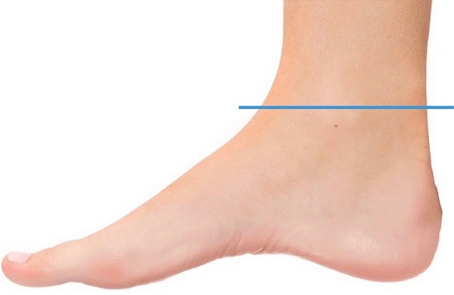
Foot: Always prudent to begin a lesson with definitions. In anatomy, the foot is the lower limb below (distal to) the ankle joint (Image A –blue line).
Orienting ourselves further, the top of foot is the dorsal surface; the bottom (sole) is the plantar surface. Inner side is the medial surface and the outer side is the lateral surface.
Image A
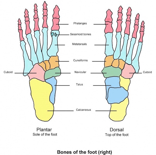
Skeleton: We begin with the foot skeleton, a foundation of 26 bones. Image B shows bones of a right foot, 26 in all. The left panel shows bones from the bottom or plantar perspective. The right panel shows foot bones from above, the dorsal view.
Phalanges: Toes contain 14 phalanges (Image B, pink) – three bones per each toe, except the big toe which, like the thumb, only contains two phalangeal bones. And, the big or great toe goes by the scientific name, hallux.
Metatarsals: Five metatarsal bones (aqua) are homologous with metacarpals of the hand.
Tarsals: Seven tarsal bones are homologous to eight carpals of the wrist, although more massive and oddly-shaped than the small carpals.The largest, the calcaneus, is the heel bone. Atop it is the talus which helps form the ankle joint.
There won’t be a quiz of tarsal bones <g>, but, just so you know, their names are (Image B):
three cuneiforms – 1, 2, & 3 (start counting on big toe side – orange)
cuboid (pink)
navicular (green)
calcaneus (yellow)
talus (blue)
Image B
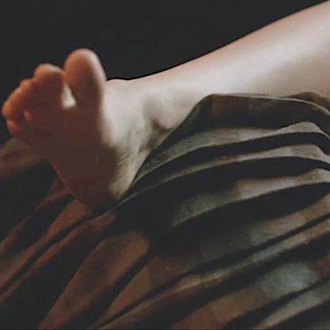
Need another Outlander hit? My pleasure. No, really! The morning after (he, he), Claire sports a pair of verra sweet feet (Starz ep 107, The Wedding)! Aw, look at those wee tootsies, shyly nesting. It was a big, big night! ’Nuf said! 😉
Oops, not quite, ‘nuf said. A lovely foot passage from Dragonfly in Amber book. Herself even mentions metatarsals!
“I’m honest enough to say that I dinna care what the right and wrong of it may be, so long as you are here wi’ me, Claire,” he said softly. “If it was a sin for you to choose me … then I would go to the Devil himself and bless him for tempting ye to it.” He lifted my foot and gently kissed the tip of my big toe. I laid my hand on his head; the short hair felt bristly but soft, like a very young hedgehog. “I don’t think it was wrong,” I said softly. “But if it was … then I’ll go to the Devil with you, Jamie Fraser.” He closed his eyes and bowed his head over my foot. He held it so tightly that I could feel the long, slender metatarsals pressed together; still, I didn’t pull back. I dug my fingers into his scalp and tugged his hair gently.
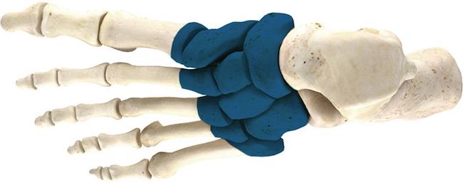
Divisions: Foot bones are divided into three regions. Such divisions aren’t whimsy, they are important in issues such as evaluating trauma, surgical amputation of part of all of a foot or evaluation of foot mechanics.
Forefoot: Includes phalanges and metatarsals (Image C – white, left side).
Mid-foot: Includes three cuneiforms, cuboid and navicular (Image C, turquoise).
Hind-foot: calcaneus and talus (Image C – white, right side).
Image C
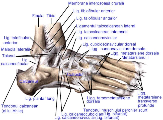
Ligaments: Now, the 26 foot bones don’t just hang out under the skin. They are firmly bound to each other and to our leg bones via dozens of ligaments (Image D)!
Ligaments are fibrous tissues binding bone to bone and they are critical for foot integrity because feet bear our weight against gravity! Loose or torn ligaments give folks many problems because these compromise the integrity of the skeletal system!
Image D shows only some of the numerous ligaments anchoring and stabilizing foot bones; here, we see lateral and dorsal ligaments. The plantar and medial ligaments are not visible.
The lesson won’t discuss these ligaments in detail because they are complex and tedious, but the image dramatically emphasizes some of the many ligaments needed to stabilize the foot skeleton!
Image D
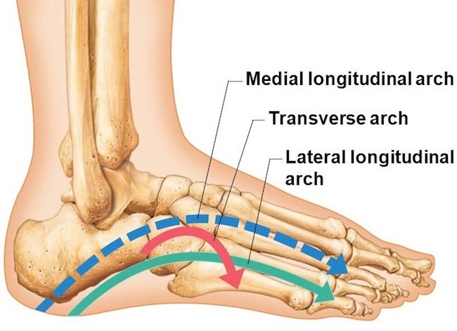
Arches: We all know the foot has an arch, but did you know it actually has three arches (some count a 4th partial arch)? Two are longitudinal and one is transverse. The arches are maintained by interlocking tarsal and metatarsal bones, supported by ligaments and very strong tendons (image E).
medial longitudinal arch (Image E – blue dashed line) extends from heel bone to first three metatarsals. Typically, it curves above the ground. When barefoot at the beach, it does not leave an imprint in sand (unless one is seriously flatfooted!).
lateral longitudinal arch (Image E – green line) is a low arch arch between calcaneus and 5th metatarsal. When barefoot, it typically leaves an imprint in sand.
transverse arch (Image E – red line) runs across the foot at the tarsometatarsal joints (defined below).
Although these arches are supported by strong ligaments and tendons, they exhibit some mobility when weight is applied to or removed from the foot. This springiness makes walking and running more economical in terms of energy.
Image E
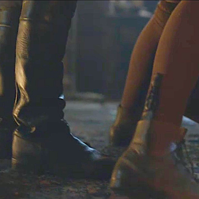
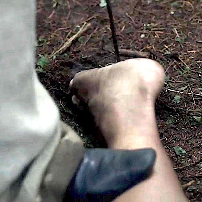
Speaking of arches, how ‘bout some booted ones! Yep, another dose to wake you students! BJR’s booted “trick or treat feet” (Starz, ep 108, Both Sides Now) will do the trick, nicely! The blackguard throws Claire over his desk preparing to further assault her. Darn! She canna reach the sgian dubh in her boot! No treat here – all vicious tricks!
Diana describes Claire’s toes just after Jamie squats in the prison window (Outlander book).
Randall bent and scooped up the gun in a quicksilver motion. As soon as the knife left my throat, I tried to sit up, but he placed a hand on my chest and shoved me flat again. He held me down with one hand, using the other to aim the pistol at Jamie. The discarded knife lay somewhere on the floor near my feet, I thought. Now, if only I had prehensile toes.… The dirk in my pocket was as unreachable as if it were on Mars.
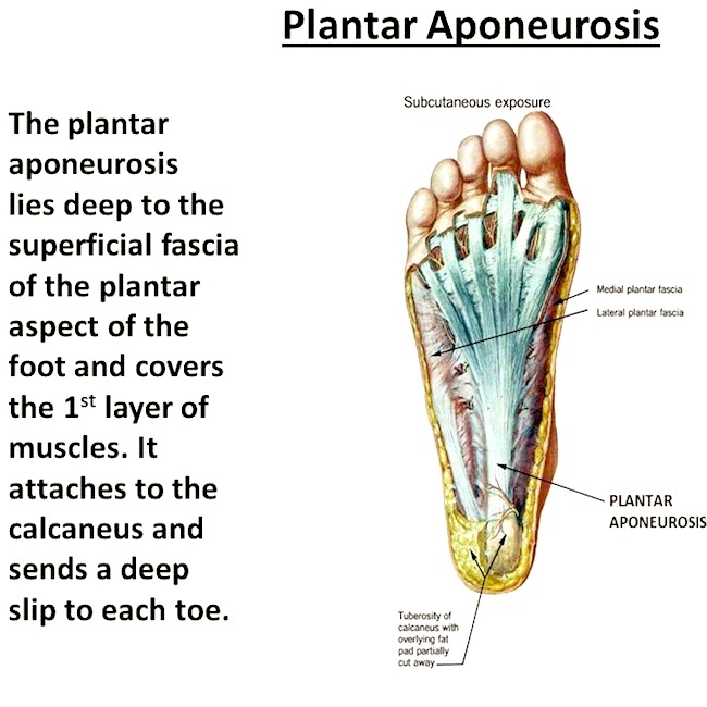
Plantar Aponeurosis: Remove plantar skin (very difficult on a cadaver) and a triangular sheet of connective tissue is revealed, the plantar aponeurosis. It is anchored to the calcaneus, flares in the mid-foot and ends as five (or more) bands radiating toward bases of the toes (Image F).
The tough, fibrous aponeurosis is made mostly of collagen fibers. As such, it is a shock absorber when the foot strikes the ground. It also stabilizes arches of the foot and allows flexion at the first metatarsophalangeal joint, which carries the majority of body weight during ambulation.
If the plantar aponeurosis becomes injured or inflamed, it may cause plantar fasciatis. A painful condition common to athletes, it causes stinging foot pain that can lead to further leg injuries if untreated.
Image F
Another break for Outlander! This is a splendid example of body heat feet (Starz, ep 109, The Reckoning). Things are on broil up at Castle Leoch! Claire’s right heel hooks over Jamie’s Fraser plaid….hum…. Talk about a foothold! Snort!
To reiterate, joints are sites where two or more bones meet; some are moveable and some are not. Moveable joints allow for motion and there are several types. Our 33 foot joints fall into the following categories:
TC Joint (1): between distal tibia, fibula and talus, a.k.a. talocrural or ankle joint
IT Joints (13): between tarsal bones
TM Joints (5): between metatarsals and tarsals
MP Joints (5) : between proximal phalanges and metatarsals
IP Joints (9): between phalangeal bones
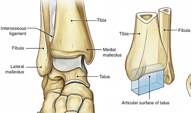
Reducing the technicality of this topic, we will only cover the superbly designed TC or ankle joint! The ankle joint is a mortise joint, a term used in carpentry. Here, the talus projects upwards and fits inside a three-sided bone box formed by tibia and fibula of the leg (Anatomy Lesson #27, Colum’s Legs and Other Things, Too!). Thus, our “ankle bones” are not separate bones, they are parts of tibia and fibula. The inner ankle bone is actually the medial malleolus of the tibia; the outer ankle bone is the lateral malleolus of the fibula. This is a highly stable hinge joint that allows movement (see below).
Image G
Before the lesson turns to movements, let’s take a quick keek at Jamie’s upbeat feet! Cheerfully dressed in nothing but a sark, he strides to the freezing mill stream to sleuth out a prob with the water wheel (Starz ep 113, Lallybroch). Seems it is producing gritty bannocks! Upbeat feet, that is, until a mess ‘o Redcoats arrive! Notice: his right foot is lifted at the ankle, a movement known as dorsiflexion. Yep! Read on to learn more about this term.
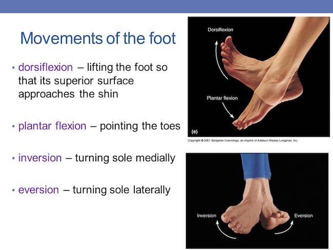
Movements: Various movements occur at the foot joints. Some are slight; others are more generous and important for ambulation. The talocrural joint (ankle joint) allows for six movements at the ankle; the first four are demonstrated in Image H:
dorsiflexion: lifting foot at the ankle
plantar flexion: pointing the foot at the ankle
inversion: turning sole medially (toward midline)
eversion: turning sole laterally (toward the side)
abduction: turning foot to side (slight)
adduction: turning foot toward midline (slight)
Psst…..Practitioners often prefer the terms, supination for inversion and pronation for eversion.
Image H
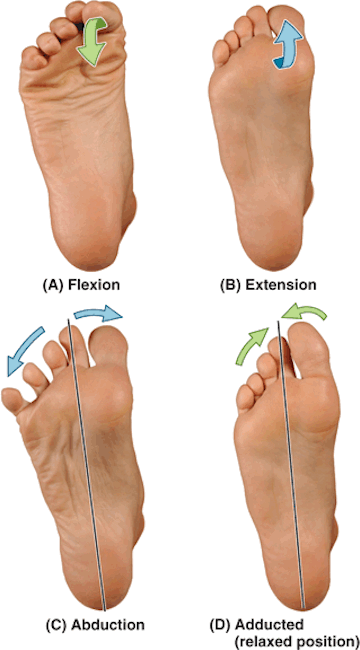
Next, there are toe movements which can occur independent of the ankle joint. These involve IP and MP joints:
flexion: curling the toes
extension: lifting the toes
abduction: spreading the toes
adduction: returning the toes to a resting position
Image I
Back for an Outlander scene and a collective gasp! Jenny takes a hot poker to the sole of a redcoat captive. Ouch! The poor man now has mincemeat feet. What ya doing Jenny?
Spill, messenger! Where is my bro??? In no uncertain terms, Big Sis declares to Claire:
Love Forces a Person to choose!
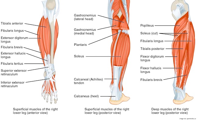
Extrinsic Muscles: First, a wee definition…long time students will remember that in anatomy the leg is the lower limb between knee and ankle joints; thigh is between hip and knee joints. Most people use the term lower leg for the anatomical leg.
Muscles acting on the foot are classified as extrinsic muscles, those originating in the leg, and intrinsic muscles, those originating in the foot.
Amazing Fact: all leg muscles, excepting one, actually act on the foot! These are so complex, they must be simplified. Yes, Image J is simplified!!!
Extrinsic muscles in Fig. J (anterior and side muscles – left panel):
tibialis anterior – dorsiflexes & supinates foot
extensor hallucis longus – extends big toe & dorsiflexes foot
gastrocnemius – plantar flexes foot (cut away in image)
soleus – plantar flexes foot
(Note: gastroc and soleus jointly share the massive achilles tendon which inserts into calcaneus. Normally, these are extremely strong plantar flexors)
Extrinsic muscles in Fig. J (deep posterior muscles – right panel):
flexor hallucis longus – flexes big toe & plantar flexes foot
Whew. That was scary! Time for another Outlander treat to lower the blood pressure. Oops, this is pretty scary, too (Starz ep 209, Je Suis Prest) – yuk! It’s eek feet for puir Angus – the lad has been careless with his feet. Nurse Claire warned him to keep them dry!
Back to anatomy. No, we are not finished with muscles. Gasp!
Intrinsic Muscles of Dorsum (top) of Foot: As stated above, intrinsic muscles arise from foot bones. There are two smallish muscles on the dorsum of the foot, not shown in Image K.
extensor hallucis brevis – extends the big toe
extensor digitorum brevis – extends toes 2-5
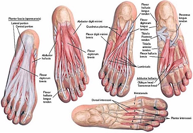
Intrinsic Muscles of Plantar Foot: Believe it or not, 18 muscles are located deep to the plantar aponeurosis. Who would have thought??? These are arranged in four layers (Image K –from left to right). If you think these look challenging, you are right. Outside the head, the foot is one of the most difficult body parts to dissect!
First structure in Image J (left panel)
Plantar aponeurosis – not a muscle
1st Layer of Intrinsic Muscles in Image J ( 2nd panel):
abductor hallucis – draws big toe towards midline of body
abductor digiti minimi (I love this name!) – draws 5th toe away from foot
flexor digitorum brevis – flexes toes 2-5
2nd Layer of Intrinsic Muscles in Image J (3rd panel)
Lumbricals – both flex & extend different phalanges of toes 2-4
Quadratus plantae – flexes toes 2-4
3rd Layer of Intrinsic Muscles in Image J (4th panel)
flexor hallucis brevis – flexes big toe
adductor hallucis – draws big toe towards foot
flexor digiti minimi brevis – flexes 5th toe
4th Layer of Intrinsic Muscles in Image J (5th panel –horizontal)
Now, given that mess of muscles, you probably appreciate how complex foot movements can be achieved. With some 20 intrinsic and 10 extrinsic muscles controlling our feet, they are quite capable, indeed!
Back to Outlander! We see Claire’s poor, weary beat feet, exposed to sand, surf, sun and formicidae (Anatomy Lesson #55, Formidable Formicidae) in Outlander ep 311, Uncharted! Trudging in wet shoes, dealing with festering ant bites, surviving snake slithers…. Diana explains Claire’s feet in Voyager book:
Squads of tiny purple crabs ran off in profound agitation at my approach. My feet sank into the mud to the ankles, and I thought better of putting on my shoes, wet as they were… My feet were bruised and sore, and punctured by fallen palmetto fronds, but the path before us looked relatively smooth.
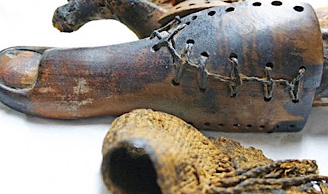
We really must take good care of our feet if we want them to last. Exercise, weight control, healthy diet, wearing supportive shoes all help ensure the feet bear our weight for a lifetime. Even to the most careful, our feet suffer many assaults: bone spurs, athlete’s foot, plantar fasciitis, corns, bunions, Morton’s neuroma, flat feet, hammer toe, warts, stress fractures, etc. Or, we lose toes because of poor circulation, trauma, or developmental issues – such problems have plagued us since ancient times. To the point, the oldest known functional prosthesis is an Egyptian wooden mummy toe (Image L). It actually articulates at the laced surfaces. So clever!
Hey, wait! How do we know it is not a daddy toe? He, he!
Image L
Speaking of toes, Voyager book describes a splendid battle with pirates aboard ship. Sadly, the scene did not make it into the TV version. But, here is a shocking tidbit from the bloody fight:
Cursing incoherently under my breath, I ran to the bottom of the ladder, and reaching up, swung the long-handled amputation knife at his foot, as hard as I could. There was a high-pitched screech from the pirate. Something flew past my head, and a spray of blood spattered across my cheek, wet-hot on my skin. Startled, I dropped back, looking down by reflex to see what had fallen. It was a small brown toe, calloused and black-nailed, smudged with dirt.
Alrightie then! <G>
Let’s consider how you can hurt your neat, sweet, elite, complete feet… In your wildest dreams, do you think shoes such as these are good for feet (Image M)? Wear high heels for long, and one guarantees that later in life, the wearer will have foot problems. The foot is not designed to walk around on the metatarsophalangeal joints (ball) of the foot, which is why wearing spikes hurt!!!
Gentle admonition: if you wear this type of footwear, you should stop.
Image M
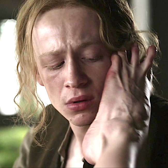
Closing this lesson with indiscrete feet: no Outlander lass is quite as indiscrete and raunchy as Geillis (Gillian) Edgars Duncan Abernathy. Puir young Ian; that dear lad’s cheek is no place for a witch’s foot (Outlander ep 313, The Bakra)!
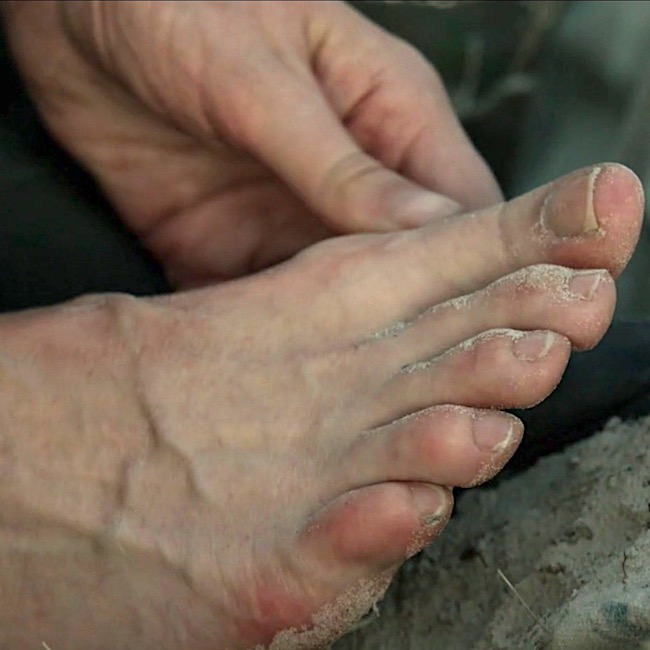
BTW, the prominent ridges passing to 2nd to 5th toes are tendons of extensor digitorum longus! Yay!
Bottom line: The complete and complex human foot is truly an anatomical work of art. Let’s vow to take good care of ours!