Anatomy Lesson #50: Care for us – Oh Pancreas!

Tommy Lee Jones, starring as Hawk Hawkins in Space cowboys (2000), said it best (Image A):

Where the hell is the pancreas, anyway?  I don’t even know what the damn thing does beside give you cancer!

Terrific question, Hawk! Welcome anatomy students to Lesson #50, the pancreas. Our goal today is to answer Hawk’s question and more: what is the pancreas, what does it look like, where is it located, what does it do, and what diseases plague it?

This is the 7th and final lesson of the immense gastrointestinal tract and associated organs! Turns out, pancreas is one of these “Klingon” organs.

Although Diana hasn’t written a great deal nor has Starz episodes showed much about the pancreas, there are bits and pieces here and there. As always, these are scattered throughout the lesson. I hope you find the pancreas as compelling as your body does. Off we go!

space cowboys movie poster
Image A

History: Herophilus (335-280 BCE), Greek anatomist and surgeon (Image B), helped found the ancient school of Medicine in Alexandria, Egypt. He also championed human dissection and was the first to describe the pancreas. Students may recognize Herophilus because he also featured in Anatomy Lesson #34, The Amazing Saga of Human Anatomy.

Image B Herophilus

Gross Anatomy: The word, pancreas, comes from the Greek pan, meaning “all,” plus the Greek kreas, meaning “flesh;” so, “all flesh.” A seemingly odd term until one views the pancreas and then, it seems sensible as it appears markedly fleshy, soft, and squishy (Image C).

pancreas
Image C pancreas

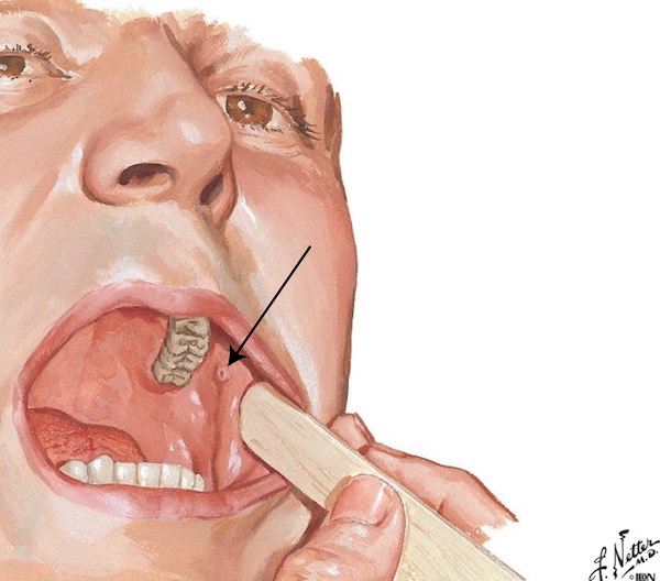
Outlander Fix #1: Answering Hawk’s first question, “Where the hell is the pancreas, anyway?”, Mary-Mary-Quite-Contrary knows (Starz episode 211, Vengeance is Mine). Or, rather, Mary’s Knife-Knows! Och, lass, that is Jamie’s dirk. Careful! Verra sharp!

mary holding a knife in outlander episode 211
The dirk finds it’s mark, pretty much where the pancreas lives! A grim ending to a vicious valet. And he isn’t the only one taking a hit from a blade in this episode. Written by our own beloved Diana, this gratifying chapter brings some very bad players to their just desserts (Starz episode 211, Vengeance is Mine), although that mad bastard captain remains at large. Come on S.3!

image of a man being stabbed from outlander episode 211

On to anatomy!

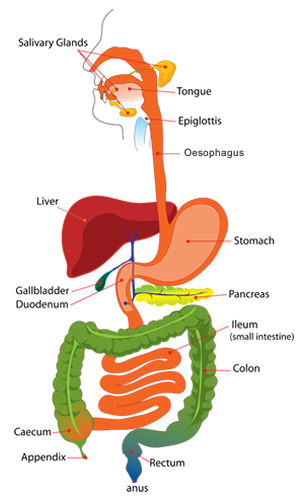
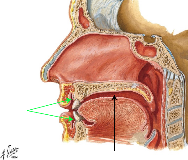
Pancreas Location: The pancreas (Image D) lies in the upper abdominal cavity behind the stomach (Anatomy Lesson #46: Splendid Stomach, Wobbly Wame). Oriented almost horizontally, it extends from the curve of duodenum to the spleen, a distance of some 6” (15 cm). In this position, it contacts stomach, spleen, duodenum, colon, jejunum, bile ducts, and major intestinal blood vessels. Pillowed among other soft abdominal organs, its location is pertinent, especially in pancreatic cancer as we shall shortly see. It lies between T12 and L2 vertebral levels.

Try this: Place thumb on the xiphoid process (tip of sternum – Anatomy Lesson #15: Crouching Grants – Hidden Dagger) and little finger of the ipsilateral (same) hand on your umbilicus. The xiphoid process is located at T9-T10 vertebral levels; the umbilicus at L3-L4. Now, place a finger of the contralateral (opposite) hand midway between xiphoid and umbilicus – this is the approximate vertebral level where your pancreas dwells. These new terms are thrown in to enlarge your anatomy vocab!
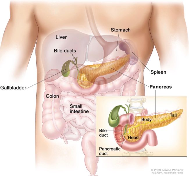
 

image of the pancreas
Image D

Divisions: Although many sources describe three divisions of the pancreas, it actually has five: head, neck, body, tail, and uncinate process (Image E). The head tucks into the C-shaped curve of duodenum. A short neck lies between head and body. The body supplies most of its 6” length. The tail tucks into the spleen (review Image D). The uncinate process (Latin meaning hook-shaped) is the smallish part that tucks in behind some large blood vessels of the gut.

You might consider these segments as rather arbitrary, but they are very useful in localizing disease entities, particularly pancreatic cancer.
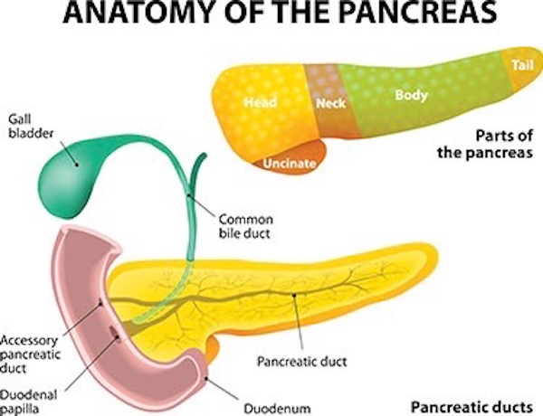
image of the parts of the pancreas

Image E

Gland: The pancreas is an organ but it is also a gland meaning it produces and releases secretions (products). It is also a mixed gland meaning it it is both exocrine and endocrine gland. Now, most glands are either exocrine or endocrine in type, but the pancreas is unusual because it is both.

Exocrine: Exocrine glands release products into ducts which carry the secretions to a destination. In this case, pancreatic exocrine secretions flow into the pancreatic duct (Image F) which joins the common bile duct (Anatomy Lesson #49, Our Liver – The Life Giver!) before entering the duodenum. Pancreatic exocrine secretions include an array of enzymes that digest various dietary substances, including:

  • carbohydrates
  • lipids (fats)
  • proteins
  • nucleic acids (DNA, RNA)

Finally, the duct system draining the pancreas produces bicarbonate to help neutralize acidic foodstuffs released by the stomach into the duodenum (Anatomy Lesson #46, Splendid Stomach, Wobbly Wame).
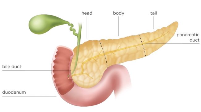
Image F

Endocrine: Endocrine gland secretions are picked up and distributed throughout the body by the blood stream –  no ducts involved! And most of such secretions are classified as hormones or hormone-like substances. The pancreas produces its hormones via small spherical islands of cells formally known as pancreatic islets of Langerhans (brilliant German pathologist). About 3 million tiny islets (Image G – violet clusters) are scattered throughout the pancreas although more are concentrated in the tail region.  Their combined mass is only about 2 grams (.0022 pounds). Yet, their effects in the body are profound!

These tiny clusters contain five different types of cells, producing the following compounds:

  • glucagon (stimulates cells to release glucose – raises blood sugar levels)
  • insulin (causes cells to absorb glucose – lowers blood sugar levels)
  • somatostatin (regulates release of glucagon and insulin)
  • pancreatic polypeptide (regulates intestinal function)
  • ghrelin, the “hunger hormone” (regulates appetite –  pancreas produces small amounts of this compound – stomach is major source)
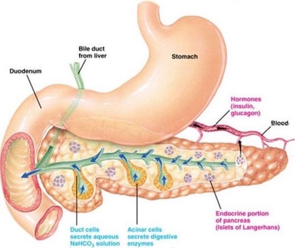
Image G

Outlander Fix #2: Yay! Claire fixes her mind on her pancreas in Dragonfly in Amber book. Yep, she does! (Psst…the image is from their Paris days – Starz episode 203, Useful Occupations and Deceptions – not from Lallybroch.)  Not a perfect match with the quote, but Claire in bed is always good, especially if Jamie is nearby. Dinna ken how Claire’s mind fixes around her pancreas, but I do trust the lass! <g>

As I began to hover on the edge of sleep, my mind fixed somewhere around my pancreas, I could dimly hear the sounds of small Jamie pattering down the hall to his mother’s bedroom—roused from sleep by a full bladder, he seldom had the presence of mind to take the obvious step, and would frequently blunder down the stair from the nursery in search of assistance instead.

image of Jamie and Claire in bed
Buh-bye Claire – Thank you for the pancreas lesson!

Microscopy: Exocrine and endocrine pancreas are readily differentiated by microscopy. Bear with me, folks-without-training-in-microscopy, as I explain. Image H is a thin slice of pancreas stained with dyes (H&E).  The red globs are pre-enzymes inside deep purple exocrine cells. After release as enzymes, they enter the pancreatic ducts and are transported to the duodenum. The pale violet blob in the center is a pancreatic islet of Langerhans. This blob contains tiny purple ovals, nuclei of the five different cell types mentioned above; collectively, these islets form the endocrine part of the pancreas. In 3-D, the pale blob is spherically- shaped.

This exercise is important to explain how anatomical pathologists diagnose disease. They learn to recognize normal pancreas (and all other organs) in the microscope. This enables them to determine if a tissue sample lies outside that expectation and, if it does, what disease does it best match. This ability is called pattern recognition. Got it? Grand!
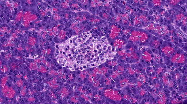
Image H

Diseases: Like other organs, the pancreas has its own palate of diseases and many have devastating effects. There are three major players:

  • Pancreatitis: means inflammation of the pancreas. Acute and chronic types. Most common cause is alcoholism. Its own enzymes start digesting pancreatic tissue – Painful!
  • Diabetes mellitus: Types 1 and 2 interfere with sugar (glucose) uptake by body cells, although causes are different.
  • Pancreatic cancer: one of the more infamous cancers, it has a very low survival rate – overall five year survival rate is only 7%.

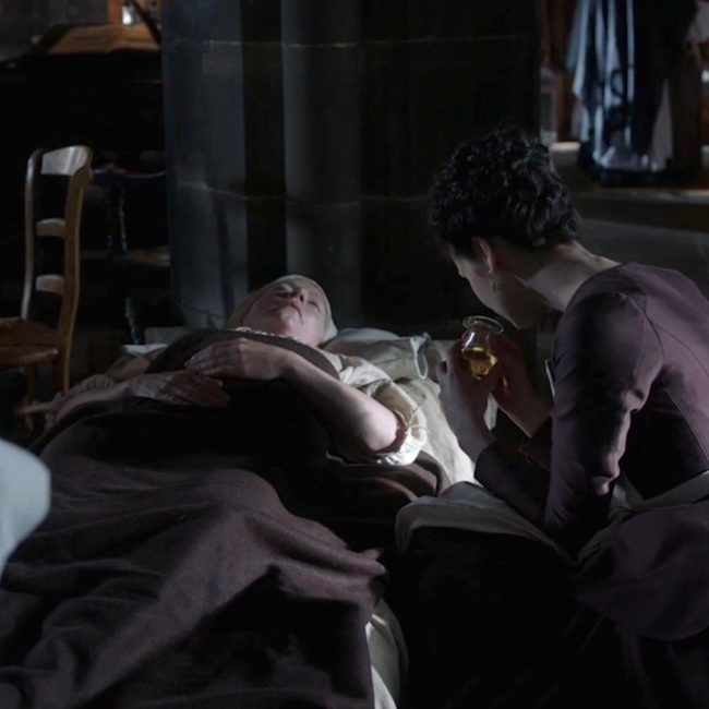
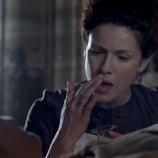
Outlander Fix #3: Claire’s own words from Dragonfly in Amber book inform us of a L’Hôpital des Anges’ patient with sugar sickness (Type 1 diabetes). Starz episode 203, Useful Occupations and Deceptions, faithfully brings Diana’s lines to life!

I bent over a pallet at the edge of the floor. A very thin woman lay listlessly under a single blanket, her eyes drifting dully over us without interest.

It wasn’t the woman who had attracted my attention, so much as the oddly shaped glass vessel standing on the floor alongside her pallet. The vessel was brimming with a yellow fluid—urine, undoubtedly.

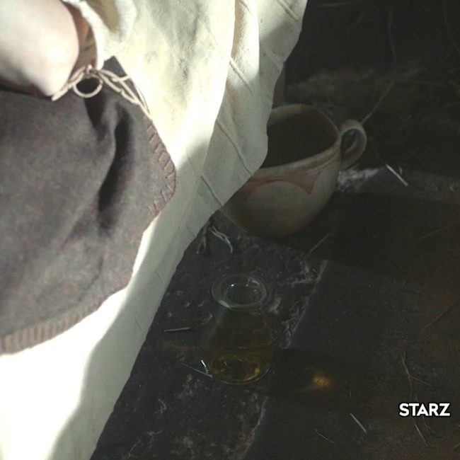
psssst: The oddly shaped glass vessel used in the episode is an Erlenmeyer flask. It wasn’t invented until 1861 <g>

I was mildly surprised; without chemical tests, or even litmus paper, what conceivable use could a urine sample be? Thinking over the various things one tested urine for, though, I had an idea. I picked up the vessel carefully, ignoring Sister Angelique’s exclamation of alarmed protest. I sniffed carefully.

Sure enough; half-obscured by sour ammoniac fumes, the fluid smelled sickly sweet—rather like soured honey. I hesitated, but there was only one way to make sure. With a moue of distaste, I gingerly dipped the tip of one finger into the liquid and touched it delicately to my tongue.

…Sister Angelique was watching with sudden interest. … “Are you thirsty, Madame?” I asked the patient. I knew the answer before she spoke, seeing the empty carafe near her head. “Always, Madame,” she replied. “And always hungry, as well. Yet no flesh gathers on my bones, no matter how much I eat.” She raised a stick-thin arm, displaying a bony wrist, then let it fall as though the effort had exhausted her.

I patted the skinny hand gently, and murmured something in farewell, my exhilaration at having made a correct diagnosis substantially quenched by the knowledge that there was no possible cure for diabetes mellitus in this day; the woman before me was doomed.

In subdued spirits, I rose…“Could you tell from what she suffers, Madame?” the nun asked curiously (Mother Hildegarde in the Starz episode). “Only from the urine?” “Not only from that,” I answered. “But yes, I know. She has—” Drat. What would they have called it now? “She has … um, sugar sickness. She gets no nourishment from the food she eats, and has a tremendous thirst. Consequently, she produces large quantities of urine.” … “And can you tell whether she will recover, Madame?” “No, she won’t,” I said bluntly. “She’s far gone already; she may not last out the month.”

Is Jamie please with Claire tasting urine and treating scrofula? Noooo….he doesn’t want his pregnant wife messing with piss and pus! Och!

History of Diabetes Mellitus: Diabetes was described three millennia before Claire sampled her patient’s urine. About 1500 BCE, Sushruta, the Indian physician, wrote the first description of diabetes mellitus, noting that ants and flies were attracted to the urine of people with a mysterious disease causing intense thirst, enormous urine output, and wasting away of the body.

In 250 BCE, Apollonius of Memphis (Egypt), coined the term diabetes meaning “I pass through.” Later, mellitus, Latin for “honey-sweet,” was added to emphasize the sugar content of the urine and to distinguish diabetes mellitus from diabetes insipidus, a disorder of the pituitary gland also characterized by intense thirst and high production of urine.

Finally, the Greek physician, Aretaeus (Image I) wrote this description in the second century AD:

“Diabetes is … not very frequent … being a melting down of the flesh and limbs into urine … for the patients never stop making water, but the flow is incessant, as if from the opening of aqueducts. It consists in the flesh and bones running together into the urine … the illness develops very slowly. The nature of the disease is chronic, and it takes a long period to form; but the patient does not live long once the disease is fully established; for the melting is rapid, the death speedy. Moreover life is disgusting and painful; thirst, unquenchable … and one cannot stop them either from drinking or making water”.
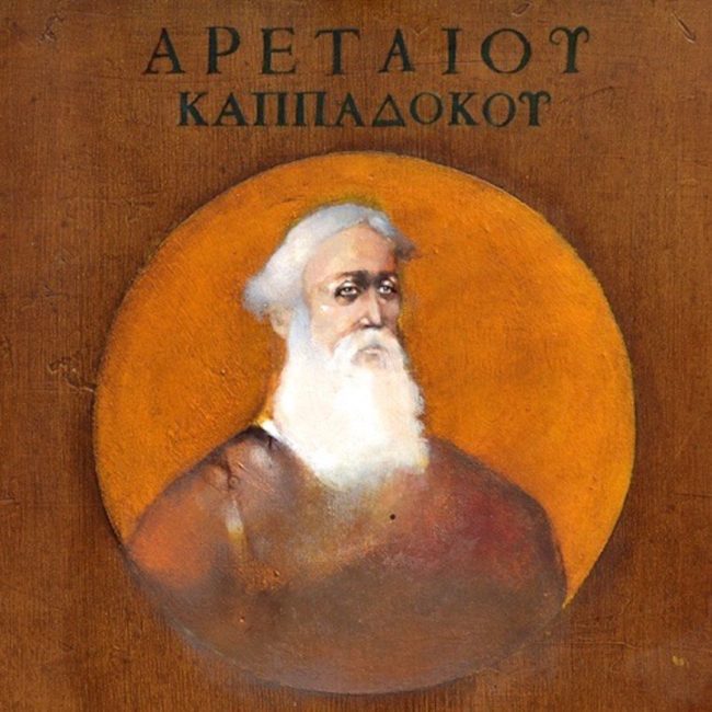
Image I

Simple Physiology: Our bodies contain millions of cells. These cells use glucose to make energy for our daily activities: heartbeat, muscle contraction, fighting redcoats. Snort! How do cells get glucose? When you eat or drink, food is broken down inside the gut into simple proteins, lipid components, and sugars, mostly glucose. Glucose enters the bloodstream and circulates. Although circulating glucose contacts body cells, they cannot absorb it on their own.

So, circulating glucose stimulates pancreatic (beta) islet cells to release insulin into the blood stream. Insulin circulates with the glucose and acts as a key to permit body cells to absorb glucose. No matter how much glucose circulates in the bloodstream, insulin is required for it to enter the cells: no insulin – no glucose uptake! The following brief  You Tube cartoon illustrates the process very well.

http://youtu.be/OYH1deu7-4E

Oh, and just so you ken, insulin produces 9 effects in the body. Glucose uptake is only one of these.

I like this cute T, but it is slightly misleading. The pancreas doesn’t really ask for sugar. Rather, pancreatic cells check blood glucose levels and as those levels rise, it releases insulin to induce body cells take in sugar. If glucose blood levels fall below normal, then the pancreas releases glucagon which mobilizes stored glucose for released into the blood stream. A very clever check and balance system.
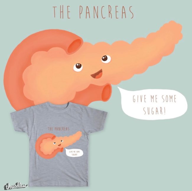

Image J shirt

Type 1 (I) Diabetes Mellitus (DM): Simply put, Type 1 DM is a chronic condition wherein the pancreas produces little or no insulin so glucose cannot enter cells. Instead, glucose stays in the bloodstream, spilling into the urine, and pulling water with it; hence, the continuous thirst and high urine production (polyuria). Even today, there is no cure for Type 1 DM but it can be managed, usually with daily doses of insulin.

The exact cause of Type 1 DM remains unknown despite decades of intense research. In many, the body’s own immune system mistakenly destroys insulin-producing (beta) cells of the pancreatic islets. Both genetics and viral infections appear to play important roles in the development of diabetes.

Type 2 (II) DM: Type 2 DM occurs when the pancreas doesn’t produce enough insulin or cells are unable to recognize the insulin and use it properly, a state known as insulin resistance. Insulin resistance is the most common cause of Type 2 DM. Here, genetics and lifestyle are important risk factors. According to the US CDC, obesity and lack of physical activity are responsible for 95% of Type 2 DM in the US!

Pancreas Transplant: Yes, these surgeries are done. A pancreas transplant is a surgical procedure to place a healthy pancreas from a deceased donor into a person whose pancreas no longer functions properly (Image K). Most transplants are done to treat Type 1 DM, and, occasionally for Type 2. Although a transplant offers a potential cure for DM, it is generally reserved for folks with serious diabetes complications because the side effects of a pancreatic transplant are significant.

The donor pancreas plus the segment of duodenum that receives the pancreatic duct are transplanted into the cecum of the large intestine (Anatomy Lesson #48, The Big Guy!). So, the recipient then has a pelvic pancreas. Such surgeries must be disclosed to new health care professionals so they are aware of unexpected changes in typical anatomy.


Image K

Pancreatic Cancer: Many of us have lost a family member, friend, or acquaintance to pancreatic cancer. This type of cancer is often detected late, spreads rapidly, and has a poor prognosis. Why? Unfortunately, early stages of this cancer are mostly asymptomatic. Later stages are associated with symptoms, but these can be non-specific, such as lack of appetite and weight loss. So, the cancer may be advanced before detected.

Pancreatic cancer is staged 0-IV depending on its spread (Image L). Early in this lesson, we learned that the pancreas is in contact with duodenum, jejunum, colon, spleen, stomach, bile ducts, and blood vessels, providing many opportunities for metastases. Also, these surrounding organs are soft, so a spreading cancer has considerable space to grow before creating an organ stramash.

As you might assume, the prognosis is better for those whose pancreatic cancer is diagnosed at an early stage. The median survival rate after diagnosis and medical treatment is still abysmal: only 6 to 12 months. Let us be grateful it isn’t one of the three most common cancers.

True story: one of my neighbors was diagnosed with pancreatic cancer in the 1960’s. She survived a Whipple procedure, a.k.a., pancreaticoduodenectomy, which is why it is called a Whipple! This horrific surgery removes head of pancreas, duodenum, gallbladder, part of common bile duct, and part of stomach. She was a lucky lady who raised three sons and survived into her 80’s!

A possible piece of good news: New Scientist magazine (15 April 2017) reports that, by chance, a drug used to treat strokes has been found to significantly prolong the lives of mice with pancreatic cancer. Turns out, pancreatic cancers are protected by a capsule of connective tissue that acts like a coat of armor. Australian researchers found the stroke drug, fasudil, weakens the capsule, making it easier for chemotherapeutic agents to reach the tumor. Drug trials in humans are planned. Fingers crossed!
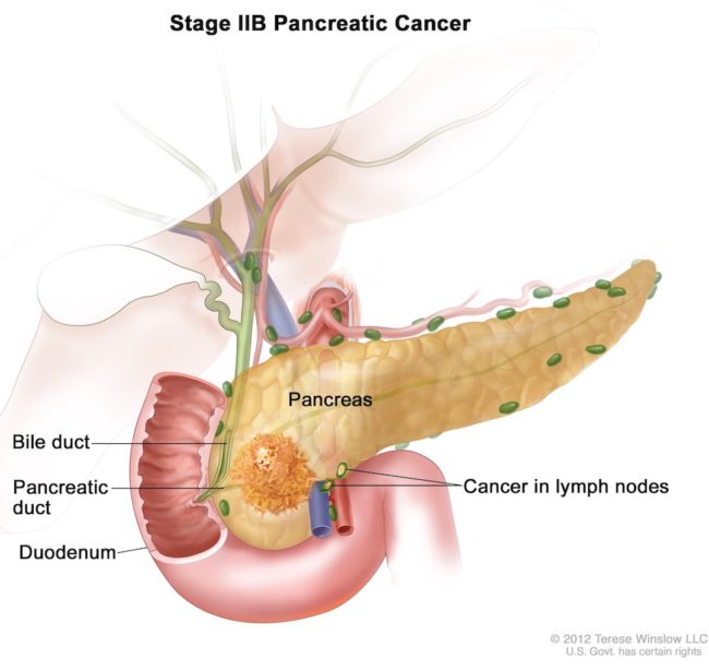

Image L

Let’s close this lesson on a happier note with Culinary Considerations.

Sweetbreads or Sweet breads??? Let’s get this out of the way, right now! Sweetbreads are thymus and pancreas (and, often salivary glands). Yes, people eat pancreas (Image M), gah! I canna like this as I have met too many of these organs on the dissection table. To me, sweetbreads are Awful-Offal! They do enjoy legions of fans, so dive in, if they work for you. You can have my share <G>
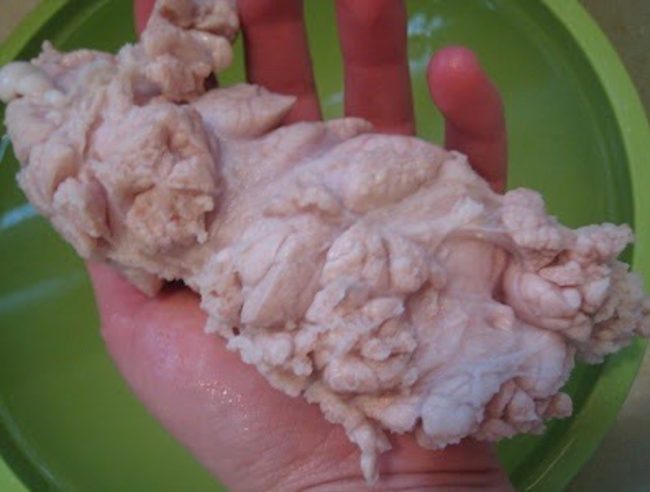

Image M

On the other hand, sweet breads (Image N) are verra hard to resist, especially the homemade variety.  Yummy, yummy, yummy, in my tummy, tummy, tummy! ‘Nuf said!


Image N

Let’s give the last word to American poet Robert Frost because he was creative enough to write a poem about sweetbreads.

Quandary

You drive me to confess in ink:
Once I was fool enough to think
That brains and sweetbreads were the same,
Till I was caught and put to shame,
First by a butcher, then a cook,
Then by a scientific book.
But ’twas by making sweetbreads do
I passed with such a high I.Q.

Dear Robert: the butcher, baker and candlestick maker – och! -butcher, cook, and science book are correct! Sweetbreads dinna include brain!

A deeply grateful,

Outlander Anatomist

Photo Creds: Starz, www.123rf.com (Image E pancreas parts), www.blog.kingarthurflour.com (Image N sweet breads)
www.cancer.gov (Image D pancreas site), www.dypatil.edu (Image B Herophilus), www.en.wikipedia.org (Image I Aretaeus), www.fanart.tv (Image A Space Cowboys), www.healthtop.com (Image C pancreas), www.mayoclinic.org (Image K pancreatic transplant; Image L Pancreatic Cancer), www.medicalwork.com (Image G pancreas mixed gland), www.medievalspanishchef.com (Image M sweetbreads), www.pancreatic.org (Image F pancreas ducts), www.shutterstock.com (Image H pancreas histo), www.threadless.com (Image J shirt)

Anatomy Lesson #44: “Terrific Tunnel – GI System, Part 1”

Hello anatomy students! Time to launch a new but thrilling anatomic topic: the GI tract. What is the GI tract (Image A)? Well, it is a complex organ system that enjoys several monikers: alimentary canal, alimentary tract, digestive tube, GI tract, gastrointestinal tract, gastrointestinal system, and gut. Most anatomists prefer the term gastrointestinal tract or system, but since that term is a bit lengthy, this lesson will shorten it to GI tract.

meaning-of-gi1

Image A

Spoiler Warning: there are lots of Outlander images and book quotes within, including one waaaay forward from book eight. A scaredy cat warning appears before hand, so, watch for it!

scaredy-cat

You can easily scroll to the relaxing cat to skip the whole thing until you’re ready to read MOBY!

relaxed-cat

The GI tract is a tube extending from mouth through anus (yes, I wrote that and I warrant that team Angus and Rupert used that word, too!). It is divided into the following regions (Image B):

  • oral cavity
  • oropharynx
  • esophagus (oesophagus)
  • stomach
  • small intestine (duodenum, jejunum, ileum)
  • large intestine
  • rectum
  • anal canal and anus

The GI system includes several additional organs that are associated with the GI tract:

  • major salivary glands
  • liver
  • gallbladder
  • pancreas

gi-tract

Image B

GI Embryology: You might wonder why the last four organs belong to the GI system. This occurs because during embryogenesis, these organs develop as outgrowths of the developing GI tube. After birth, they retain connections with the parent GI tract via ducts (hollow tubes). Parental offspring!

Embryogenesis of the GI tract is extraordinarily complex but consider this simplified version (Image C – gold layer):

  • The human embryo starts as a flattened sheet of layered cells which folds and expands due to cell division (and death) directed by chemical growth factors.
  • By 18 days, the future gut expands at the head end as the foregut and a second hollow, the hindgut, develops at the rear. The open midgut lies between the two hollows.
  • By 22 days, the gut assumes a more tube-like configuration. The early liver appears.
  • By 30 days, the nascent pancreas appears.
  • Over the next days and weeks, the foregut differentiates into pharynx, esophagus, stomach, part of small intestine, liver, gallbladder, and pancreas.
  • Midgut develops into the rest of small intestine, and part of large intestine.
  • Hindgut gives rise to the remaining large intestine, rectum, anal canal, and part of anus.

embryology-of-gi1

Image C

The Tube (not the one in London!): Think of the mature GI tract as a long, hollow tube, albeit one that is variously folded and dilated. The tube shown in Image D has a wall made of zirconium but, consider that the space inside the tube is an extension of the outer world. Yes? Same is true with the GI tract: from mouth through anus, the lumen (space) inside the GI tract tube is an extension of the outer world. Even as ingested materials disappear after swallowing, anything in the lumen technically remains outside the body until it crosses the wall of the GI tract. Make sense? Yay!

tube

Image D

Length: The adult GI tract is also very long. Just how long is it? In adults, it measures 8.3 m (27’+) – almost the height of a three-story building! Much of its length is highly folded so it nicely tucks within the abdominal cavity. A Body Worlds exhibit (Image E) shows an expanded view of the GI tract and its associated organs. German labels reflect the language of the show’s creator, Gunther von Hagens. Lay the tube out straight, and it approaches 27’ in length.

The length isn’t a fluke of nature, rather, it is required for digestion of foodstuffs, absorption of nutrients, and preparation of residues for elimination. There are also many more functions provided by associated organs of the GI system. More about such functions soon.

bodyworld1

Image E

So, with those issues behind us (har har), the lesson begins and ends with the oral cavity or mouth. The mouth includes many components: inner lips, inner cheeks, gums, hard and soft palates, floor of mouth, tongue and teeth (Image F). Let’s start with the lips.

Anatomy Of Oral Cavity Structures Of The Oral Cavity, Pharynx, And Esophagus. Biology - Human Anatomy Library
Image F

Lips: Visible flaps at the mouth opening, the soft pliable lips are important additions to the GI tract because they augment food intake, chewing, and articulations as in speech (Image G). Hopefully, the lip-flash doesn’t freak you out!

People generally think of the lips as the part to which lipstick is applied. But, anatomists hold that the lips are more extensive.

lips

Image G

Anatomical lips include not only the lipstick canvas, known as the vermillion zone (Image H – pink arrows), but also the hair covered flaps above and below the vermillion zone (Image H – green arrows) as well as the moist and red inner surface of these flaps (Image H – blue arrows).

The hairy outer lip is part of the face which was presented in an earlier lesson (Anatomy Lesson #14, “Jamie and Claire” or “Anatomy of a Kiss”). The inner lips belong to the oral cavity and are lined with mucous membrane or mucosa (Anatomy Lesson #42, “The Voice – No, not that One!”). The vermillion zone is the transition area between these two surfaces.

image-h

Image H

Lips are equipped with orbicularis oris (Image I), a major muscle mass, rendering them highly mobile. For decades, orbicularis oris was described as a sphincter muscle encircling the opening of the oral cavity, a description that remains rampant on today’s Internet. Modern dissections have shown this description to be insufficient. Rather, orbicular oris is divided into four quadrants, each containing both circular and direct (diagonal) muscle fibers, lending an amazing ability to purse, pucker, and pout. For a more thorough consideration of orbicularis oris, visit Anatomy Lesson #14, “Jamie and Claire or Anatomy of a Kiss.”

Although not shown in Image I, lips are also provided with seven pairs (that’s 14 total!) of muscles that elevate (lift), depress (lower), and/or retract (pull back) the lips. Very mobile structures, the lips!

lip-muscles1

Image I

Jamie provides a fantastic example of pursed lips as he endures rambling threats and ignoble insults from BJR (Starz episode 109, The Reckoning). No choice – Cap. Mad Man holds a knife to his beloved Claire! Puckering requires simultaneous contraction of direct and circular orbicularis oris muscle fibers. ?“Nobody does it better … Nobody does it half as good as you. Baby, you’re the best!” ?

ep-109-lips1

Lastly, lips are anchored by webs of mucous membrane: a frenulum of the upper lip and a frenulum of the lower lip (Image J).

figure0047b2

Image J

Lips are highly tactile structures imbued with numerous sensory receptors which detect light touch, deep pressure, pain, vibratory sense, heat, and cold. These many tactile attributes help our lips close, grip objects, and engage in kissing and other acts of intimacy (Image K). Ha!

kiss1

Image K

Because lips are highly innervated and possess startling mobility, they enjoy a prehensile-like ability to grip things, even items they should avoid gripping (Starz episode 101, Sassenach). Small Saucy Sassynach!

ep-101-claire-cigarette1

Outlander kisses would be neither as passionate nor as (ahhhh!) satisfying sans nimble muscles and sharp tactile sensibilities of the lips. Smack down!

Jamie and Claire share many kisses, especially during Outlander Season 2 (Starz episode 210, Prestonpans). But a quote from Dragonfly in Amber book says it all – a bit of a love poem by Catullus:

Then let amorous kisses dwell

On our lips, begin and tell

A Thousand and a Hundred score

A Hundred, and a Thousand more.

ep-210-kiss1

I love writing about our fav couple’s lips, but we must move on. Sigh!

Oral Cavity: Before describing structures of the mouth, it is important to further define the oral cavity. The oral cavity is divided into two parts. The oral vestibule is a slit-like space between teeth and inner lips and between teeth and cheeks (Image L – green arrows). The oral cavity proper is the larger slit-like space behind the teeth (Image L – black arrow).

image-l

Image L

Oral Cavity Proper: However, with jaws open, the oral cavity dramatically expands into a rather large cavern (think foot-in-mouth). Gazing into the oral cavity proper, it starts at the vermillion zone and ends at the paired palatoglossal arches, two mucosal folds descending from the soft palate to the tongue (Image M). See the paired palatine tonsils (we have three pairs of tonsils)? These lie outside the oral cavity.

figure0047a

Image M

Wanna see Jamie’s proper oral cavity? Of course ye do! Jamie lets out a big-old howl after that bratty MacDonald lad slices and dices his left side (Starz, episode 110, By the Pricking of My Thumbs). Ouch, that hurts! Jamie is in for it now – his usually not-a-silent wifie willna appreciate such blatant butchery. Say ahhhhh, lad!

ep-110-oral-cavity

Palates: The roof of mouth is divided into two palates: an anterior hard palate, so named because its mucosa overlies bone, and a posterior soft palate which lacks bony underpinnings. Image N shows the palates as if you sat on the tongue and stared upward. The soft uvula is a midline structure suspended from the soft palate. The two palatoglossal arches drop right and left from the uvula.

Here is an interesting fact: The hard palate is characterized by permanent folds known as rugae. These ridges are stable and unique for each person suggesting they could be used for identification purposes. Their human function is not clear but is probably related to mastication (chewing).

Try This: With mouth open, use tip of tongue or finger to feel where hard palate and soft palate meet. Carry a flashlight/torch to a mirror. Open your mouth, but shine the light directly into the mirror; light reflects off the mirror and into your mouth. Identify your uvula, the dangly bit hanging from the roof of the soft palate. Several small muscles control it during swallowing, speech, etc. My fav muscle is the itsy bitsy teenie-weenie tensor veli palatini. <G>

palates

Image N

Mucous Membrane: Consider the mouth’s interior. Every surface, except teeth, is covered with mucous membrane, a red, shiny, wet, and sensitive surface (Image O). The membrane appears red because it has a rich blood supply. It is wet because saliva bathes its surfaces. It is shiny because saliva reflects light. It is sensitive because it is supplied with numerous sensory receptors.

With some exceptions, the mucous membrane of the entire oral cavity is non-keratinized (Anatomy Lesson #5, “Claire’s Skin” – “Ivory, Opal and White Velvet”), meaning its cells do not contain keratin. However, mucous membrane of gums near the teeth, top of tongue, and hard plate contain keratinized cells. Why? Because they are subject to abrasion during mastication. Keratin is an intracellular protein which hardens cells, making them resistant to abrasion. Make sense? Yay!

Remember when Angus Mhor administers a methodical beating to Jamie in Castle Leoch’s great hall (Rupert does the dirty deed in Starz episode 102, Castle Leoch)? Diane writes about Jamie’s oral mucosa in Outlander book. I’ve said it before and I’ll say it again, if you’re not reading Diana’s books, you are missing out on gobs of anatomy!

“Is your mouth cut inside too?” “Unh-huh.” He bent down and I pulled down his lower jaw, gently turning down the lip to examine the inside. There was a deep gash in the glistening cheek lining, and a couple of small punctures in the pinkness of the inner lip. Blood mixed with saliva welled up and overflowed.

Try this: Return to the mirror with a flashlight. Open mouth and inspect its lining. See that the mucous membrane surfaces are moist, shiny, and red. We all known how tender they are. With tip of tongue explore the surfaces of cheeks, floor of mouth, and inside of lips. All surfaces are smooth and moist excepting the rougher hard palate, gums near teeth and dorsum of tongue.

mouth

Image O

Minor Salivary Glands: Image P demonstrates a little known fact: deep to the mucous membrane, the entire mouth is riddled with thousands of minor salivary glands; these microscopic entities are much smaller than Image P implies. Tiny ducts (channels) lead from the glands, pierce the mucous membrane, and open into the oral cavity. Minor salivary glands produce and secrete saliva to moisten oral surfaces. Some of these glands specialize in producing mucus, a thick secretion rich in glycoproteins (protein molecules with carbohydrate side chains) which renders the saliva slippery.

Try This: Return to the mirror, grasp and evert one cheek (Ha, ha. No, not that one!). Can you see tiny surface humps and bumps? These are caused by aggregations of minor salivary glands.

figure0048a

Image P

Tongue: The human tongue is an organ. What is an organ? In anatomy, an organ isn’t a musical behemoth equipped with large keyboard and foot pedals; rather, it is a collection of tissues joined into a structural unit that serves a common function(s).

Ably demoed by Miley (Image Q), the tongue is rather large. We use it to help chew (as in mixing food with saliva), swallow, speak, and taste. And, to be bratty, of course!

miley-cyrus-sticking-tongue-out_112444

Image Q

The mature tongue is divided into (Image R):

  • Body – oral part (anterior 2/3)
  • Apex – the tip (part of the body)
  • Root – pharyngeal part (posterior 1/3)

The tongue is an extraordinary organ, arising from two different embryonic anlage (rudimentary parts) which fuse during development: one part gives rise to body and apex, and a second part produces the root. The two parts fuse leaving a remnant, the terminal sulcus (Latin meaning end groove), an inverted, V-shaped groove (Image R – dashed lines) on dorsum of tongue.

Take another look at Miley’s tongue (Image Q); the part you see is body and apex. The tongue root is not visible because it drops backward and downwards, out of view.

The tongue body has two different surfaces. A protected ventral (under) surface is covered with thin mucous membrane. The dorsum (top) of tongue is covered with mucous membrane but it is rough. Do you remember why?

image-r

Image R

Tongue Papillae: The dorsum of tongue is rough because the surface is covered with a mess of tiny bumps, the papillae (Latin meaning nipple). Image S is a vertical section through back of tongue showing surface papillae. Human tongues have four different types of papillae: fungiform, filiform, foliate, and circumvalate (the big guy marked papilla in Image S). Foliates are present on the sides of our tongues – they do not appear in the image

Papillae derive their names from their shape. Fungiform look like mushrooms, filiform resemble filaments, foliate are leaf-like, and the humongous circumvalate (valate) are bound by a deep circumferential moat.

BTW: See the blue ovals near the right edge of Image S (black arrow)? These are lingual tonsils . We have a large mass of tonsils embedded in each side of our tongue just posterior to the sulcus terminalis. Yes, we do!

Taste Buds: All papillae except the filiform type harbor taste buds, units embedded in the covering cells of the mucous membrane. The cells of filiform papillae are keratinized to help grip food particles and to protect the tongue dorsum during mastication. The human tongue has between 2,000 and 8,000 taste buds. But, here is an interesting Fascinating Fun Fact: taste buds are also found in the mucosa of soft palate, esophagus, inner cheeks and epiglottis (Anatomy Lesson #42, “The Voice – No, not that One!”)! Yep, ’tis true

Viewed vertically, taste buds resemble sections of a peeled orange (Image S – lower left), and are composed of supporting and sensory cells. A tiny taste pore opens into the oral cavity. Food molecules, delivered as a salivary cocktail, reach the taste pore, sensory cells detect the molecules and depolarize (change in electrical charge). this stimulates nearby nerve fibers which carry the impulse to the brain where it is interpreted as flavor. Whew!

Try this: Return to the mirror with a flashlight. Open your mouth and peer closely at the tiny papillae covering the tongue dorsum. Look carefully, you will likely see larger fungiform papillae scattered among the smaller, more numerous filiform type. You will not see the foliate because they are far back at the sides of tongue. If you are truly adventurous, place forefinger on the tongue dorsum and push it backwards as far as possible. Feel the rough bumps on either side? These are the circumvalate papillae. Rah!

image-s

Image S

Flavors: Since 1901, students have been taught that human taste buds detect four different flavors: salty, sour, bitter, and sweet. Recent lobbying by researchers have added a fifth flavor, umami (savory). And, for over a century, taste bud distributions were mapped out on the tongue dorsum to show where these flavors are detected (Image T). This myth has been debunked as research shows we can detect all five flavors anywhere on the dorsal tongue albeit more intensely in some regions than others. And, the detection of flavors is far more complex than the five types provided by taste buds, as we shall consider at lesson’s end.

taste-map

Image T

Ventral Tongue: The ventral (under) surface of tongue isn’t exactly beautiful, but it is covered with a thin mucous membrane similar to inside of cheeks and lips. This surface exhibits several folds and a midline lingual frenulum, a fold of mucous membrane which anchors the tongue to limit its movements (Image U). Bluish longitudinal squiggly ridges are created by large, underlying lingual veins.

figure0047b-2-1

Image U

BOOK SPOILER WARNING!

Skip the next paragraphs and scroll down to the relaxed cat if you don’t want to read a quote from Diana’s eighth book…

scaredy-cat

During embryogenesis, the lingual frenulum is quite long. Programmed cell death within the frenulum causes it to shorten (Fact: embryonic development involves gobs of programmed cell death). Occasionally, the programming goes awry and the frenulum fails to shorten leaving its owner tongue-tied (Image V – left side).

Known as ankyloglossia (Greek for fused tongue or fixed tongue), a simple case requires a clip through part of the lingual frenulum (Image V) to release the tongue from its tether (Image V – right side). This procedure is known as a frenotomy.

Diana provides! Claire performs this surgery on a character (no name!) from book eight, Written in My Own Heart’s Blood, known by faithful readers as MOBY:

I’m going to do just a frenotomy, at least for now. That is a very simple operation; it will literally take five seconds…. I had a tiny cautery iron, its handle wrapped in twisted wool… I had a fine suture needle, threaded with black silk, too, just in case. The frenulum is a very thin band of elastic tissue that tethers the tongue to the floor of the mouth, and in most people it is exactly as long as it needs to be to allow the tongue to make all the complex motions required for speaking and eating, without letting it stray between the moving teeth, where it could be badly damaged … the frenulum was too long and, by fastening most of the length of the tongue to the floor of her mouth, prevented easy manipulation of that organ.

Go Diana!

Try this: Return to “mirror-mirror-on the wall”. Open mouth and inspect undersurface of tongue and its anchoring lingual frenulum.

tongue-tied

Image V

relaxed-cat

We’ll leave the tongue with a timely quote from Dragonfly in Amber, Diana’s second book:

He didn’t break away from the kiss, but held himself motionless, gently exploring my lips, the tip of his tongue caressing, barely stroking. I touched his tongue with my own, and held his face between my hands.

Yep! a verra useful organ, the tongue (Starz, episode 107, The Wedding). Snort!

ep-107-tongue

Teeth: Moving on… Human teeth are designed to grip, cut, tear, and crush food in preparation for swallowing and digestion. Chewing grinds food into smaller bits so we can swallow a food bolus more easily and to expose more food surface to digestive enzymes. Ever tried to swallow a big chunk of poorly-chewed food? Even a spoon-full of sugar won’t help it go down. Stuuuuck!

Permanent (adult) teeth fall into four different groups (Image W): incisors, canines, premolars and molars. Incisors cut, canines tear, and premolars and molars crush food.

Anatomy Lesson #26 “Jamie’s Chin – Manly Mentus,” discussed teeth in detail, but to briefly review, an adult human is potentially equipped with 32 teeth (things do go awry). The maxillae (upper jaw) contain eight teeth per side. Ditto for the mandible (lower jaw).

Baby Teeth: As you know, youngsters have deciduous (baby or milk) teeth, meaning they are shed with age. There are 20 deciduous teeth, five per side in the maxillae and five per side in the mandible. Baby teeth have no premolars and only two molars per side, top and bottom, hence, a different overall count.

Claire gives Jamie an anatomy lesson about good teeth in Dragonfly in Amber book:

Eat those first, though; they’re good for you.” He shared the Highlanders’ innate suspicion of fresh fruit and vegetables, though his great appetite made him willing to eat almost anything in extremity. “Mm,” he said, taking a bite of one apple. “If ye say so, Sassenach.” “I do say so. Look.” I pulled my lips back, baring my teeth. “How many women of my age do you know who still have all their teeth?” A grin bared his own excellent teeth. “Well, I’ll admit you’re verra well preserved, Sassenach, for such an auld crone.”

figure0052b

Image W

Major Salivary glands: Because we have minor salivary glands, there must be “The Majors.” Actually, we have three pairs of major salivary glands but all reside external to the mouth. Then, why discuss them with the oral cavity? Because, as stated earlier, major salivary glands began existence as outgrowths of the embryonic oral cavity. As such, they are equipped with ducts that drain their secretions into the mouth.

What are those secretions? Well, saliva, of course. But, you should know that saliva isn’t just a slimy mixture of mucus, water, and ions, it also contains an enzyme that begins digesting starches before they leave the oral cavity. Known as salivary amylase and earlier as ptyalin (from Greek ptualon meaning spittle), this enzyme cleaves carbohydrates into smaller molecules. Once again, mom’s adage to chew your food slowly applies not only to breaking it down for easier swallowing but to mixing it with ptyalin to begin carbohydrate digestion. Smart lass, your mam.

The paired major salivary glands include parotids, submandibulars, and sublinguals (Image X). Parotid glands wrap around the ramus of each mandible (lower jaw bone), just anterior to the ear. Did you know, during a mumps infection, the offending virus takes up residence in the parotid glands? Submandibular glands lie in floor of mouth near the angle (back corner) of the mandible. Sublingual glands are located in the floor of the mouth but under the tongue. In case you forgot parts of the mandible, you can read about them in Anatomy Lesson #26, “Jamie’s Chin – Manly Mentus.

figure0057a-copy

Image X

Parotid ducts pierce the cheek mucosa and empty into the oral cavity adjacent to the upper second molars (Image Y – black arrow).

image-y

Image Y

The floor of mouth is equipped with a pair of sublingual folds, pleats of mucous membrane (Image Z). Each submandibular duct opens onto a sublingual caruncle, an anterior knob on each sublingual fold. Multiple, small sublingual ducts open onto each sublingual fold.

Try This: Place tip of tongue against cheek mucosa near the upper second molar tooth. Can you feel a small blip in the mucous membrane? This is the opening of a parotid duct, also known as Stensen’s duct. Place tip of tongue in floor of mouth and wiggle. Do you feel a long ridge of mucosa on each side? This is the sublingual fold. Return to the mirror, open mouth and lift tongue. Look for the sublingual folds on each side of the floor of mouth. At the front of each fold is a sublingual caruncle. Opening onto each caruncle is the submandibular gland duct, also known as Wharton’s duct. You probably won’t see openings of the sublingual ducts because they are small.  Just so you know, stones can form in salivary gland ducts just as they do in kidney and gallbladder.

figure0047b-3-1

Image Z

Saliva serves purposes other than moistening mucous membranes of the mouth and providing enzymes. These new uses are a bit more, social or antisocial as the case may be.

Gleeking: Ever hear of gleeking? Gleeking is the ability to propel a stream of saliva out through the submandibular duct orifices. Saliva accumulates in the submandibular glands and is forcefully expelled as the tongue compresses the glands. Some folks can gleek on both sides, others on just one. Take a peek at this amazing YouTube example! Talented gleeker!

Enticing Thoughts: As you know from Pavlov’s dog studies, eating stimulates salivary glands, but enticing thoughts may also flood the entire mouth with saliva.

Consider Malignant Marley, BJR’s own personal Icky-Igor, in Outlander book and Starz episode 115, Wentworth Prison:

Marley, who had begun to pant rather heavily during the search, stopped and wiped a thread of saliva from the side of his mouth. I moved as far away as I could manage, disgusted.

Eeeewww, poor Claire!

ep-115-marley


Great Expectorations or Spitting #1: Spitting is another great use of saliva! Again, not your typical biological function, but it has its purposes! Starz episodes provide three excellent examples of such mouth showers. Claire is the first spitter of the series, giving that redcoat bastard a wet blast right between the eyes (Starz, episode 101, Sassenach)! Well, he did have her between a rock and a hard place! Hee hee!

ep-101-spitting

Spitting #2: Next, Murtagh hawks an awesome, audacious loogie at Dougal (Starz episode 109, The Reckoning). He has no use for this MacKenzie war chief and does not ken why it takes Jamie until another 20 episodes to get rid of this Revolting Relative!

ep-109-spitting

Spitting #3: Black-Jack gets a well-aimed juicy one from Jamie (Starz, episode 116, To Ransom a Man’s Soul). And, why not? Jamie has little to lose because Claire is safe and BJR will proceed to extract his measure of agony no matter what. Just look at Jamie’s puir hand! Gah!

ep-116-spitting

Drawing this lesson to a close, here is a final tidbit. Taste buds detect the five basic tastes and their combinations, but more flavor detection comes from hundreds of nasal receptors that activate as food chemicals are breathed out through the nose (Anatomy Lesson #28, “The Savvy Sniffer – Claire’s Nose Knows!”). Detection of flavors is actually a complex combination of memory, movement, sight, smell, sound, touch and taste (Image AA). Amazing!

scan

So, remember this take-home message: the entire oral cavity and its associate elements are designed for the intake, mastication, taste, and breakdown of foodstuffs. These essential steps await further processing by the terrific tube! Very important, the oral cavity. I am grateful for mine every time I take a bite of something yummy. Mr. Bean’s complex poem says it all:

Food – by Mr. Bean

Food is good.

Hee, hee! Next Anatomy Lesson will continue with the GI Tract. TTYL!

A deeply grateful,

Outlander Anatomist

Photo creds:  Starz, Gray’s Anatomy, 39th ed. (Image I), National Geographic, Dec. 2015 (Image AA), Netter’s Atlas of Human Anatomy, 4th ed. (Images H, J, L, M, O, P, R, S, T, W, X, Y, Z), www.anatomy-bodychart.us (Image F), www.beyondthedish.wordpress.com (Image C), www.bodyworlds.com (image E), www.brettelliot.com (Image B), www.channelingerrik.com (Image of scaredy cat), www.histology.leeds.ac.uk (Image T), www.keyword-suggestions.com (Image N), www.mayoclinic.org (Image V), www.onedio.co (Image G), www.singtheidol.com (Image Q), www.smithsonianmag.com (Image O), www.smt.sandvik.com (Image D), www.superteachertools.net (Image K), www.youtube.com (Image A),