Anatomy Lesson: Sam’s Surface

Greetings all Anatomy Students!

What do I mean by Sam’s surface? I refer to his surface anatomy, also know as topographical anatomy. This branch of anatomy identifies structures from features expressed at the body surface. It does require anatomical knowledge by the user and is non-invasive. 

Over the years, I have received many images of Sam and Jamie from followers asking me to apply arrows and identify structures. This lesson is a compilation of some of these images. I hope you enjoy as much as I do. 😉

So, let’s get started!

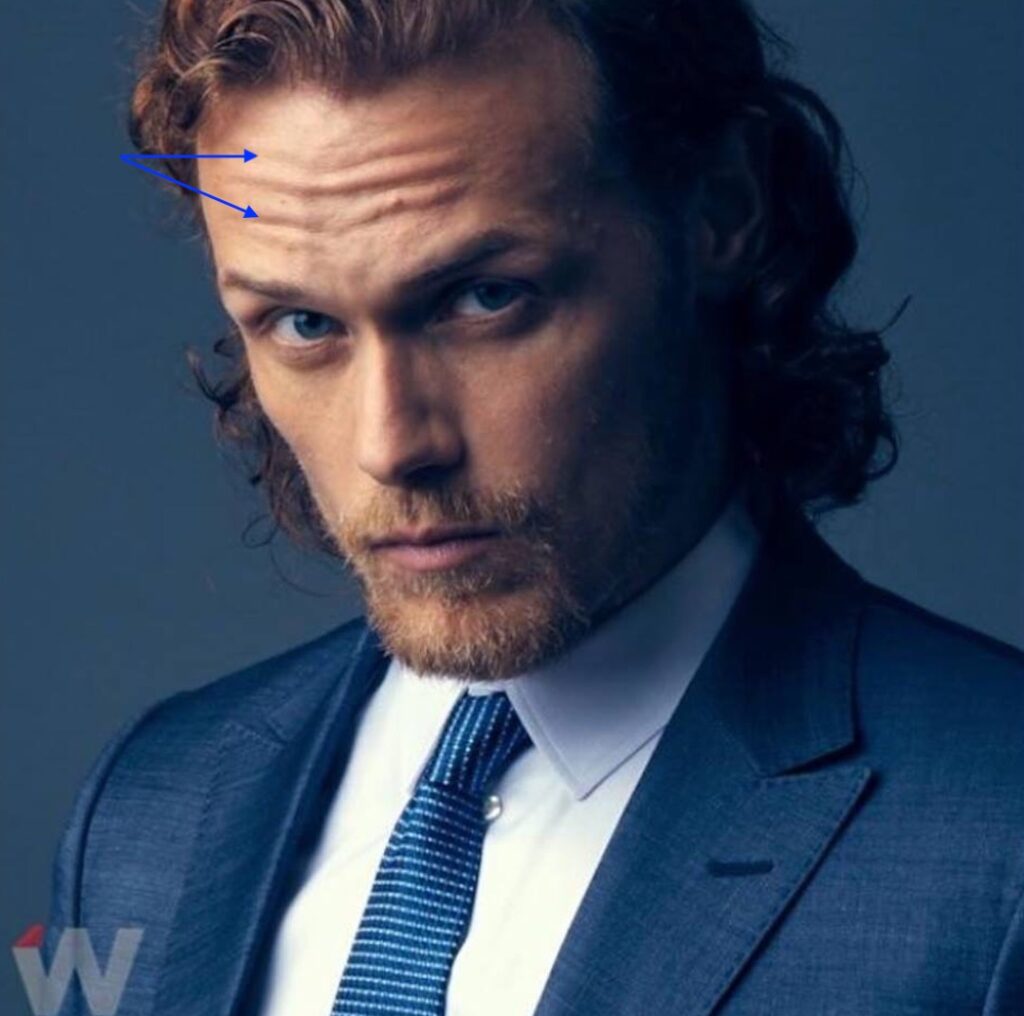
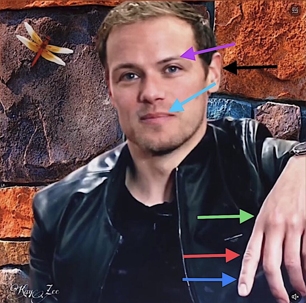
Frontalis: Paired muscles of facial expression, frontalis fibers run vertically in the forehead. When they contract, they lift the brows and produce horizontal wrinkles of forehead skin, conveying concentration and concern. Perfect example from model Sam (blue arrows).

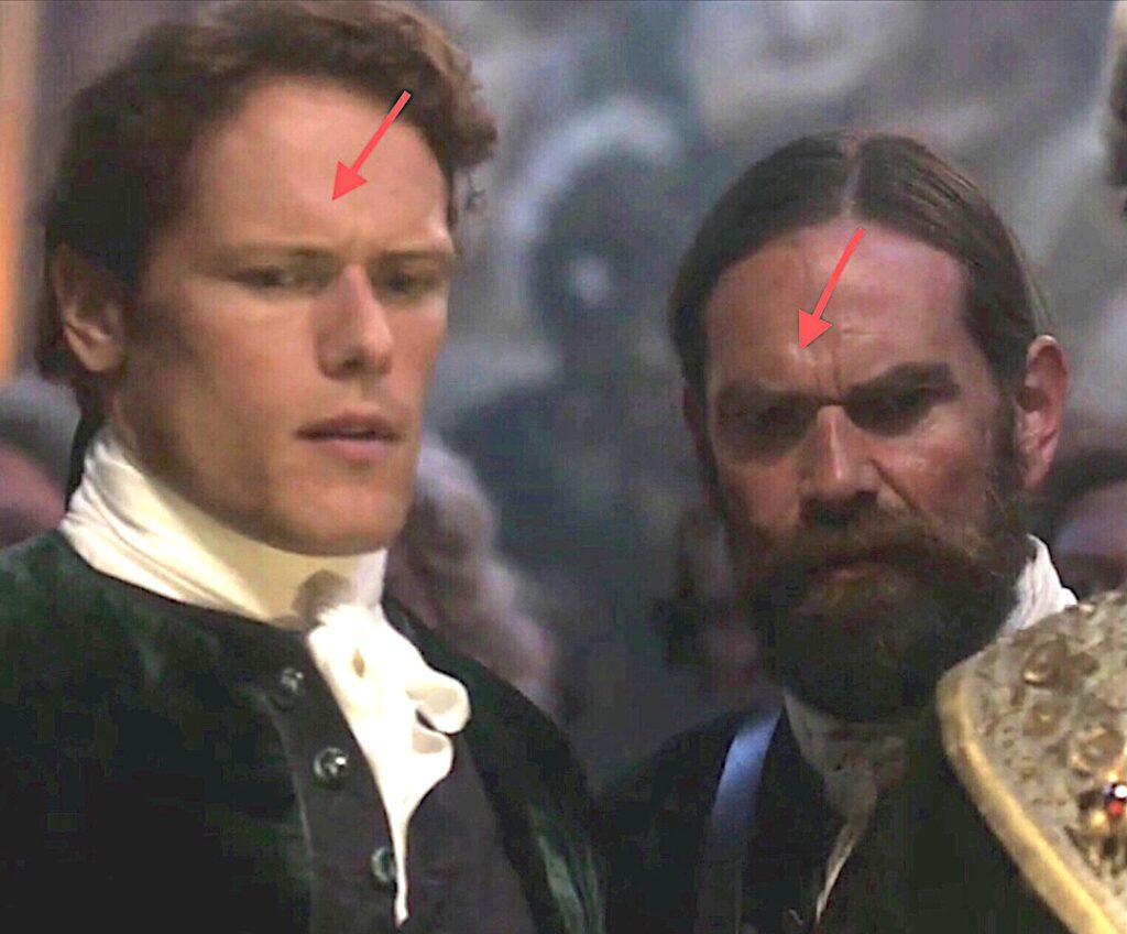
Corrugator Supercilii: Paired muscles of  facial expression from the nose side to the middle of each eyebrow. Upon contraction, corrugators draw the brows together and down producing small vertical wrinkles between the brows and a small bulge of skin above the brows. Jamie and Murtagh show  corrugator prowess (red arrows) as they watch the King of France during his a “private” moment (Outlander, episode 202 “Not in Scotland Anymore!” 😱

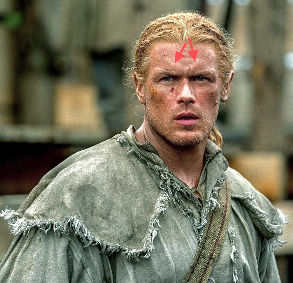
And, this image from outlander episode 708 “Turning Points,” shows an outstanding example of corrugator supercilii (red arrows) as Jamie realizes he almost shot his son during the second Battle of Saratoga! 🥺

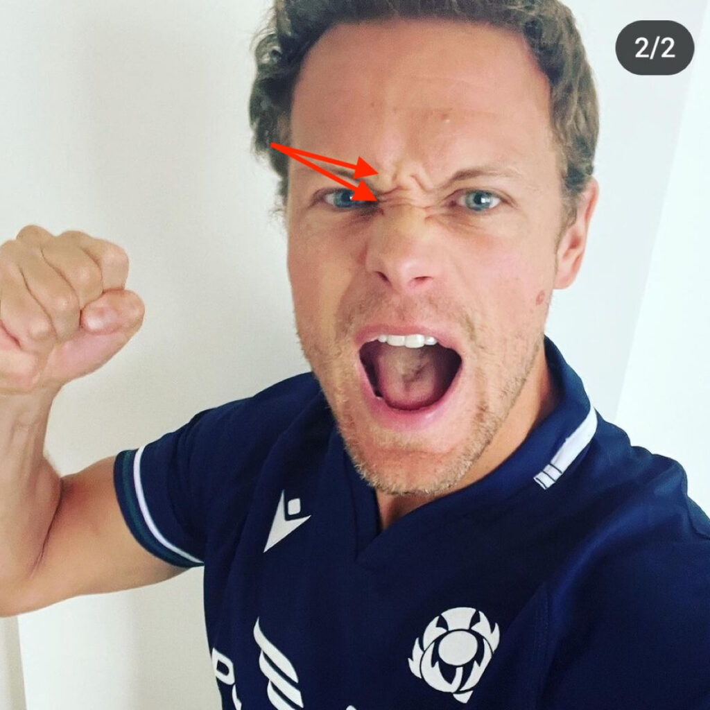
Procerus: Procerus are muscles of facial expression wrinkling the skin over the bridge of the nose and flaring the nostrils. Results? They help create an expression of anger. “Who are you calling a procerus?” threatens Sam (red arrows). 😆

Supercilium: As everyone kens, eyebrows add to facial expression. Anatomically, the eyebrow is the supercilium (super silly, huh?). Sam has naturally thin, beautiful brows (purple arrow). I recall him being asked at during an interview if he plucked them, to which he responded, “Never.”

Vermilion Border: The vermillion border is intersection between facial skin and the rim of lips (turquoise arrow). Sam’s border forms a lovely Cupid’s Bow in the midline.

Moving on to the hands….

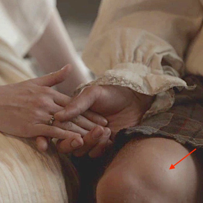
Metacarpophalangeal Joint: This joint (green arrow) is formed where the metacarpals of the palm meet the proximal phalange, the first and largest finger bone.

Proximal Interphalangeal Joint: This is the intersection between proximal phalange and middle phalange (red arrow).

Distal Interphalangeal Joint: This joint occurs between middle phalange and distal phalange (blue arrow).

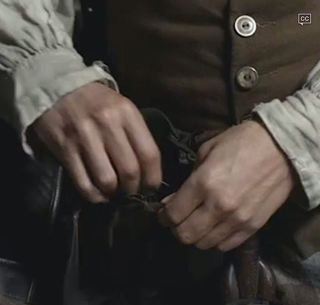
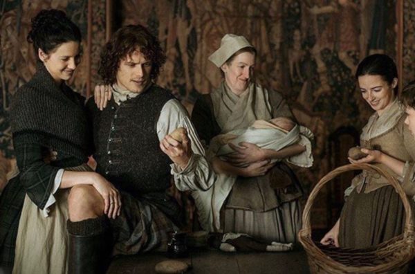
Attention: The wonderful image below is the property of @KayZee. ❤️

Didn’t everyone notice Sam’s beautiful hands as he discusses Claire with Murtagh before the wedding in Outlander Episode 107, “The Wedding.” 😍

Moving on to the torso…

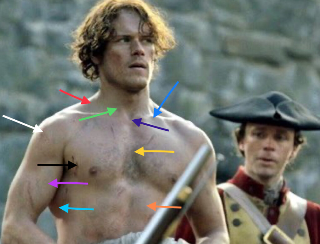
The next image is JAMMF between floggings – Outlander episode 106, “The Garrison Commander!” 😱 Lots of arrows on this one and the list is loooong but so worth it! 

Trapezius: Trapezius (red arrow) is  a massive flat triangular-shaped muscle that is paired. Together, they lift the shoulder joint and pull the shoulder joint back and down.

Clavicle: Commonly known as the collar bone (turquoise arrow), the paired clavicles are 6″ S-shaped bones between sternum and each shoulder joint. Clavicle is also the most commonly fractured bone of the body (one of my grandsons fractured his last year). It holds the shoulder joint away from the sternum allowing for greater mobility of the arm.

Fun Fact: Dogs and cats don’t have clavicles so their shoulder blades shift to lie at sides of the torso; ours lie over our backs. Thus, their front limbs move forward and back whereas, ours can rotate almost 360°. The human shoulder joint is the most movable joint of the human body, all because of the clavicle! 🤩

Sternocleidomastoid: Paired strap-like muscles (green arrow) joining sternum (breast bone) and clavicle to skull behind ear. Acting alone, each muscle flexes the neck toward the shoulder and rotates the chin toward the shoulder. Together, the muscles draw the chin toward the sternum.

Fun Fact: Sternocleidomastoid muscles are one of over  20 pair of muscles acting on the neck! 🤓

Suprasternal Notch: This bony landmark (purple arrow) indicates the top of the sternum. Intrathoracic pressure can be measured via the soft tissues above this landmark.

Sternum: The sternum (yellow arrow) is an unpaired bone which forms the front of the chest. It provides attachments for clavicles and first seven pair of ribs. It also supports and protects vital organs such as heart and lungs.

Rectus abdominis: This paired muscle (orange arrow) forms the belly on either side of the midline. Each muscle is long and flat, extending from sternum and ribs to pubic bone. Acting together, they bend head toward pelvis. 

Deltoid: The deltoid (white arrow) is shaped like an inverted triangle and overlies the shoulder joint, giving the shoulder its rounded contour. It helps raise the arm forward, to the side and backward. It is subdivided into anterior (front), middle, and posterior (back) sections.

Pectoralis Major: The term pectoralis is derived from the Latin meaning “breast.” Gyms refer to them as ‘pecs.” Pec major is paired and the largest muscle (black arrow) of the chest They draw the clavicle downward. They also raise the arm forward, pull arm against torso, or rotate arm toward sternum.

Biceps Brachii: The biceps (pink arrow) are the large muscles at the front of the arm. They create the fabulous bulge that Popeye made famous. In Latin, biceps means “two heads,” so named because the muscle originates from two different parts of the scapula. Biceps flexes the elbow joint and rotates the palm forward/upward. It also flexes the shoulder joint and draws the humerus (arm bone) against the torso.

Fun Fact: Contrary to popular opinion, biceps is not the prime mover of the elbow joint! A deeper lying muscle, brachialis, is the prime mover. 💪🏻

Latissimus Dorsi: Latissimus dorsi (aqua arrow) is a large flat muscle of the back. It inserts on the humerus pulling it backwards, against the body, and towards the sternum. Sam’s latissimus is massive! 🥳

and

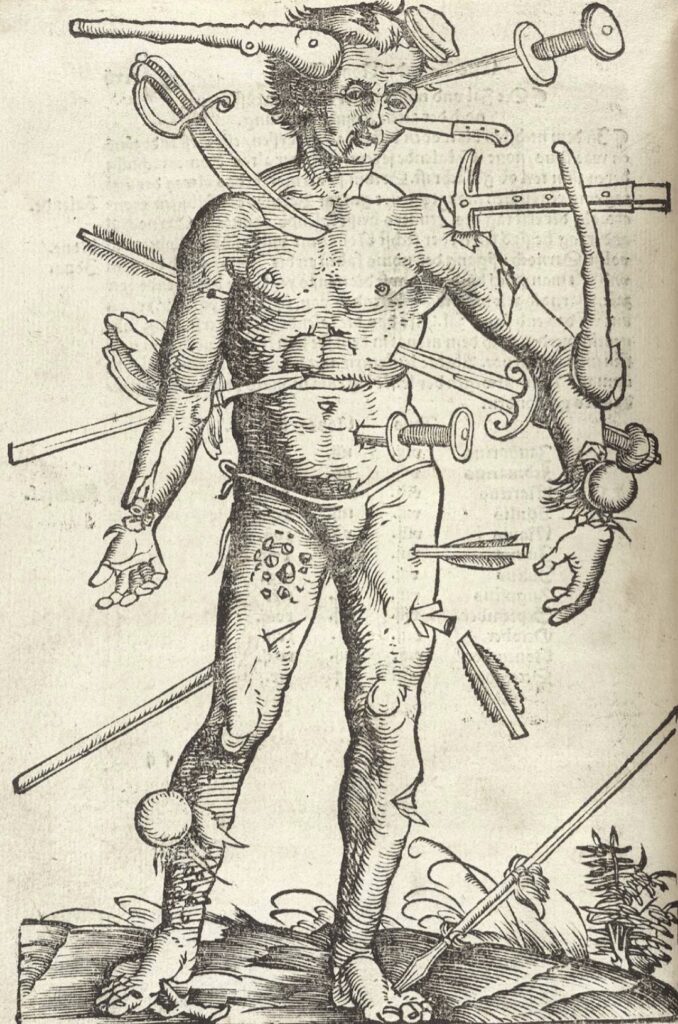
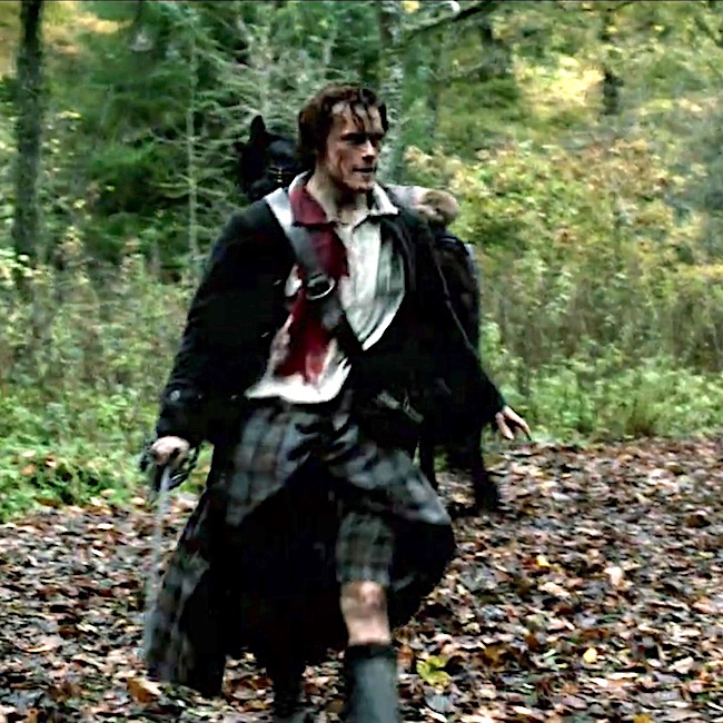
The previous image of Sam as Jamie reminds me of “The Wound Man,” a surgical diagram that appeared in European medical texts of the fourteenth and fifteenth centuries, up until the 1700s. It showed various battle injuries and diseases that a medical practitioner might encounter. Cures were listed on nearby pages. A horrifying image, for sure! 😳

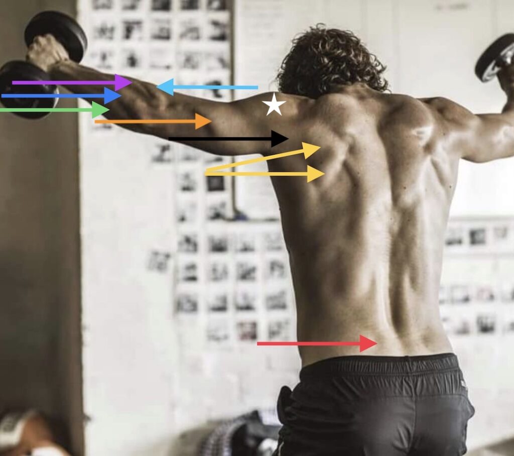
Next, Sam’s back is a marvelous roadmap of topographical anatomy.

Infraspinatus: This muscle (yellow arrows) extends from scapula (shoulder blade) to humerus (arm bone). It externally rotates and stabilizes the shoulder joint. Along with three other muscles, it helps form the rotator cuff of the shoulder joint.

Triceps Brachii: The triceps (orange arrow) derives its name from Latin meaning three heads because it takes origin from the scapula and two different areas of the humerus. It ends on a forearm bone, the ulna. It extends (straightens) the elbow joint.

Brachioradialis: This muscle (aqua arrow) attaches humerus to radius, a forearm bone. It helps flex the elbow joint.

Extensor Carpi Radialis Longus: This forearm muscle (violet arrow) reaches from humerus to second metacarpal bone. It extends (straightens) wrist and abducts hand (moves hand toward thumb).

Extensor Digitorum: Extensor digitorum lies next to extensor carpi radialis longus (blue arrow). It extends all four fingers (not thumb). Straighten your bent fingers. Extensor digitorum did that!

Extensor Carpi Ulnaris: This forearm muscle (green arrow) reaches from humerus to fifth metacarpal bone.  It extends the wrist and adducts the hand (moves hand away from thumb).

Posterior Deltoid: The deltoid  was explained above but now we add a caveat. Sam’s posterior deltoid (black arrow) is unusual because a distinct groove separates it from  middle deltoid (white star). Most people do not exhibit this distinct separation.

Erector Spinae: This massive muscle  (red arrow) is paired; it has several parts based on origin and insertion of the muscle fibers. Working together, erector spinae straightens the back; working alone, it rotates the back.

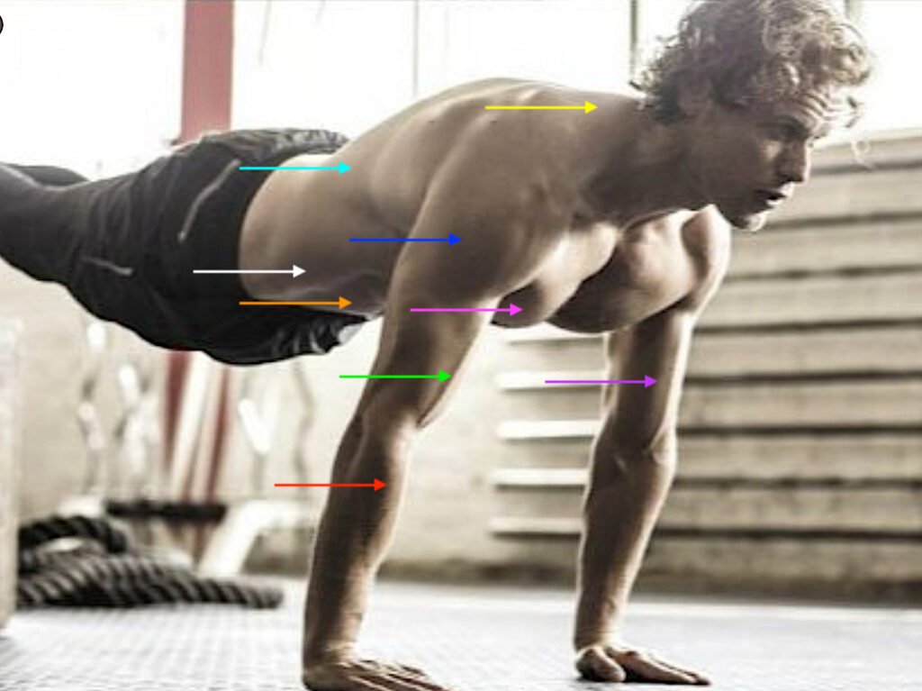
Next is Sam in a full plank position with elevated feet. This one has a number of repeated structures, but is still delightful to view. 🤩

Trapezius: Yellow arrow – see above

Deltoid: Blue arrow – see above

Pectoralis Major: Pink arrow – see above

Biceps Brachii: Violet arrow – see above

Latissimus Dorsi: Aqua arrow – see above

Rectus Abdominis: Orange arrow – see above

External Abdominal Oblique: The EAO is the largest flat abdominal muscle found at front and side of abdomen. It is also paired. It attaches to ribs above and pelvic bones below. Its fibers run from the sides downwards like your hands tucked into jean pockets. Acting alone, the EAO rotates the torso; acting together EAO pulls chest toward pelvis (as in curl ups). It also compresses the abdominal cavity. This muscle is important for posture and torso movements.

Brachioradialis:  red arrow- see above

Cephalic Vein: A vein of the arm – see below

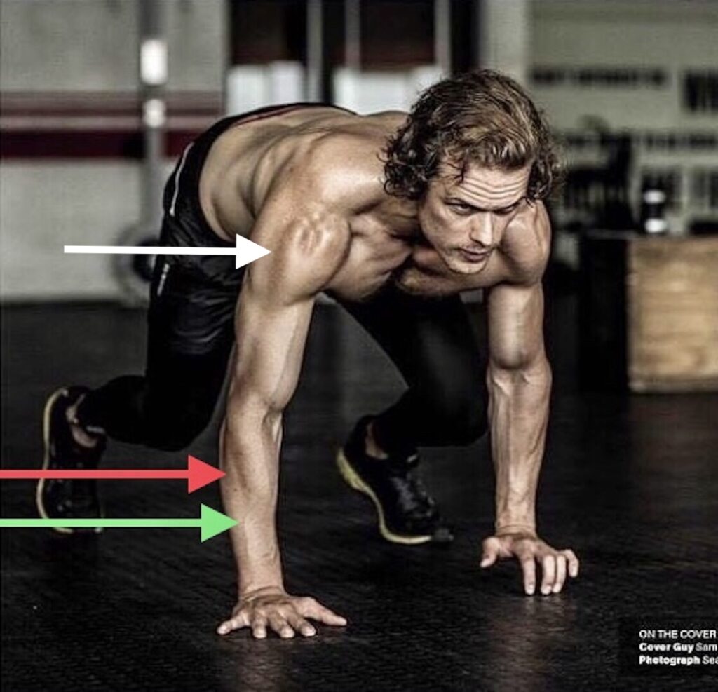
This image of Sam in a sprint position is awesome because it shows:

Posterior Deltoid: Already described above, the white arrow indicates the unusual and distinct groove between middle deltoid and posterior deltoid.

Extensor Digitorum: Green arrow – see above

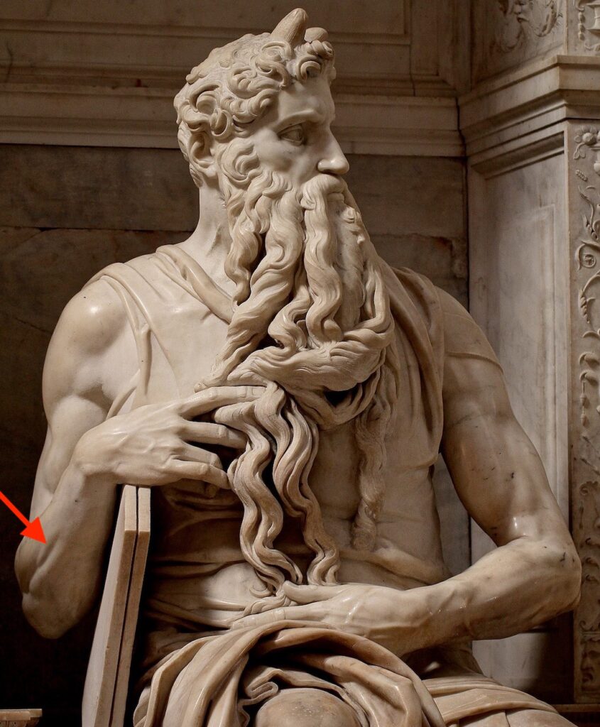
Extensor Digiti Minimi: This wee muscle (red arrow) isn’t prominent unless the forearm is highly muscular and subcutaneous fat is low. EDM reaches from humerus to wee finger and extends (lifts) it.

Some X followers already have seen my tweet of this image of Michelangelo’s “Moses” sculpture. But, for those who haven’t, it shows the master’s  attention to wee extensor digiti minimi! 🥰

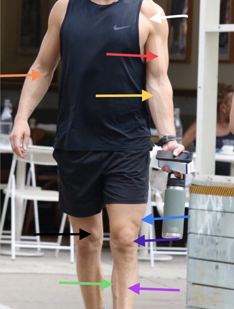
Next is a full body view of Sam’s surface anatomy. This one shows a few arms veins that is a phlebotomist’s dream!

Just a note that venous pattern throughout the body is extremely varied so much so that hand vein patterns can be used to identify a person.

Median Antebrachial Vein: The median antebrachial vein (gold arrow) and its tributaries gather blood from hand and forearm and return it to the basilic vein (not shown).

Cephalic Vein: This large vein (red arrow) gathers blood from hand and forearm and returns it to a large vein (axillary vein) deep to the collar bone. Its name means “head” in Latin because its path through the arm points toward the head.

Deltopectoral Groove: Cephalic vein is traced through the deltopectoral groove (white arrow), a groove between anterior deltoid and pectoralis major muscles. 

Median Cubital Vein: This vein (orange arrow) located in the cubital fossa (elbow hollow) forms a bridge connecting cephalic and basilic veins.

Fun Fact: Median cubital vein is the preferred site for blood draws because it is large and doesn’t tend to roll or move when a needle is inserted. The area also has fewer pain endings.

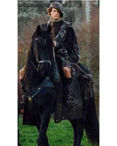
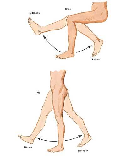
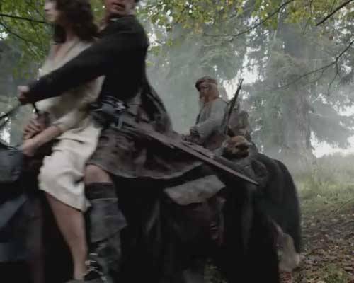
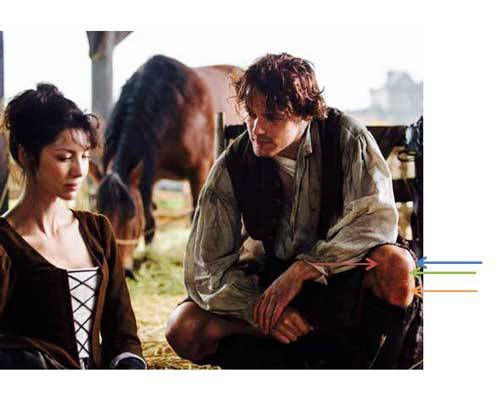
Moving to the lower limb! 🤗

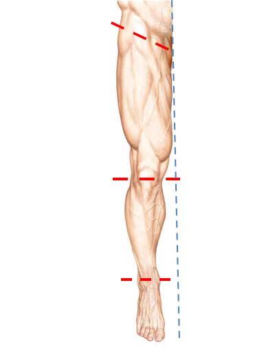
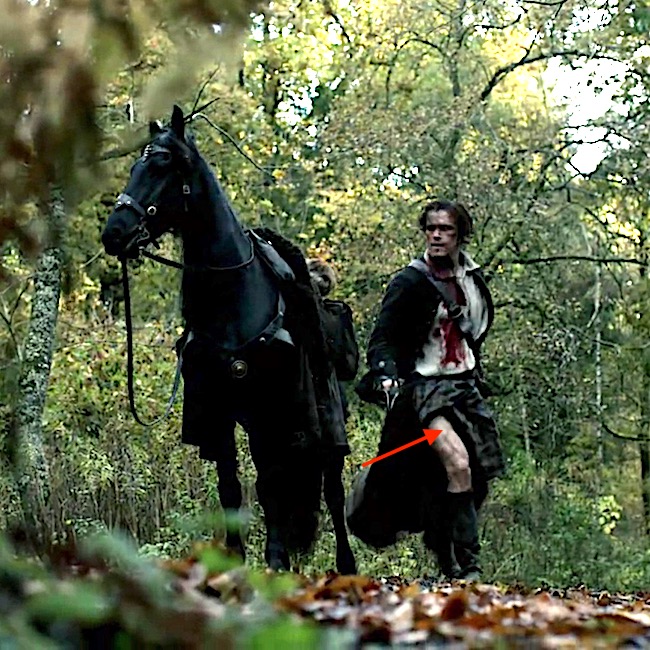
Just So You Know: Anatomists define the thigh as that part of the lower limb between hip and knee and the part between knee and foot is the leg.

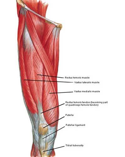
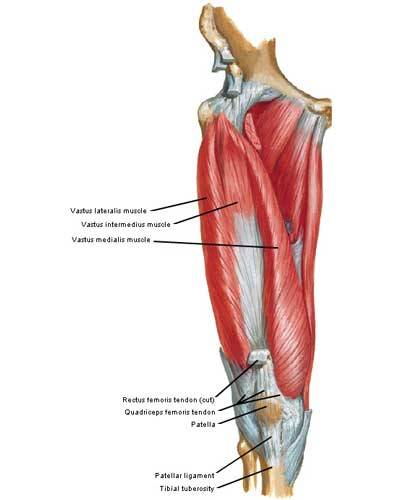
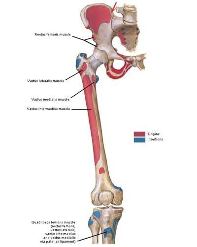
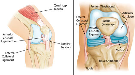
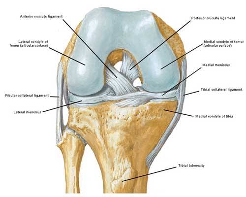
Vastus Lateralis: Aptly named, vastus lateralis (blue arrow) is vast on Sam-our-Man! Vastus lateralis is part of the quadraceps group of four (some say five) thigh muscles. It arises from the femur and inserts on the patella. It then joins the other quad tendons to form a common tendon that inserts on the tibia (largest leg bone). It is the largest and most powerful muscle of the quadraceps group. Together with the other quadraceps muscles, it extends (straightens) the knee joint and keeps patella in proper alignment.

Fun Fact: The vastus lateralis is the recommended site for intramuscular injection of infants under 7 months old and those unable to walk or with loss of muscle tone and mass.

Vastus Medialis: Also a member of quadraceps, this muscle (black arrow) arises from the femur and inserts on the patella and then, tibia. It has the same function as vastus medialis (see above).
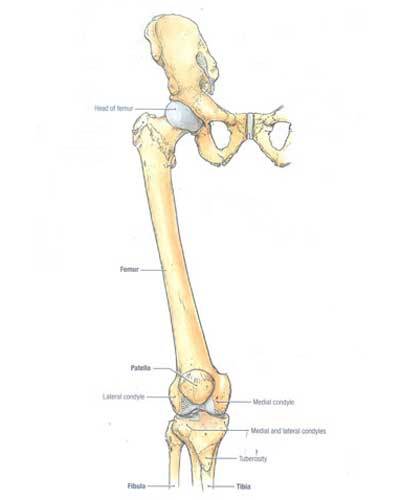
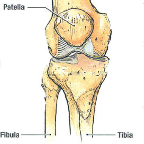
PatellaAlso known as the knee cap, patella (purple arrow) is the largest sesamoid bone in the body, meaning it is enclosed in ligament or tendon.

GastrocnemiusGastrocnemius has two heads arising from different parts of the femur. These join together to help form Achilles tendon which inserts into the calcaneus (heel bone). Sam’s medial head (green arrow) is very apparent in this image. It is a powerful muscle that plantar flexes (points) the foot and flexes the knee joint.

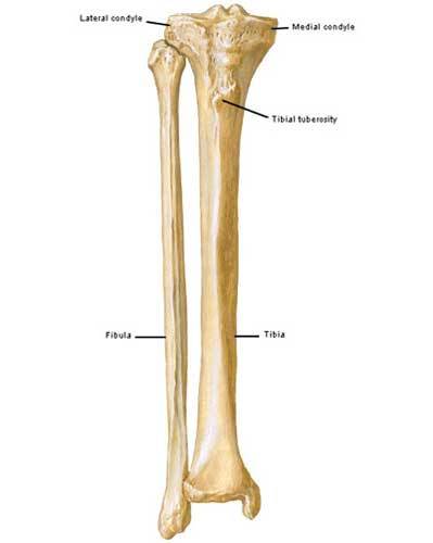
Tibia: Tibia (violet arrow) is also known as the shin bone. It is the larger of the two leg bones. Together with the femur, tibia forms the knee joint and with the fibula (smaller leg bone), it forms the ankle joint.

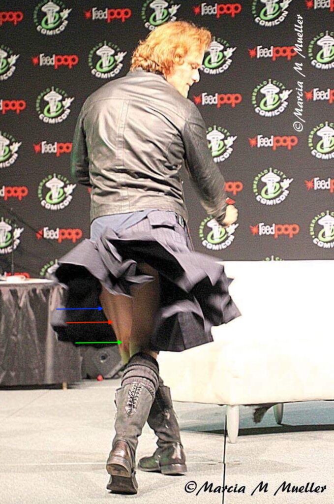
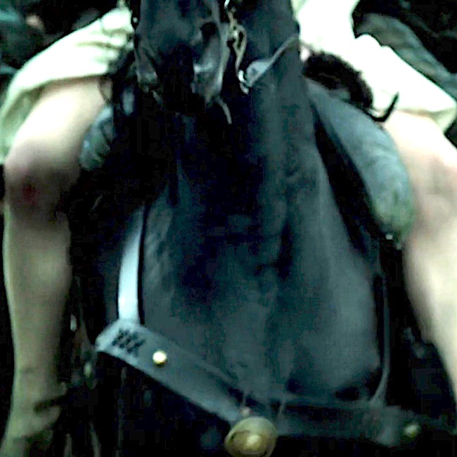
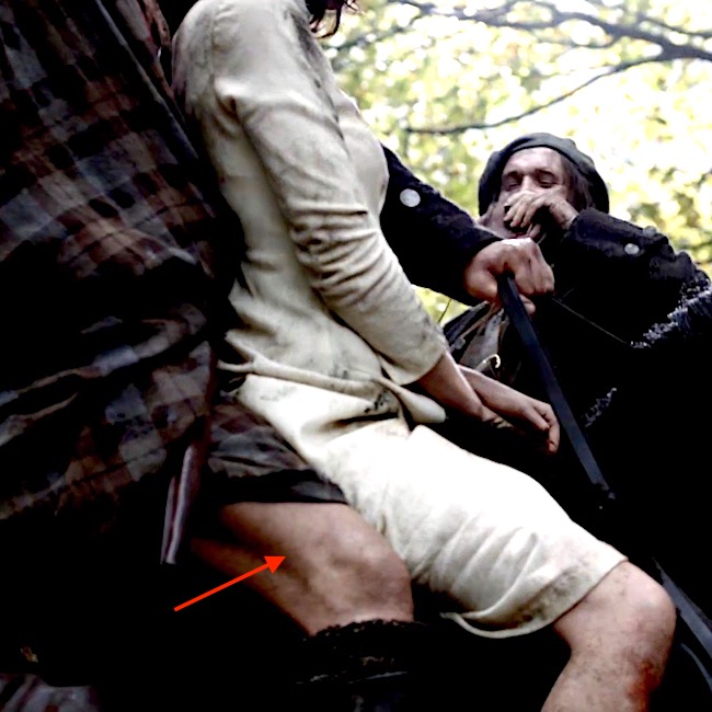
Next is a famous image of Sam flipping kilt for the “girls” at Emerald City ComicCon, March 6, 2017.  Plenty of thigh muscle on display here! 😜

This amazing image is property of Marcia M Mueller. 👏🏻

Biceps Femoris: Biceps femoris (so named because it has two heads). The long head (red arrow) arises from the ischium (part of pelvic bone) and the short head (green arrow) arises from the femur (thigh bone). Both heads join into a single tendon that inserts on the fibula. Biceps femoris is a powerful flexor of the knee joint.

Not so Fun Fact: Avulsion (tearing away) of the biceps femoris tendon is common in sports that require explosive bending of the knee as seen with sprinting! 😱

Vastus Lateralis:  Blue arrow – See above
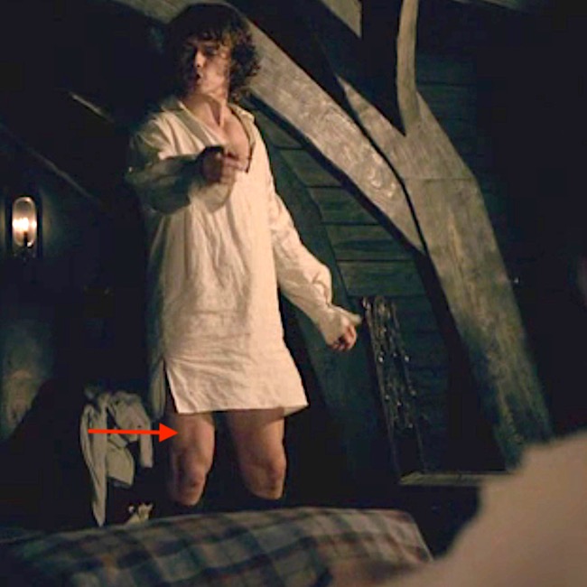
Quiz time!

Try to identify the structures in this last image of Sam. Do your best. Answers appear after the image. Good luck!

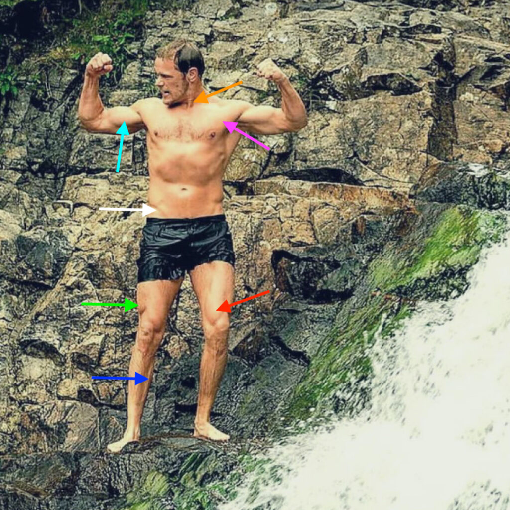
    • Orange arrow – sternocleidomastoid muscle
    • Violet arrow – pectoralis major muscle
    • Aqua arrow – Biceps brachii muscle
    • Green arrow – Vastus lateralis muscle
    • Red arrow –  Vastus medialis muscle
    • Blue arrow – medial head of gastrocnemius muscle
    • White arrow – Inguinal groove, (aka Adonis belt)  *** Extra credit for this one because we didn’t discuss! 😃

Well done, students! 🏆

The deeply grateful,

Outlander Anatomist

Follow me on:

Photo Creds: Sony/Starz; www.menshealth.com, www.thewrap.com, www.Wikimedia, @marciammueller, @samheughan, @kayzee

Fun Fact: patella

These canna be small potatoes! Nope, it’s the patella!

ep-208-patella

Patella pa·tel·la,

sing. n patella; pl. n patellae

From Latin patina meaning shallow dish

First use: late 16th century

Anatomy def: The kneecap or kneepan; a shallow, conical-shaped bone which articulates with the femur and covers the knee joint.

Outlander def: Knee porn! “nuf said!

Learn about patella and knee joint by reading Anatomy Lesson #7, “Jamie’s Thighs or Ode to Joy!”

Read about the knees in Dragonfly in AmberQuoting one of many great examples:

Jamie laced his hands together, elbows braced on his knees, and rested his chin on his linked knuckles. “This wilna be like France,” he said quietly. “Fighting there, ye risk no more than your life in battle. Here …” He hesitated, then went on. “Jenny, this is treason. If it goes wrong, those that follow the Stuarts are like to end on a scaffold.

Herself even specifies the knee cap. Again, from Dragonfly in AmberClaire describes this sad scene:

”Mother Hildegarde wrapped her in a length of white satin,” I said, looking down at my fists, clenched in my lap. “Her eyes were closed. She hadn’t any lashes yet, but her eyes were slanted. I said they were like yours, but they said all babies’ eyes are like that.” Ten fingers, and ten toes. No nails, but the gleam of tiny joints, kneecaps and fingerbones like opals, like the jeweled bones of the earth itself. Remember man, that thou art dust.…

See a great patella on a handsome fella in Starz episode 208, The Fox’s Lair. Claire canna keep her hands off Jamie’s knee <G>!

A deeply grateful,

Outlander Anatomist

Anatomy Lesson 7: “Jamie’s Thighs – Ode to Joy!”

Anatomy Lesson #7, Jamie’s Thighs, is a true ode to joy! I hope Ludwig won’t be offended by my reference to his 9th, but the title perfectly suits this lesson! Perfect timing with the upcoming US Thanksgiving Holiday.  I’m down on my knees giving thanks because Jamie’s knees also appear in this lesson! 🙏🏻

Are you ready? Let’s go!

Just to prove my goodwill, take a gander at Jamie’s knees in the image below. May this tide you over as our lesson proceeds!

image

Definitions: Let’s begin with a few basic tidbits, otherwise much of this lesson might remain obscure!

  • Joint – Site where bones meet and move relative to each other
  • Muscle – Muscles attach to bones (some exceptions); these are voluntary meaning we can contract them at will – a.k.a. skeletal muscle
  • Contraction – As muscles contract, a joint moves in various ways (Image A)
  • Origin – Attachment site of muscle to bone – site moves little or not at all during contraction
  • Insertion – Attachment site of muscle to bone – site moves during contraction
  • Flexion – Muscle contraction closes (approximates) bones of a joint
  • Extension – Muscle contraction opens (straightens) bones of a joint

image

Image A

Lower limb: Our lower limbs are the body parts from hip joints to toes. Anatomists don’t use the terms upper and lower leg. Rather, the lower limb is divided into (Image B):

  • Thigh – segment between hip and knee joints
  • Leg – segment between knee and ankle joint
  • Foot – segment beyond ankle joint

BTW, the dashed blue line in Image B represents a vertical midline through the body. A medial structure lies closer to this midline – lateral structure lies further from this midline. That’s the gist of it!

Try this: Locate your thigh, leg, foot and midline.

Test Q: Are your tiny toes medial or lateral to your middle toes?

image

Image B

Test A: They are lateral. Great job!

Outlander Time! The first time we rencounter Jamie’s thighs is in Outlander book where Claire straddles-his-saddle on the way to Castle Leoch. Herself writes:

My companion seemed to be having little trouble, in spite of being unable to use his right hand. I could feel his thighs behind mine, shifting and pressing occasionally to guide the horse. I clutched the edge of the short saddle in order to stay seated; I had been on horses before, but was by no means the horseman this Jamie was.

And yet another quote from Outlander book – this one from randy-dandy, Jamie:

But then that ride through the dark together….with that lovely broad arse wedged between my thighs…

Well! Starz Claire doesn’t sport a broad arse, but it surely is curvaceous and pert and we all ken where it was wedged during this ride!  Mmphm!

image

Back to anatomy and more about bones!

Femur: The thigh contains one bone, the femur. I love superlatives, so here’s the first one for this lesson: the femur is the longest bone of the human body (Image C – front of right femur). The top of each femur ends in an angled neck and head that fits securely into the acetabulum, a deep socket of the hip bone Together, they form the acetabulo-femoral (hip) joint, a ball and socket joint.

Fun Facts: You may recall from Anatomy Lesson #2, When Claire Meets Jamie or How to Fall in Love While Reducing a Dislocated Shoulder Joint!:  although the glenohumeral (shoulder) joint is very moveable it is also less stable due to a shallow ball and socket joint! Conversely, each hip joint is very stable but less moveable due to a deep ball and socket joint. The hip joint needs to be stable to support our weight against gravity. Very interesting!

At the knee, the femur ends in two sturdy knobs, medial and lateral condyles (Greek for knuckle); both knobs help form the knee joint (Image C).

image

Image C

Tibia and Fibula: The leg contains two bones, tibia and fibula (Image D – front of right leg bones). The larger tibia is medial to the smaller, more lateral, fibula. The top of tibia ends in two flat surfaces, the medial and lateral condyles; lower down, it bears a midline knob, the tibial tuberosity. Only tibia helps form the knee joint; fibula plays no role.

image

Image D

Patella: The small patella or knee cap is critical to knee anatomy. Patella is the largest sesamoid bone of the body (there are others) meaning it is enveloped in tendon. Its deep surface slides in a groove between medial and lateral femoral condyles as the knee joint extends and flexes (Image E – front of right knee joint).

image

Image E

Thigh Muscles: Yes! Thigh muscles are massive because they support much of our weight and help maintain our bipedal stance against gravity.

There are 13 thigh muscles divided into three compartments. To keep this lesson shorter than a bloody master’s thesis, we will cover only quadraceps femoris (Latin meaning four-headed muscle of the femur) with a wee bit about iliopsoas muscle.

Quadraceps: The quads (as trainers call them) are four muscles at the front of each thigh:

    • rectus femoris
    • vastus lateralis
    • vastus intermedius
    • vastus medialis

Image F (front of right thigh) shows the three of the quad muscles. A fourth, vastus intermedius, is visible only after removal of rectus femoris  (Image G).

image

Image F

image

Image G

Origins: Each quad muscle has a different origin (Image H –  red marks). Rectus femoris originates from hip bone and crosses both hip and knee joints . The three vasti (pl.) muscles take origin from different sites on the femur and cross only the knee joint.

Insertion:  All four muscles  fuse into a common quadraceps tendon that engulfs the patella and then continues as the patellar ligament to insert (Image H – blue mark) on the tibial tuberosity.

Actions: Acting together, all four heads of each quad extend the knee joint but only rectus femoris also flexes the hip joint.

Because quads are the only muscles that extend the knee joints, they are crucial for walking, running, jumping and squatting. Quads are also called anti-gravity muscles because they contract as we rise from a seated position or lower our bodies in reverse, holding our weight against gravity (think of thighs during snow skiing or Jamie lowering Claire onto the marriage bed. Yum!).

image

Image H

Iliopsoas: One last muscle… Although recti femoris (pl.) are decent hip flexors, iliopsoas muscles are the strongest. Like the quads, iliopsoas is a compound muscle formed by fusion of iliacus and psoas (pronounced soas) muscles.

Iliacus arises from the hip bone and psoas arises from the lumbar vertebrae (Image I – right front). They fuse into a single tendon that inserts into the femur. These muscles draw the femur closer to the torso, assisted by rectus femoris and a few other minor hip flexors. Or, with thighs held stationary, they draw torso closer to thighs (think sit-ups).

Image I

Try this: To test your quads, place back against a wall.  Drop the tush while walking your legs away from the wall until you assume a squat position (Image J). Thighs and legs should be at 90° to each other (don’t drop lower – this is bad for knee joints!). Now hold your torso in place for 30 seconds and then gradually straighten (extend) the knee joints. If you feel wobbly, then you may need quad work as these muscles quickly lose mass and strength due to inactivity, sedentary jobs or aging. Quads can be strengthened by wall squats or by any exercise that adds resistence while extending the knee joint!

image

Image J

Knee Joint: Now on to the knee jointlargest joint of the human body (Image K – right side and front)! Here medial and lateral femoral condyles (knuckles) ride atop a flat plateau formed by the medial and lateral tibial condyles – no stable joint here!

All bony surfaces of knee joint are covered with articular cartilage, a bloodless, firm connective tissue that allows for smooth movement. This odd joint also has medial and a lateral meniscus (different type of cartilage) that create two shallow sockets atop the tibial plateau – one for each femoral condyle; these also act as shock absorbers.

As mentioned above, the patella glides between the femoral condyles during flexion and extension.

image

Image K

Knee Ligaments: The knee is stabilized by very strong ligaments designed to secure the tibia and femur (Image L – right knee in full flexion – patella absent):

  • medial (tibial) collateral ligament
  • lateral (fibular) collateral ligament
  • anterior cruciate ligament
  • posterior cruciate ligament (and, there are others!)

Overall, the knee joint is at risk due to our sports-crazy cultures and because supporting the body weight while in motion is a challenge for these relatively flat surfaces. Hence, the über-strong ligaments.

image

Image L

Now, for the fun stuff!  I promised in Anatomy Lesson #3 that I would be returning to the scene where Jamie dismounts to grab Claire (Starz episode 101, Sassenach).  Yep, that’s the one!  Let’s relive that scene wherein we first spy Jamie’s thigh – aye, readers, there’s a first time for everything.  Hah!

Wait fer it…………….”Lost yer way?”

Wait fer it……….. Jamie’s shifts his weight. Um…..he looks a wee bit fashed!

Wait fer it.…. With a high kick Jamie’s right thigh clears the steed’s neck. Yay, iliopsoas!

Wait is over! Jamie’s drops.  Gad, this lad has loooooong legs!!!!  Thank gawd that kilt didna do its job here! (That impressive thigh muscle is vastus lateralis!)

A firm landing and further evidence of those thighs and knees!

This just keeps getting better and better! Geez, Jamie is a perfect anatomical model!

Three of four quad heads are visible as he strides towards Claire. The red arrow points to the head of rectus femoris! (Claire! why aren’t ye laid out in a swoon?)

OK, Claire, now you are in fer it! Ye’re gonna get what ye deserve!  Oops, sorry, wrong  Starz episode.  (Thinking 109, The Reckoning). Snort!

Okey dokey then, are you ready for a pop quiz? Let’s go for it!

Can you identify one of Jamie’s quads in the next image (Starz episode 7, The Wedding)?

Q: Name the major bulge (no, not that one! <G>) on the inside of his thigh (red arrow).

A: Right vastus medialis muscle. Good job!

Q: The light is dim, but name the quad at the red arrow.

A: Left vastus lateralis muscl– just peeking out from under his wedding sark. He is sooo modest!

Back to anatomy!

Knees: Now for those gorgeous knees!  I know gazillions of you have been awaiting another glimpse of Jamie’s knees!  Sorry to mess them up with a quiver of arrows, but just so you ken:

  • Blue arrow marks his lateral femoral condyle
  • Red arrow is his medial femoral condyle
  • Green arrow marks his patella
  • Orange arrow is his tibial tuberosity.

So now you can name all the knuckles and knobs o’ Jamie’s knees!

Claire, lift your eyes lass! He squatted down to show off his gorgeous gams! He kens he is one damn fine-looking Scot – grubby sark or no!image

Almost done, so hang with me!

IT Band: Lastly, thigh muscles are wrapped in a strong sleeve of connective tissue, the fascia lata (Image M – right lower limb). Fascia lata thickens at the side to form the iliotibial (IT) tract or IT band (Anatomy Lesson #1, Jamie’s Tush).

image

Image M

Now, for more applied anatomy! The red arrow marks a thick ridge of tissue at the back of Jamie’s thigh (Starz episode 101, Sassenach). Yep, that’s his right IT band! You can see it plain as Rupert swigging that raw whisky!

Fascia lata Function: You all should understand the importance of the fascia lata and IT band. The heart faces a long haul to pump venous blood against gravity from the foot back to the chest. Thigh muscle contractions help milk venous blood back toward the heart. Fascia lata aids this process by compressing contracting muscles against the deep thigh veins.

Danger!  If thigh muscles get seriously injured, they swell and the inelastic fascia lata does not stretch to accommodate the swelling. When this happens, it is a medical emergency because swollen muscles restrict the return of venous blood to the heart and diminish the flow of oxygen-rich arterial blood to the lower limb – and, as you ken, tissue dies if deprived of oxygen!

This condition is known as compartment syndrome and the next photo (Image N) shows the result of medical intervention. This is the right thigh of a former students (he gave permission to post). After injuring his thigh muscles, the swelling threaten necrosis (death) of his thigh muscles so surgeons slit his fascia lata (near the IT band) to relieve pressure and re-establish blood flow. Now, when he contracts his right quads, they bulge through the slit in the fascia lata! An excellent visual of how the fascia lata compresses the thigh muscles!

image

Image N

Okey dokey, with this example, we come to the end of our thigh and knee lesson! But, I’ll not leave you bereft. Here is one last image designed to increase your admiration and respect for Jamie’s anti-gravity quads and iliopsoas muscles!

In this, ahem, modest scene from Starz episode 7, The Wedding, Jamie bears Claire’s entire weight (9 stone or 126 lb – Herself records in Outlander). Then, he slowly lowers their combined weight (about 23 stone/322 lb?) to the bed afore he flips her over! That takes a whole lot of quad strength!  Are you impressed?  <G>

Ok, mukkers, that’s it for now. Please stay tuned for our next lesson.  Still gobs of anatomy to cover as we work our way through the first eight episodes of Outlander!

The lessons are deliberately slow-paced to keep us occupied until Starz episode 109 makes its way into our hearts and minds!

Oh, almost forgot. You can now follow me on Facebook, Tumblr, IG and Twitter.

The deeply grateful,

Outlander Anatomist

Image credits: Starz, Grant’s Atlas of Anatomy, 10th ed., Netter’s Atlas of Human Anatomy, 4th ed., Clinically Oriented Anatomy, 5th ed, www.AuthenticFX.com, www.gouletballet.com, Wikipedia, OA archival photos, AAOC Website, Tumblr.com