Anatomy Lesson #58: Needles, Nerves and Penicillin!

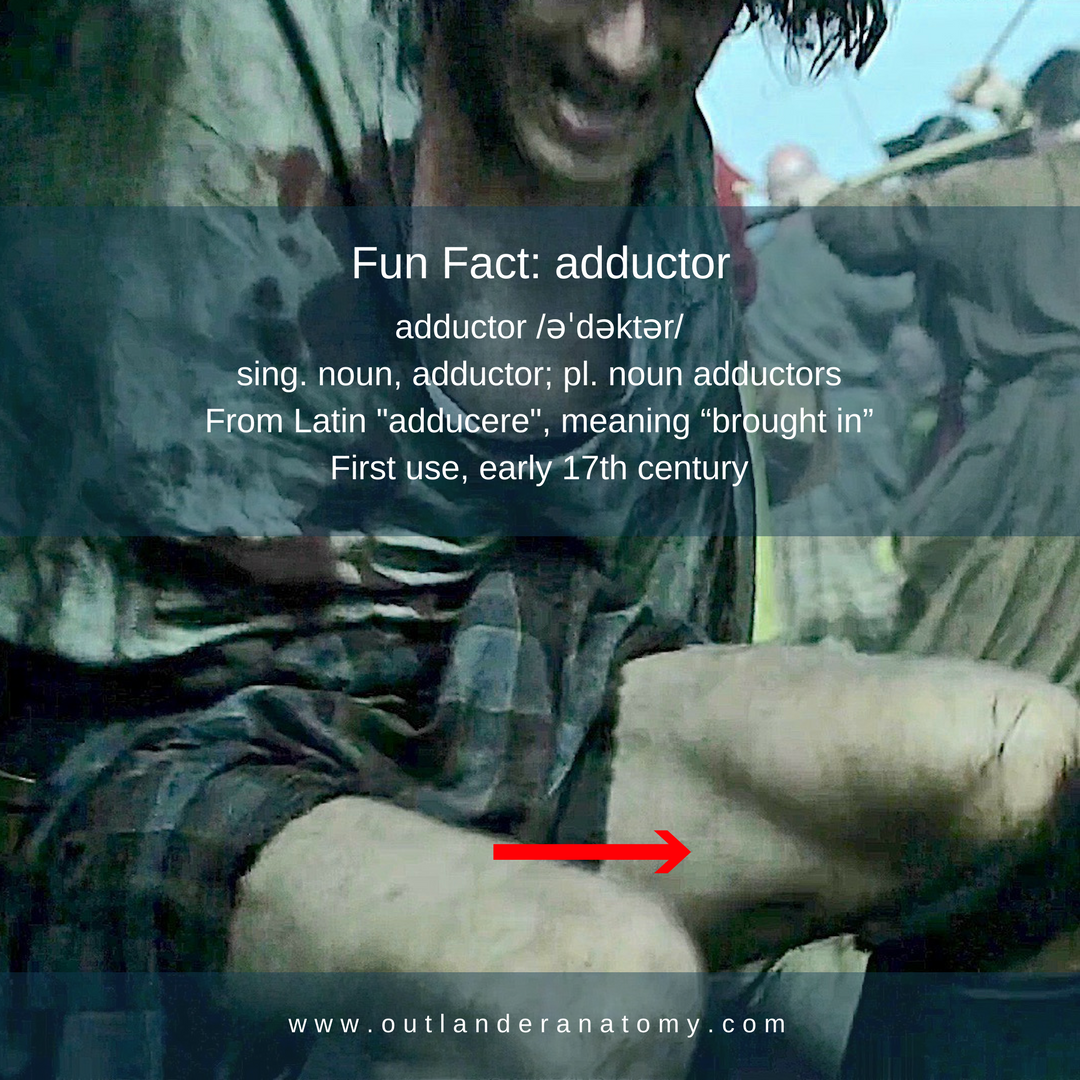
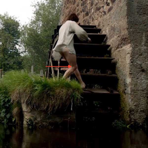
Welcome, all students to Anatomy Lesson #58, Needles, Nerves and Penicillin. What do these words share in common? Why, Jamie, of course!  In Anatomy Lesson #56, Achy Brachial Artery, we learned that birdshot from Laoghaire’s fowling piece almost ruptured one of Jamie’s major arteries. We also learned how Claire analyzed and repaired those wounds – based on human anatomy, of course!

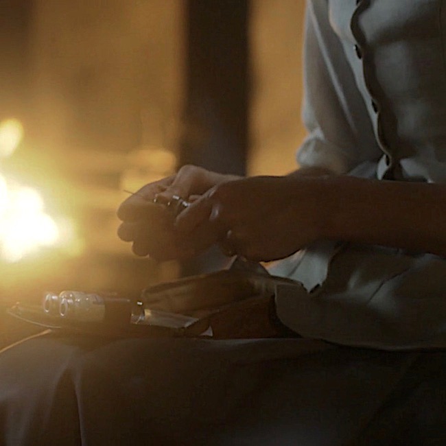
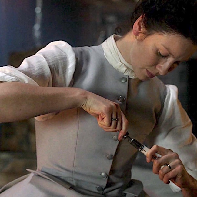
Today’s lesson will examine Claire’s final cure for what ails her man. In Outlander episode 308, First Wife, we see her open a small medical bag equipped with needle, plunger and bottles of white fluid. What is she doing? Diana explains in Voyager book:

I laid the small, flat case on the table and flipped the latch. “I’m not going to let you die this time either,” I informed him, “greatly as I may be tempted.” I carefully extracted the roll of gray flannel and laid it on the table with a soft clinking noise. I unrolled the flannel, displaying the gleaming row of syringes, and rummaged in the box for the small bottle of penicillin tablets.

Translation: Dr. Claire busily prepares a hefty dose of penicillin for our fever-ravaged, red-heided hero!  So, let’s begin the lesson with penicillin.

What is Penicillin? Penicillin is an antibiotic, from the English word, anti- meaning “against,” and the Greek word, bios meaning “life.”  In a nut shell, antibiotics are substances which retard the growth of and/or destroy bacteria (germs).

How does Penicillin Work? First, understand that human cells are surrounded by a fragile cell membrane, but bacterial cells are surrounded by a sturdy cell wall. Penicillins destroy bacteria by preventing synthesis of said cell walls or by promoting processes to break them down. They do not harm human cells because our cells lack cell walls. Make sense? Grand!

From Whence Cometh, Penicillin: Before penicillin, there were no effective treatments for common infections such as pneumonia or rheumatic fever; hospitals were filled with folks suffering blood poisoning contracted from nothing more than a mere cut or scratch!

In 1928, Alexander Fleming (Image A), a British bacteriologist, returned from summer vacation in Scotland (yay!). Checking his lab specimens, he found that a mold (Penicillium notatum = P. notatum), probably from an open window, had contaminated his colonies of a common bacteria (Staphlococcus aureus or S. aureus).
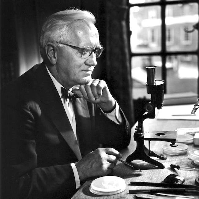
Image A

Examining the specimens via a truly primitive microscope (see Image A), he found the mold inhibited growth of S. aureus! Image B shows a modern version of what he observed: zigzagging ribbons of bacterial growth and a “halo” of no growth around the Penicillium colony. He correctly deduced his “mold juice,” which he later dubbed penicillin, inhibited bacterial growth!
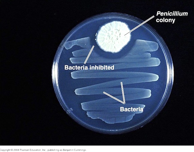
Image B

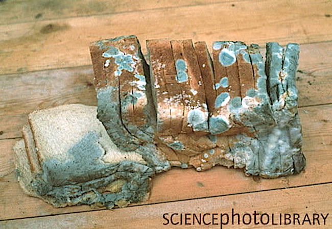
Stop! Now, we might imagine this was an entirely modern discovery, but others in the early 1900s had made similar observations although not much came of these. Even more amazing? Ancient Egyptians applied a poultice of moldy bread to promote healing of infected wounds. Very doubtful that ancient Egyptian bread looked anything like this loaf of sliced, white bread (Image C), but you get the idea. Icky!

Image C

Penicillin to the Rescue! Fleming published his experiments in 1929, but his paper focused mostly on uses for penicillin in the lab with only a brief remark about possible therapeutic benefits.

Ten years later, an Australian scientist and Oxford pathologists proved penicillin was an effective antibiotic. But, the mold was cranky and fastidious to grown and the yield so low, only small quantities could be produced.

Enter WWII with its urgent need for battlefield antibiotics! Various pharmaceutical companies teamed with US Department of Agriculture to mass produce penicillin (the chemical engineer who designed 10,000 gal. production vats was a woman!).

Simultaneously, a global search was launched to find a new mold that could be grown easier and yield larger quantities of penicillin. One hot summer day, a laboratory assistant arrived with a cantaloupe covered in  ”pretty, golden mold” which she (yes, another woman!) had found at a Peoria (IL) market. This mold was Penicillium chrysogeum, yielding 200 times the volume of penicillin as P. notatum!!! In 1954, using P. chrysogeum, US production of the drug jumped from 21 billion units to more than 6.8 trillion! And, penicillin was made available to allied forces (Image D).

A final note: Australia was the first country to make penicillin available for civilian use. Go, Aussies!

Penicillin does have drawbacks; it is ineffective against viruses and some types of bacteria (gram-negative), some people (0.03%) are allergic, and some bacteria have developed resistance due to over prescription or patients not following dosage instructions. Despite these drawbacks, penicillin can effectively treat the following short list of diseases (there is a long list, too):

  • Strep infections such as strep throat, scarlet fever
  • Staph infections such as post traumatic infections
  • Syphillus
  • Gonorrhea
  • Diphtheria
  • Lyme Disease
  • Typhoid Fever
  • Necrotizing Fasciitis (Flesh-eating disease)

If you remain curious about the fascinating history of penicillin, your thirst might be quenched by reading this article: https://www.acs.org/content/acs/en/education/whatischemistry/…/flemingpenicillin.html

Although I rarely move ahead of TV Outlander, this apropos quote from Diana’s book, The Fiery Cross, describes Claire’s work with penicillin. Herself wrote about molds from varied sources including the melon. Way to “break the mold,” Diana!

I twiddled the quill, rolling it between thumb and forefinger. I had kept a faithful account of my experiments with penicillin—the growing of cultures on media ranging from bread to chewed pawpaw and rotted melon rind, painstaking descriptions of the microscopic and gross identification of the Penicillium molds…
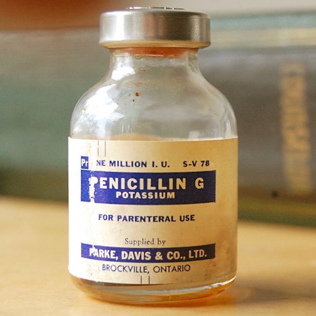
Image D

OK, now, back to Outlander episode 308, First Wife!

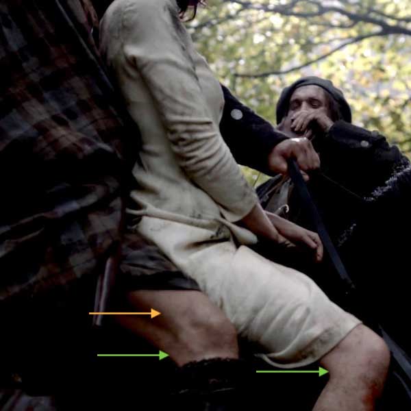
Nefarious Needles: With penicillin behind us (har, har) we broach the topic of needles. Ahhhh, yes, needles – a cruel device, with which most folks share a love-hate relationship! Claire prepares the penicillin and draws it into a syringe. Here, from Voyager book:

“What in God’s name are those?” Jamie asked, eyeing the syringes with interest. “They look wicked sharp.”

Psst … To me, he looks more wary than interested. Hah!

And more about needles:

I didn’t answer, occupied in dissolving the penicillin tablets in the vial of sterile water. I selected a glass barrel, fitted a needle, and pressed the tip through the rubber covering the mouth of the bottle.

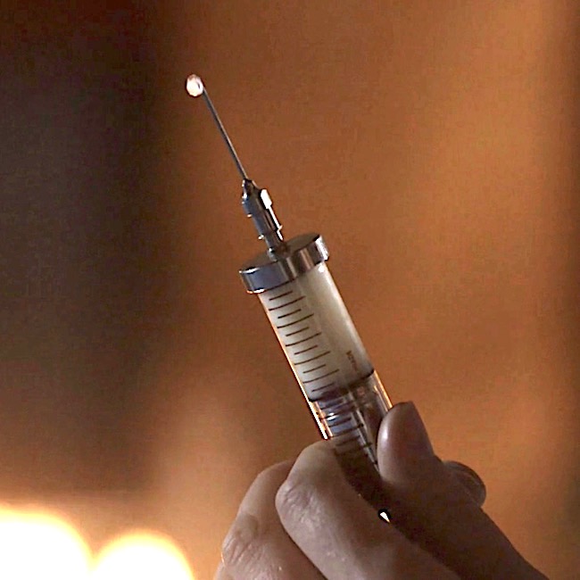
Holding it up to the light, I pulled back slowly on the plunger, watching the thick white liquid fill the barrel, checking for bubbles.

And more!

Then pulling the needle free, I depressed the plunger slightly until a drop of liquid pearled from the point and rolled slowly down the length of the spike.

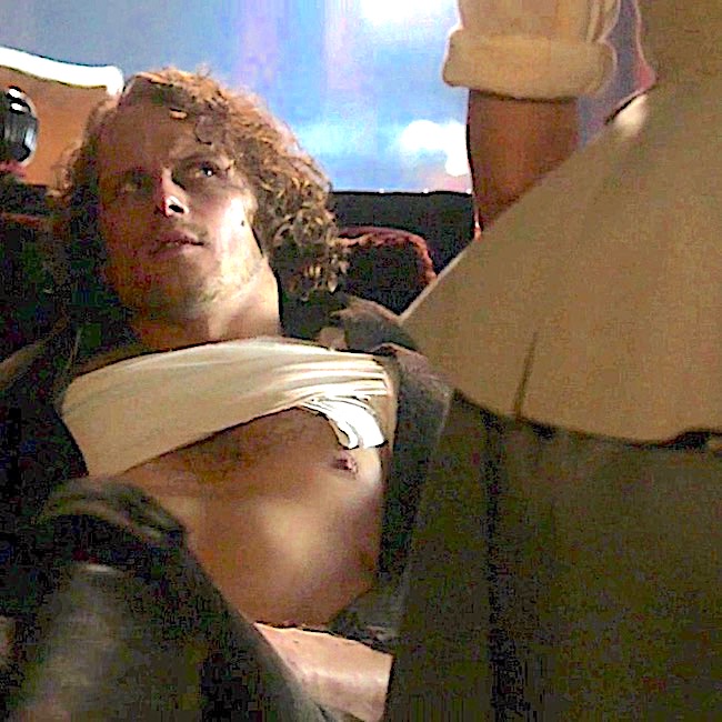
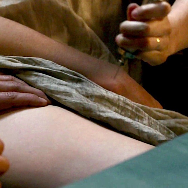
Claire’s penicillin is a thick, white liquid, poorly received by Jamie’s remarkably regal rump. We might ask, why Claire didn’t carry oral penicillin for her return trip to 1700s Scotland? It was available in her time. I really don’t ken the answer, but the injectable type promises readers and viewers a much more rousing and carousing tale! <G>

Understand, the first penicillins were injected, not ingested (Image E – puir wee laddie). Why? Because, when swallowed, stomach acid breaks down penicillin rendering it impotent. But, in the 1950s, types of oral penicillin which resisted stomach acid were developed. Today, many forms of penicillin include: capsule, chewable tablet, tablet for suspension, extended-release tablet, powder for suspension, powder for solution, capsule and syrup.
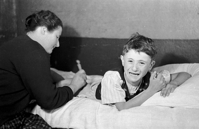
Image E 

Claire also brought her own 20th century syringes because they had not been invented in Jamie’s time. Almost a century later (1840s), Irish physician Francis Rynd invented the hollow needle to deliver the first modern-era subcutaneous (under-the-skin) injections! …”I’ve Got You Under My Skinnn!” ? Och!

Before we move on, let us honor resourceful Pre-columbian Native Americans who created early hypodermic needles and syringes using “hollow bird bones and small animal bladders!” Incredibly inventive and creative – using only nature’s gifts to presumably administer healing substances! No Spoilers: This relatively unknown fact should please book readers who will recognize something similar which Claire employs in The Fiery Cross.

I’ve said it before and I’ll say it again: if you haven’t read/listened to the books, you are missing one long joyous, glorious voyage!

More penicillin adventures from Voyager book. Claire stabs Jamie in the backside with her wee needle spike. Hah!

“Roll onto your good side,” I said, turning to Jamie, “and pull up your shirt.” He eyed the needle in my hand with keen suspicion, but reluctantly obeyed. I surveyed the terrain with approval. “Your bottom hasn’t changed a bit in twenty years,” I remarked, admiring the muscular curves.

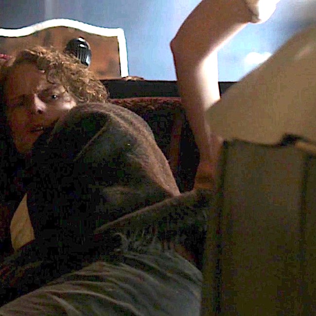
… I jabbed deftly and pressed the plunger slowly in.

“Ouch!” Jamie rubbed his posterior resentfully.

“It’ll stop stinging in a minute.” … Finally he said, “I thought ye stuck pins in ill-wish dolls when ye meant to witch someone; not in the people themselves.”

“It’s not a pin, it’s a hypodermic syringe.”

“I dinna care what ye call it; it felt like a bloody horseshoe nail. Would ye care to tell me why jabbing pins in my arse is going to help my arm?”

Splendid, splendid passage!

Here’s a few pearls from the above quote to consider: Why did Claire push the plunger slowly? Because, penicillin is a thick liquid which flows slowly and because it is viscous, it requires a large bore needle (18 gauge). The bigger the needle, the more it hurts while traversing the skin (yikes)! Because it is injected into muscle, the fibers are pushed apart by the fluid and that hurts, too!  I well remember penicillin shots from my youth. Not fun!

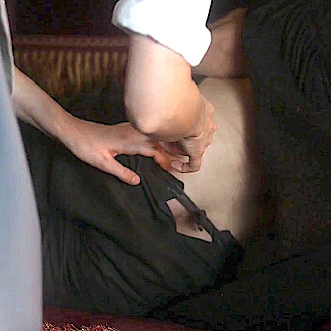
Crucial Question: Let’s get to the bottom of things (snicker)…..is Claire free to poke that needle anywhere in Jamie’s bottom? Absolutely not!!! Clarie carefully selects the area where she deftly jabs her needle spike.

Anatomy to the Rescue: Now, wait just a sec! Why does it matter where Claire sticks the needle? After all, it hurts regardless, right? Let’s find the answer by studying anatomy of the backside.

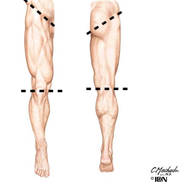
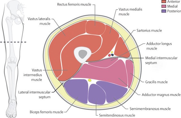
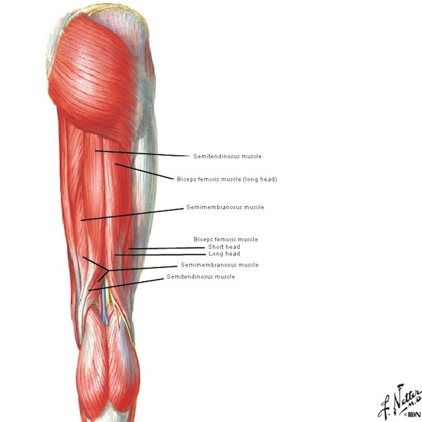
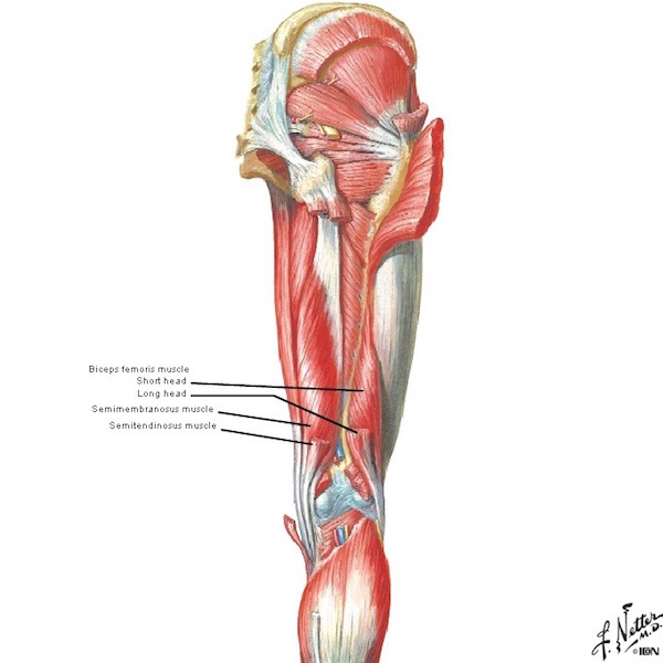
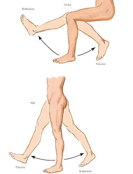
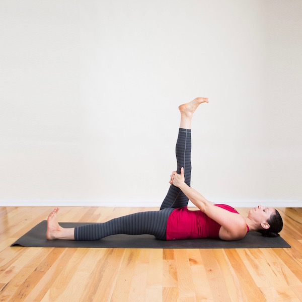
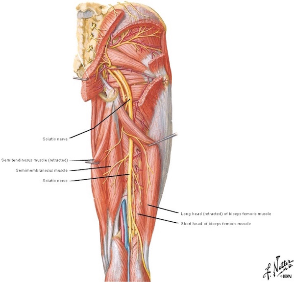
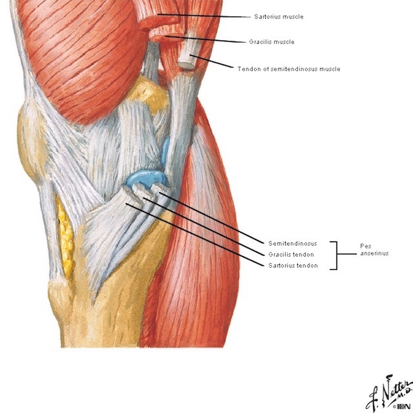
Remove skin and subcutaneous tissue and the buttock region looks like the left side of Image F: Each massive gluteus maximus muscle (Anatomy Lesson #1,  Jamie’s Tush) covers the back of one hip. Because the muscle is thick and meaty, injection into its fibers is a perfect destination!

Try this: Stand erect and place your palms on each buttock with fingers facing downward. Now, clench your bottom and feel it lift and tighten (hopefully)? Gluteus maximus muscles at work!

Sciatic Nerve: Butt, wait (he, he)! Remove the gluteus maximus and what lies underneath (Image F – right side)? One sees a host of smaller hip muscles and a very large yellow structure emerging from under cover of the right piriformis muscle . This is the sciatic nerve, largest and longest nerve of the human body – many adult sciatic nerves are as big around as an adult thumb! And, just to keep us crazy, although most sciatic nerves emerge from below the piriformis, there are five other variations that occur with less frequency.

Each sciatic is formed by contributions from five levels of the spinal cord (L4, L5, S1, S2, S3 – Anatomy Lesson #10, Jamie’s Back or Aye, Jamie’s Back!) as it traverses one buttock and descends through the back of thigh. In case you don’t know, L = lumbar and S = sacral.
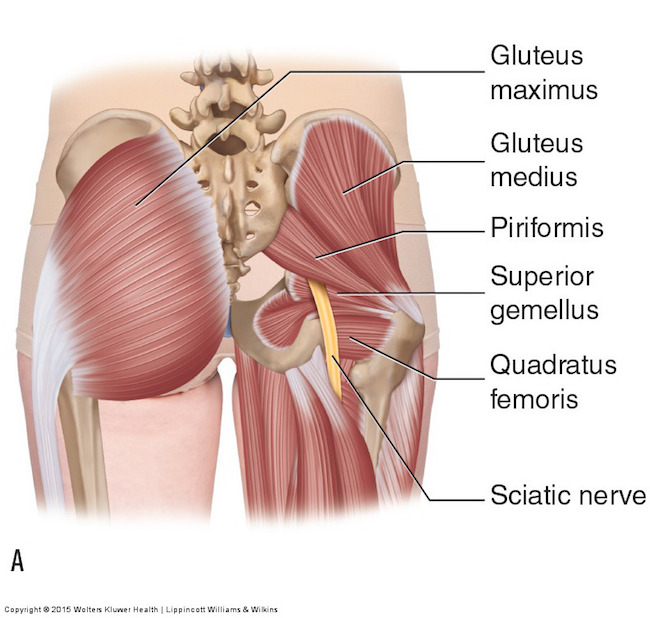
Image F 

Protect the Sciatic Nerve! Heath practitioners who inject the buttock know this cardinal rule: Never inject penicillin (or other drug) into or near a nerve as this may result in permanent neurological damage! Such damage can include permanent paralysis, muscle inflammation, gangrene or necrosis (tissue death). Major arteries must also be avoided.

True Story: Years ago, I knew a woman with foot drop, meaning she could not lift her foot at the ankle joint (called dorsiflexion). Why? Because a careless health care provider injected penicillin into the wrong area of her gluteus maximus and it destroyed that part of the sciatic nerve responsible for innervating leg muscles which are required to lift the foot. And, yes, the injury was permanent!

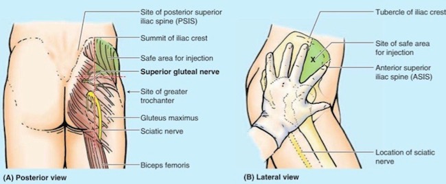
Safe Zone: So, is there a safe zone for injections? Well, of course there is, otherwise, Claire would not have put her beloved Jamie at risk.  

Let’s pretend this is Jamie’s bum. Hey, this is Jamie’s bum! <G> Black dots show regions corresponding to right and left buttocks. The blue rectangles are considered safe zones for intramuscular injections, including penicillin.


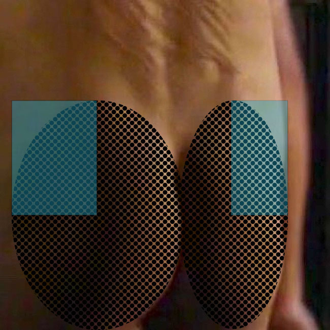
Using a graphic image might not be as much fun, but surely is informative. The buttock to be injected is divided into halves by a vertical line then divided into halves again by a horizontal line (Image G – left side). Injecting near the center of the upper outer quadrant, shown in blue, is considered the safe zone and avoids injuring sciatic nerve or nearby artery and vein (superior gluteal).

A newer technique uses a diagonal line as shown on the right side of Image G. Injecting above this line is also considered a safe zone. This line is harder to visualize because it requires a more thorough understanding of surface (topographical) anatomy.

Either technique is typically done in the mind’s eye of the practitioner, although, drawing actual lines on the buttock can also be done to verify safe zones.
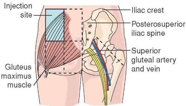
Image G 

This excellent You Tube video, created by Dr. Nabil Ebraheim, explains both approaches.

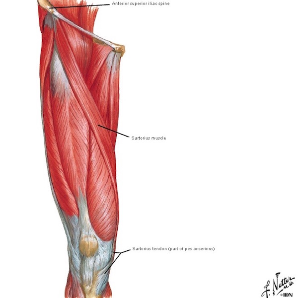
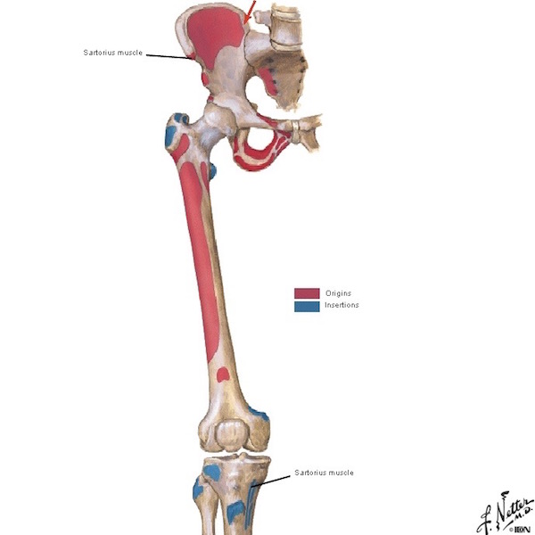
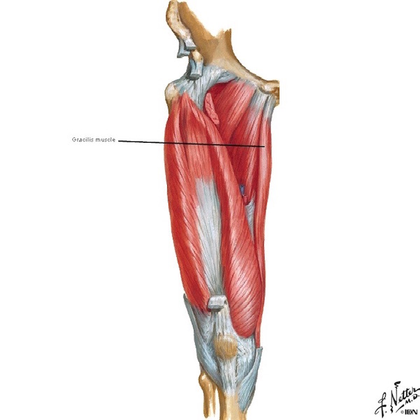
The meaty quadraceps femoris muscles (Anatomy Lesson #7,  Jamie’s Thighs or Ode to Joy!) of the thigh present another acceptable site for penicillin injection. This explains why, much later on the Artemis, Claire injects herself in the thigh with penicillin after Jamie canna do the deed (Outlander episode 311, Uncharted).  Our braw hero is afeared of wicked needles!

But, arguably the safest approach involves injecting into the gluteus medius muscle which peeks above the gluteus maximus. The safe region is indicated as the green area in Image H. This technique avoids the huge and very important sciatic nerve!

Ubiquitous Germs! One final issue and then our lesson ends. How might Jamie get an infection from second-wife wounds? Both birdshot and his skin would be likely sources for the bacteria causing his raging fever! Claire explains it quite well in this passage from Voyager book:

I took a deep breath. “Well, do you remember my once telling you about germs?”

He looked quite blank. “Little beasts too small to see,” I elaborated. “They can get into your body through bad food or water, or through open wounds, and if they do, they can make you ill.” He stared at his arm with interest. “I’ve germs in my arm, have I?” “You very definitely have.” I tapped a finger on the small flat box. “The medicine I just shot into your backside kills germs, though. You get another shot every four hours ’til this time tomorrow, and then we’ll see how you’re doing.”

I paused. Jamie was staring at me, shaking his head. “Do you understand?” I asked. He nodded slowly. “Aye, I do. I should ha’ let them burn ye, twenty years ago.”

Nay, Jamie doesn’t mean this wee salvo aimed at first wife. Snort!

Thank you Diana, Jamie and Claire for helping us to understand needles, nerves and penicillin!

Everywhere: Bacteria, molds and yeast are ubiquitous, covering virtually every non-sterile surface on earth. Recent tests have shown, seven of the 10 germiest (is this even a word?) spots in our homes are found in kitchens – with the dirtiest being kitchen sponges or dish cloths! Imagine, these are more germy than bathrooms!

Our lesson closes today with this visual of bacteria and molds found on one person’s hands (Image H). The hand was pressed into growth medium filling a petri dish. After some time, colonies of mold, yeast and bacteria grew from the hand print. Beautiful in an weird way, but definitely GROSS! The bottom line? Our mums were right. Let’s wash our hands

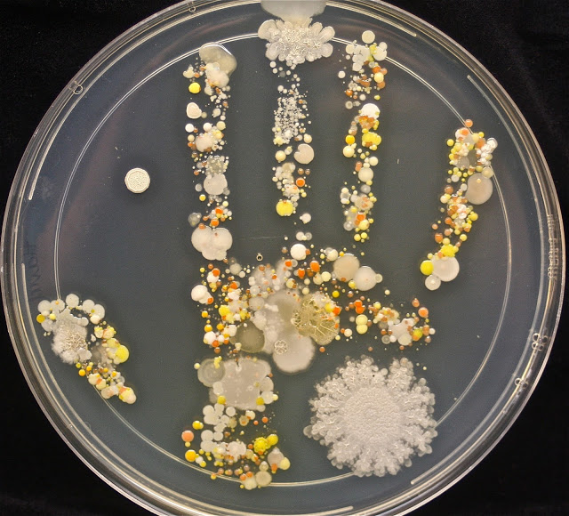
Image H by Tasha L Sturm, microbiologist and photographer

The deeply grateful,

Outlander Anatomist

Follow me on:

Photo Creds: Starz, www.pbs.org/newshour Image E; www.Karsh.com  Image A;  www.learnmuscles.com Image F; www.free-stock-illustration.com Image C; www.medium.com Image B; www.prezi.com Image D; https://everythingmicro.blogspot.com Image H; www.teachmeanatmy.info Image G

Fun Fact: adductor

picture of jamie fraser on battle field from outlander

Starz Outlander episode 301, The Battle Joined

Anatomy def: Brought in? Adductor means brought in? How do such words apply to anatomy?

Easy peasy: adductors are muscles whose contraction draws a body part toward the midline or toward another body part. Six adductor muscles draw the thigh toward the midline: gracilis, adductor brevis, adductor longus and adductor magnus, aided by pectineus and obturator externus.

Outlander def: Jamie’s thigh-high kilt <G> reveals powerful left thigh adductors forming a well-defined mass of the inner thigh (red arrow). A grinning Murtagh dispatches a red coat and helps Jamie rise from a seated position: “ye are welcome!” Adductors contract to lend balance and assist Murty’s lift: git up, man, there’s a battle raging!!! 

Learn about the thigh and its powerful muscles in Anatomy Lesson #7, Jamie’s Thighs or Ode to Joy

Read about thighs in all Diana’s works as Herself often writes about them – not specifically about adductor muscles, but about various thigh muscles, sinews, bones, skin and scars! Consider this wonderful bit from Voyager book; takes you right back to that intimate C + J reunion after 20 agonizing years apart:

I reached out and stroked his thigh, touching the long scar that ran down it. “I wish I could have been there to take care of you,” I said softly. “It was the most horrible thing I ever did—leaving you, knowing … that you meant to be killed.” I could hardly bear to speak the word. 

“Well, I tried hard enough,” he said, with a wry grimace that made me laugh, in spite of my emotion. “It wasna my fault I didna succeed.” He glanced dispassionately at the long, thick scar that ran down his thigh. “Not the fault of the Sassenach wi’ the bayonet, either.” 

See Jamie’s adductor muscles in Starz Outlander episode 301, The Battle Joined. They are admirably awesome (red arrow)!

A deeply grateful,

Outlander Anatomist

Anatomy Lesson #43 – “Hamstring – You Make My Heart Sing!”

Hi, anatomy students! So great to meet with you again. Today’s Anatomy Lesson #43 uncovers (ha) the posterior (back) thigh. We already met the anterior thigh waaaay back in Anatomy Lesson #7, “Jamie’s Thighs or Ode to Joy!” where we studied the quadraceps. Thus, we have much more to learn about the thigh.

Our object is to study muscles of the posterior thigh with two other thigh muscles thrown in for good measure. Some folks don’t find muscles all that compelling (unless they are Jamie’s, of course!); mayhap this lesson will change their minds. Anatomical tidbits are added to hold your attention.

As always, Outlander books and Starz episodes are blatantly scattered throughout. Let’s start with a great view of Jamie’s posterior thighs (Starz episode 112, Lallybroch). Och! Lad, best tuck in those flapping shirt tails! Oops, nothing to tuck into. Where’s a kilt when you need one?

ep-112-thigh-03

Ah, there’s that plaid (Starz episode 109, The Reckoning). Gird up your loins, Jamie! Hee, hee.

ep-109-kilt

Lower Extremity: This lesson starts with essential definitions. Anatomists define the lower limb or lower extremity as the appendage from hip joint through toes. Folks (including me, at times) refer to the lower extremity as the upper leg and lower leg but, in anatomy, this is not so. The thigh is that part of the lower limb between hip and knee joints (Image A – between dashed lines). The leg lies between knee and ankle joints and the foot is distal to (beyond) the ankle joint.

figure0467-thigh-01

Image A

Thigh Overview: Today’s lesson focuses on posterior thigh so let’s define this area. The human thigh is divided into compartments based on connective tissue septa (partitions) and actions; these are best visualized in cross-section (Image B). By convention, cross-sections are interpreted is as if you stand at a person’s feet and look toward the head.

A plane crossing the femur, reveals three compartments, each bearing different muscle groups: anterior (Image B – red), medial (Image B – pink; OK, OK – it’s dusty rose!) and posterior (Image B – lavender). Anterior means front; medial means toward the midline; posterior means back. We will study three muscles of the posterior compartment, one of the anterior and one of the medial.

thigh-x-section

Image B

Posterior Thigh Muscles: Three muscles make up the posterior thigh compartment; all are typically long and strong.

  • Semitendinosus
  • Semimembranosus
  • Biceps femoris

Try This: Open the space between thumb and forefinger and grip your posterior thigh about mid-femur. Your entire hand should grasp a sturdy muscle mass containing the three posterior compartment muscles. Appreciate the same anatomy with your other thigh or someone else’s. <g>

Topographical Anatomy: Looking at topography or surface anatomy of an intact posterior thigh, is it possible to see grooves and ridges created by its muscles? Yes, if the subcutaneous fat layer is not too thick and if the thigh is well-muscled. Posterior thigh (Image C – right lower extremity) bears three muscles in succession: Blue arrow marks semimembranosus, red arrow marks semitendinosus and green arrow shows biceps femoris.

figure0467-thigh-01-c

Image C

Not easy to see? OK, let’s look again using a male body builder (Image D). He is not mooning us, rather, he demonstrates posterior muscles of the lower extremity. Same color coding as Image C and not a challenge to recognize the long columns of posterior thigh muscles. Impressive!

Understand that semitendinosus (red arrow) and semimembranosus (blue arrow) fill the medial (inner) side of posterior thigh and biceps femoris (green arrow) occupies the lateral (outer) side.

hamstrings-01

Image D

Book Quote: Speaking of long columns of thigh muscles, Diana obligingly (yay!) provides a description of Jamie’s own fine muscular columns, here from Dragonfly in Amber. In case you don’t recognize the scene, these are Claire’s poignant thoughts the eve before she passes through the stones (sob!), back to Frank (gah!). Dinna ken if  “seasoned oakwood in the columns of his thighs” refers to anterior or posterior thigh muscles but, knowing Claire, it was very likely both. Snort!

I touched each soft hollow, the hidden places of his body. Felt the grace and the strength of each curving bone, the marvel of his firm-knit muscles, drawn lean and flexible across the span of his shoulders, smooth and solid down the length of his back, hard as seasoned oakwood in the columns of his thighs.

Book quote and photo (Starz episode 204, La Dame Blanche) aren’t a match, but will do in a pinch…the pinch being a love bite on Jamie’s thigh. Man, you are in BIG trubble! Yep, seasoned oakwood it is. Ha, ha!

ep-204-thigh-01

Remove the skin and, voila, three posterior thigh muscles pop into view (Image E – posterior right thigh).

Semitendinosus: Superficial semitendinosus courses down the medial side of posterior thigh. The muscular part ends roughly midway down the femur, transforming into a long, round tendon and curving behind the knee joint to end in the tibia. Did you know, part of semitendinosus tendon can be harvested during knee reconstructions to replace the ACL or anterior cruciate ligament (Anatomy Lesson #7, “Jamie’s Thighs” or “Ode to Joy!”)? Yep, a very useful muscle.

Semimembranosus: Semimembranosus (Image E) is a broad, flat muscle so named because it has an unusually membranous tendon of origin. Although it lies deep to semitendinosus, it is the most medial of the posterior thigh muscles. Semimembranous also drops across the knee joint to end in tibia. Semimembranosus is variable; it may be reduced, enlarged, duplicated or absent.

Biceps Femoris: Longtime students will ken that we studied biceps brachii back in Anatomy Lesson #20, “Arms! Arms! Arms! – Redux.” Well, like the arm, posterior thigh also has a biceps muscle, biceps femoris. Biceps derives from Latin, meaning two heads and femoris reflects its close association with femur. Indeed, biceps femoris (Image E) does have two heads of origin: the long head courses the length of the thigh and the short head is roughly half as long. Both muscle parts unite as a single tendon ending in the fibula. The short head of b. femoris may be absent but otherwise, the muscle demonstrates few variations.

Clinical Correlation: Recently, a professional soccer player suffered major posterior thigh pain. Imaging revealed a large, anomalous (duplicated?) right semimembranosus which was crossed over and compressed by the tendon of semitendinosus. This unusual anatomy created what is known as entrapment syndrome of the posterior thigh, which compresses the popliteal artery (see below) restricting blood flow. This is especially acute during exercise and hence, pain.  Muscles object when deprived of oxygen….. “blood of my blood.”  We know the drill.

figure0477a-thigh-03

Image E

Remove gluteus maximus and posterior thigh muscles spring into view (Image F). Remove semitendinosus and long lead of b. femoris and, ta da,  semimembranosus and short head of b. femoris say hello! Image F reveals the long, flat membranous tendon of origin for semimembranosus. Short head of b. femoris arises from lower half of femur.

figure0477b-thigh-04

Image F

Moment of Silence, please!: In Image E, the tops of posterior thigh muscles are not visible because gluteus maximus covers them. G. maximus is a large, flat quadrangular-shaped muscle honored in Anatomy Lesson #1, “Jamie’s Tush” and was the original inspiration for Outlander Anatomy lessons! Thus, it is only fitting that we interrupt this lesson to pay homage to lovely body curves created by these, the body’s most massive muscles (Starz, episode 107, The Wedding). Gasp, thud!

ep-107-thigh-06

Muscle Attachments: Next, let’s suss out bony attachments of the posterior thigh muscles – know these and muscle actions make sense. Image G of the posterior hip bone and femur is busy but nicely shows muscle origins (red) and insertions (blue). Origin means the body site (almost always a bone) giving rise to a muscle. Insertion means the site where the muscle ends by attaching to a different bone. Muscles usually cross one or more joints (there are exceptions) – a site where bones meet to allow for movement. Make sense? Dandy! Succinctly put:

  • Origin:
    • Proximal (nearer the body center)
    • Fixed point (least moveable site)
  • Insertion:
    • Distal (further from the body center)
    • Movable (moves with muscle contraction)

Origins and Insertions: Long head of biceps femoris, semitendinosus, and semimembranosus, all take origin from the ischial tuberosity (Image G) of the respective hip bone, cross the hip joint and pass down the posterior thigh. As noted, the short head of b. femoris takes origin from the lower half of femur (Image G). BTW, the sturdy ischial tuberosities are known at gyms as “sits bones” and, indeed, we do sit on them!

After coursing down the posterior thigh, each muscle claims a different insertion site.

Semimembranosus crosses knee joint to end in medial tibia (Image G – inner leg bone).

figure0473-thigh-02

Image G

The tendon of semitendinosus swings behind the knee joint and inserts into anteromedial (front/inner) surface of tibia (Image H).

Long and shorts heads of biceps femoris unite as a single tendon which crosses the knee joint to end in the head of fibula (outer leg bone) Image H.

figure0472-use

Image H

Actions: Now for the nitty-gritty: What actions do these muscles perform? Well, they are critical for successful lower limb ambulation: walking, running, climbing, kicking, squatting, jumping, etc. Semitendinosus, semimembranosus and long head (not short head) of b. femoris cross the hip joint so as they contract, the hip joint extends or straightens (Image I – bottom right). All three muscles (including short head) cross the knee joint so as they contract, the knee flexes or bends (Image I – top right). All three muscles counteract forward bending at the hips to keep us from doing a face plant. Whew, busy muscles!

figure0525b

Image I

Hamstrings: Now, here’s an interesting tidbit: Anatomists call semitendinosus, semimembranosus and long head of b. femoris true hamstrings; “ham,” from Old English hom, meaning the hollow or bend of the knee and “string” referring to tendons located there. To receive this “anatomical blessing,” a posterior thigh muscle must meet the following criteria:

  • Take origin from the ischial tuberosity (ergo, short head of b. femoris doesn’t quality)
  • Cross the hip joint causing extension (short head of b. femoris doesn’t quality)
  • Cross the knee joint causing flexion
  • Same innervation (short head of b. femoris has a different innervation)

Hamstringing is an dreadful event wherein a victim is incapacitated by severing hamstring tendons at the back of the lower thigh (Image J – in cartoon form, thank goodness!). This laceration was crippling, painful and often caused death by exsanguination from severed vessels at back of the knee (see below). Hamstringing was a time-honored (eek!) method of permanently crippling animals and humans so they could not reengage in future warfare.

Hamstringing is a very old practice. Identified as houghing in the King James Version of the Bible, both Joshua and David ordered hamstringing of chariot horses. Carthaginians hamstrung their Roman enemies as did Germanic tribes. Romans hamstrung elephants and the practice has been revived most recently in Zimbabwe!

hamstringtcg

Image J

After he is stabbed in the torso by the mouthy MacDonald, Jamie doesn’t fail us! Pulling a dirk with his left hand, he prepares to strike his attacker (Starz episode 110, By the Pricking of My Thumbs). Who’s the cowering coward behind the tree? The Duke, of course. Off with his head!

ep-110-thigh-04

And, strike he does! Down goes MacDonald as Jamie’s blade slices into the unguarded right biceps femoris tendon, hamstringing his opponent. Given the state of 18th century medicine, this MacDonald willna be farming his croft. Ei, Ei, Ohhhhh….. Well, Jamie is a warrior – even if he is cute!

Try This: Want to feel your own hamstrings? Sit in a chair or on a sofa with knees bent and right foot turned outward. Place right fingers beneath outer right thigh (near knee). Pull right heel back against chair rung or couch base. Feel the tendon? This is biceps femoris. Relax and place fingers of left hand behind inner lower thigh, this time with turn foot inward. Pull heel back again. Feel two tendons? The superficial one belongs to semitendinsus and the deep is semimembranosus! A+ for you!

ep-110-thigh-05

“Down by the old mill stream,” the tendon of Jamie’s right biceps femoris muscle is clearly visible as he shifts weight to the mill wheel. See the taut tendon behind his right knee? Oops, lost some of you! Please focus on the hamstring. Don’t see it? Bummer! <G>

ep-112-thigh-04

Let’s try again. How about both Jamie and Claire this time (Starz episode 101, Sassenach)? “Here’s to you lass, for tipping us to the villains in the rocks, and gieing us a wee bit of fun,” boasts and toasts Rupert. Green arrows mark tendons of biceps femoris muscles. Can you spy a second band of taut tissue, just forward of Jamie’s biceps tendon? This is the iliotibial tract or IT band (orange arrow – Anatomy Lesson #7, “Jamie’s Thighs or Ode to Joy!”). Yep, keep your eyes peeled for all sorts of Outlander anatomy!

BOOK QUOTE: Both Claire and Jamie have something to say about rubbing thighs during that intimate ride together, although Claire’s is slightly more high-brow. From Outlander book:

My companion seemed to be having little trouble, in spite of being unable to use this right hand. I could feel his thighs behind mine, shifting and pressing occasionally to guide the horse. I clutched the edge of the short saddle in order to stay seated; I had been on horses before, but was by no means the horseman this Jamie was….

but then that ride through the dark together – with that lovely broad arse wedged between my thighs…with a bum like that… What does it matter if she’s a f-face like a ah-ah- sheep?”

Baa, baa, bleats Claire! 🙂

ep-101-thigh-01

It took more than a bit of sleuthing to find an Outlander image showing inner hamstring tendons. Probably the best comes from The Wedding (Starz episode 107). Squee! Shift eyes, ahem, to Jamie’s left inner thigh; green arrow marks a ridge created by tendons of semitendinosus and semimembranosus. Flexed knee brought to you by power of the mighty hamstrings!

Although a bit later in their relationship, this quote from Dragonfly in Amber book says it all!

I scooped out a good bit of the salve and spread it down the long muscle of the thigh, pushing Jamie’s kilt above his hip to keep out of the way. The flesh of his leg was warm; not the heat of infection, only the normal heat of a young male body, flushed with exercise and the glowing pulse of health. I massaged the cream gently into the skin, feeling the swell of the hard muscle, probing the divisions of quadriceps and hamstring. Jamie made a small grunting sound as I rubbed harder. “Hurt?” I asked. “Aye, a bit, but don’t stop,” he answered.

ep-107-thigh-05

Hamstring Injury: Moving on… anyone who has suffered a hamstring pull understands just how much we rely on these muscles. You can scarcely engage in a single lower-limb activity without feeling the burn. Yeow! This is a short list of things which contribute to hamstring injury:

  • Sitting for long periods of time
  • Sitting on a hard surfaces, putting pressure on the hamstrings
  • Kicking any ball (football, soccer)
  • Hurdles
  • Splits
  • Poorly conditioned folks suddenly trying things they once did as teenagers!

Hamstring Stretch: Because we are so dependent on hamstrings, it is important to keep them supple by stretching. This can be done sitting, standing or reclining (Image K). Anatomically, the reclining stretch is safest because shoulders, head, spine and hips are supported by the floor; sitting and standing stretches can result in hunched backs, arched necks and hips out of alignment. Here are some pointers:

  • Keep hips and shoulders flat and aligned
  • Contract abdominal muscles to support the lower back
  • Flex both ankles
  • Grip hands behind the thigh (NOT behind the knee)
  • Gently lift one limb until resistance is met – then stop! Don’t force the lifted limb
  • Don’t arch the neck or back
  • Alternatively, flex the down knee to release tension on the raised hamstrings
  • Alternatively, pass a towel behind the up thigh and gently pull on towel to lift

hamstring-stretch

Image K

Videos can be helpful, so the next one demonstrates a good hamstring stretch. As with the above photo, hands go behind the thigh with the interesting addition of flexing alternating with not flexing the ankle joint. Some yoga practitioners grip the great (peace) toe, which works wonderfully if one is a seasoned hamstring stretcher, otherwise avoid!

Sciatic Nerve: A note about the sciatic nerve, the largest of the body (Image L). This behemoth (in adults, it rivals the size of my thumb!) courses down the back of the thigh hidden by gluteus maximus and hamstrings. It gives off twigs to innervate all the hamstring muscles, crosses the knee joint and supplies most of the leg and foot. It is a very busy nerve, hence its size. Sciatic nerve pain is a common affliction but must wait for another lesson. So sorry!

Popliteal Vessels: Behind the knee lies the popliteal fossa, a potential space filled with fat and blood vessels. The large popliteal artery and vein descend through this space to supply knee, leg and foot. These are at risk during hamstringing. If severed, the victim will bleed out, very quickly!

figure0484-sciatic-nerve

Image L

Switching gears, do you recall this terrified wee fellow? He served as Colum’s tailor-of-the-day (Starz episode 103, The Way Out). His wife was a MacKenzie, but kinship won’t protect him from Colum’s dirk! The MacKenzie chieftain was mightily p.o.ed at the tailor because his newly minted frock coat was too long. Did the tailor mean to hide Colum’s legs (Anatomy Lesson #27, “Colum’s Legs and Other Things too!”)? Well, erm, aye, he did, but he hadn’t planed to lose his larynx over a Heiland fashion statement (Anatomy Lesson #42, “The Voice – No, not that One!”).

What in the world does a tailor have to do with the posterior thigh? Read on!

ep-103-tailor-01

Perhaps you recall at lesson start, I mentioned we would cover two other muscles, one from anterior and one from medial thigh compartments (review Image B). Here we go!

Sartorius: The sartorius muscle spirals down the anterior thigh. Its name derives from the Latin sartor meaning, tailor. Why? Because, back when garments were hand sewn, many tailors assumed a cross-legged posture so the knees could support a garment under construction (Image M). Sartorii (pl.) are engaged in adopting the cross-legged position, hence the name.

tailor

Image M

The striking sartorius muscles are very apparent in this female body builder (Image N). Thin, flat and superficial, we rarely see sartorii except in folks who are heavily muscled and/or express little subcutaneous fat (Anatomy Lesson #5, “Claire’s Skin – Opal, Ivory and White Velvet”).

sartorius-01

Image N

Spiraling down the thigh but superficial to the quadriceps (Image O), sartorius is the body’s longest muscle (love superlatives)! Sartorii can be pulled during activities which require a forcefully push off, as with sprinting, jumping and running. Thus, sports, such as hockey, rugby, football or basketball, place one at higher risk of injuring this muscle, although improperly-performed squats can also do the trick. Pain can occur anywhere along the length of sartorius with groin and inner knee as common sites of complaint.

figure0474a-sartorius

Image O

Sartorius belongs to the anterior thigh compartment. It takes origin from a knob of hip bone, the anterior superior iliac spine or ASIS (Image P). Crossing both hip and knee joints, the muscle arcs across the front of the thigh to end in the anteromedial tibia.


figure0472

Image P

As each sartorius contracts, hip and knee joints flex and knee rotate laterally (to the side) as in yoga’s lotus pose (Image Q). Sartorii don’t appear in Image Q because the feet cover them.

lotus-pose

Image Q

However, this image of a yoga practitioner entering half-bound lotus pose nicely shows the left sartorius (Image R – double arrow); its tendon creates a skin groove just above the arrow.

half-bound-lotus

Image R

And, Jamie reveals a near perfect sartorius groove (of course) as he scolds new wifey: Lassie, I tell you the truth about Ned Gowan and that strumpet! Ned paid a high price for your splendid wedding dress; a dirty job against which he fought, valiantly. Ha!

See the thigh groove created by Jamie’s left sartorius (Starz episode 107, The Wedding)? Look closely, a red arrow marks the groove directly behind his vast vastus medialis, (Anatomy Lesson #7, “Jamie’s Thighs” or “Ode to Joy!”). Got it? Yay!

ep-107-thigh-03

Gracilis: Graceful gracilis belongs to the medial thigh compartment  (review Image B). Derived from the Latin gracilis meaning slender, this is the most superficial muscle of the medial thigh (Image S). It is thin and flat, broad above, narrow and tapering below. Like true hamstrings and sartorius, gracilis also crosses both hip and knee joints.

figure0474b-gracilis

Image S

Gracilis takes origin from the pubic bone and inserts on anteromedial tibia (Image T). It helps adduct the thigh (draw toward body midline) and is a weak flexor of both hip and knee joints. Interestingly, gracilis is widely used in reconstructive surgery as a replacement for facial muscles, hand muscles or the external anal sphincter. Loss of the gracilis is not disabling as larger, stronger muscles of the medial compartment are able to compensate.

Clinical Correlation: A few years back, surgeons teaching in my anatomy course presented a case wherein they replaced the external anal sphincter with gracilis muscle, performed on a young woman who had suffered a horrific injury in an auto accident. So shocking and graphic, one of our first year medical students fainted, undoubtedly from vasovagal syncopy wherein heart rate and blood pressure suddenly drop due to extreme emotional distress. Yes, that brought this memorable lecture to a halt! But, the student soon regained consciousness and composure and this gave the surgeon a teaching moment to explain what had happened. I am pleased to report that not only student recovery but the reconstructive surgery were both successful. Surgeons I worked with were very resourceful docs and imaginative thinkers. Their motto: A chance to cut is a chance to cure! Very confident folks, the surgeons.

figure0472-gracilis

Image T

Pes Anserinus: Now, perhaps you spy an insertion theme: tendons of sartorius, gracillis and semitendinosus all converge and insert as a unit into anteromedial tibia. At insertion, they form a webbed configuration known as the pes anserinus, Latin for goose foot. The name, “goose foot,” arises from a three-pronged appearance assumed by the three tendons, each from a different thigh compartment (Image U).  The order of insertion from front to back can be recalled using the acronym for sergeant: SGT for sartorius, gracilis, semitendinosus. Now, this seems especially appropriate because sergeant comes from the Latin servier meaning “one who serves”; an apt moniker for these three hardworking “guy ropes” as they are sometimes called!

All three pes anserinus muscles are primarily knee flexers but studies show that the peculiar three-pronged insertion provides rotatory stability to the knee, meaning together they stabilize external (outward/lateral) rotation of the knee. Hallelujah! Why? Remember, the knee joint is inherently unstable because its constituent bones do not enjoy a ball and socket type of interaction (Anatomy Lesson #7, “Jamie’s Thighs or Ode to Joy!”). Ergo, the joint is provided with numerous ligaments and tendons, including pes anserinus, to ramp-up stability. Go anatomy!

figure0488b

Image U

Goose Foot: Did you see a goose’s foot in the pes anserinus? No? OK, then, let’s look at a goose’s foot! How about now (Image V) – see the three pronged toes joined by webbing? Still not convinced? Understand that early anatomists didn’t have magazines, internet, social media, TV or movies for creative word imagery so they drew on the natural world to devise a new language describing body parts and passions (Lesson #34, “The Amazing Saga of Human Anatomy”). Many of their words may seem quaint but, some 500+ years ago, they were cutting edge!

pes-anserinus-01

Image V

That’s it for the hamstrings and its two closely allied pals. Remember, these muscles are important in virtually all types of ambulation. The next time you stride through a mall seeking new fashions, kick a soccer ball or chase down an errant child, please give a brief nod of thanks for these wonderful muscles!

Do you love art? I love art. Created by Steel Neal and installed at Cooper Square, NYC, “The Agony of Man” is a 1200 pound, 3 times life-size rendition of the human form (Image W). It is constructed entirely of salvaged scrap metal: I-beams, railroad track, rebar, boilers, New York City garbage cans. Teeth (Anatomy Lesson #26, “Jamie’s Chin – Manly Mentus”) were repurposed from road resurfacing equipment. Bony orbits (Anatomy Lesson #30, “Aye, Eye – The Eyes – Part 2!”) were salvaged from original park benches at Madison Square Park. Ribs (Anatomy Lesson #15,“Crouching Grants – Hidden Dagger”) are from the original concrete island of Worth Square.

Sartorii? Yes, there they are, spiraling across the front of thighs – these are made from an extinct type of rebar salvaged from the original foundation of the Union Square Subway Station. Indeed, an evocative rendering of human agony!

agony-of-man

Image W

How does human agony fit into a lesson about “hamstring – you make my heart sing?” Well, because we, the wretched, are suffering from drrrrrouuuuughtlander (Photo X)! Gasp! Ron.com, must have a glass of OUTLANDER soon! Dying here!

droughtlander

Image X

All together now:

Hamstring!

You make our hearts sing!

You make everything,

groovy!!! (shamelessly lifted from the Troggs)

A deeply grateful,

Outlander Anatomist

Photo Creds: Starz, Netter’s Atlas of Human Anatomy, 4th edition (Images A, C, E, F, G, H, I, L, O, R, S, T), Outlander Anatomy photo collection, www.christineshipjoint.blogspot.com (Image M), www.getbig.com (Image N), www.keyword-suggestions.com (Image U), www.mnn.com (Image W), www.popsugar.com (Image K), www.sartoriusmusclepainigi.wordpress.com (Image V), www.seannal.com (Image D), www.sensation-yoga-poses.com (Image Q), www.thelancetnorway.com (Image B), www.wowwiki.wikia.com (Image J), www.yogatrail.com (Image P)