Anatomy Lesson #65: Anatomy of a Hanging

Greetings anatomy students! Hanging is a ghastly topic, but it has a substantial presence in Outlander and is pertinent to anatomy. So, here we go!

The Oxford Dictionary defines hanging as “specifically to put to death by suspension by the neck.” But, in the past, hanging also meant death by crucifixion or impalement! 

An interesting fact…. The appropriate terminology is the person was hanged, not the person was hung.

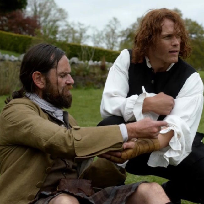
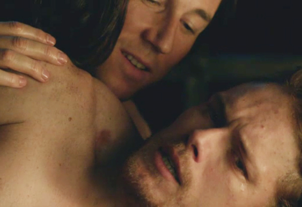
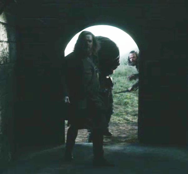
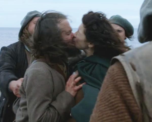
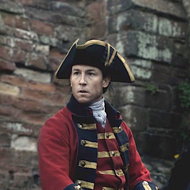
Outlander’s first traditional hanging appeared in episode 115, Wentworth Prison; crusty Taran MacQuarrie of the Watch and Jamie Fraser face their ends with stirring gallows’ humor: Jamie is afraid “My wife will never forgive me for getting myself foolishly hung!” Hanged,  Jamie…. the word is hanged!

But, Taran aways knew he was fated to die at the end of a rope – his name is read next.

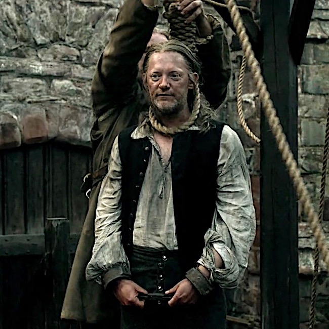
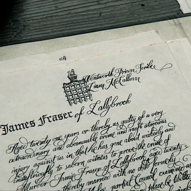
Jamie’s turn! Scribe, Liam McCallum, recorded Jamie as guilty of “a very extraordinary and abominable crime and most atrocious injury against us!” Snort!

Jamie struggles, but is subdued and resigns himself to this ignoble fate. Such an unworthy end for Laird Broch Tuarach!

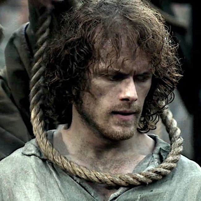
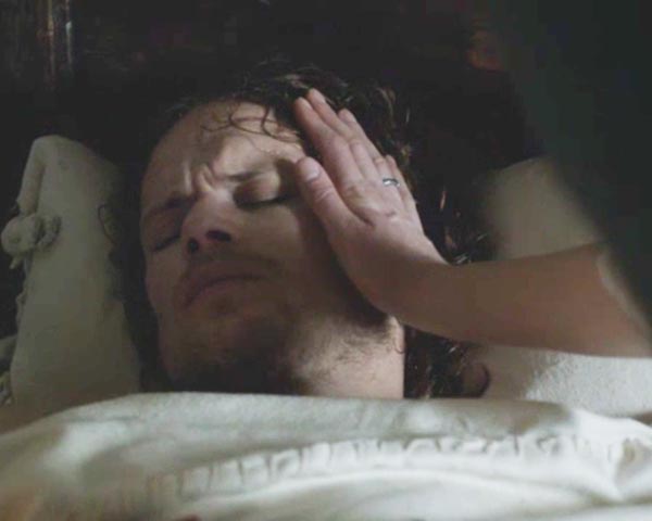
Then one of Lucifer’s avenging angels swoops in to spare Jamie for a different fate! But, this outcome awaits another lesson. 

No further traditional  hangings, until season four. Episode 401, America the Beautiful, starts off with a bang… Well, with a hangman’s noose, truth be told.

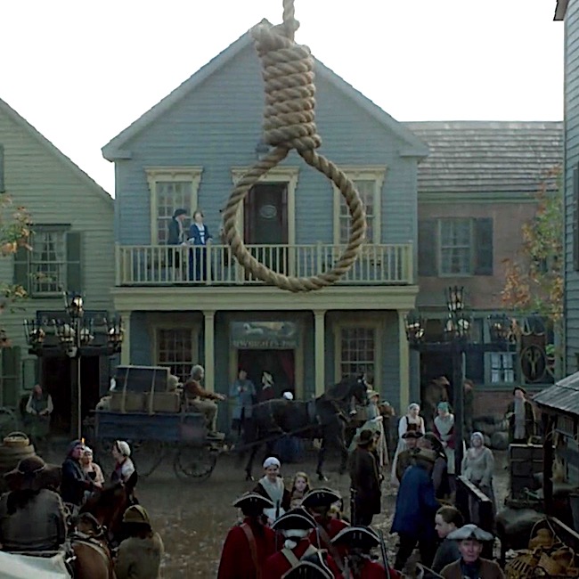
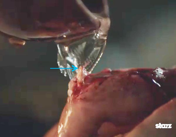
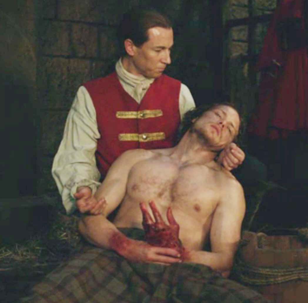
In a drunken state, Gavin Hayes lay with a marrit woman (psst…Jamie warned him!). When the outraged hubby came at him with a pitchfork, Gavin kicked him down the stairs and now, the hangman’s noose awaits. He should have listened to Mac Dubh! (In Drums of Autumn book, Gavin was guilty of stealing six pounds, ten shillings).

Hayes asks Jamie for two favors: 

#1. Whisky! 

Herself described in Drums of Autumn:

“It was what he asked of me,” he said. “And the best I could manage for him.” 

“Brandy or whisky?” asked Fergus, evaluating Hayes’ appearance with a practiced eye. 

“The man’s a Scot, wee Fergus.” Jamie’s voice was as calm as his face, but I heard the small note of strain in it. “Whisky’s what he wanted.” 

“A wise choice. With luck, he won’t even notice when they hang him,” Fergus muttered

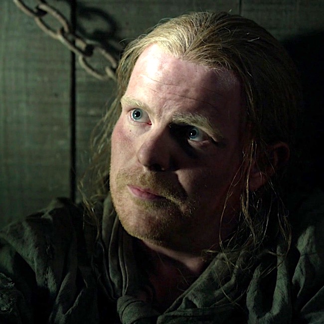
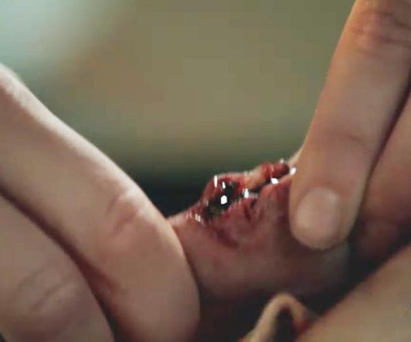
#2. May his last sight be the face of a friend.

Again, from Drums of Autumn: 

If Hayes was still sober enough to see anything, the last thing he saw on earth would be the face of a friend. 

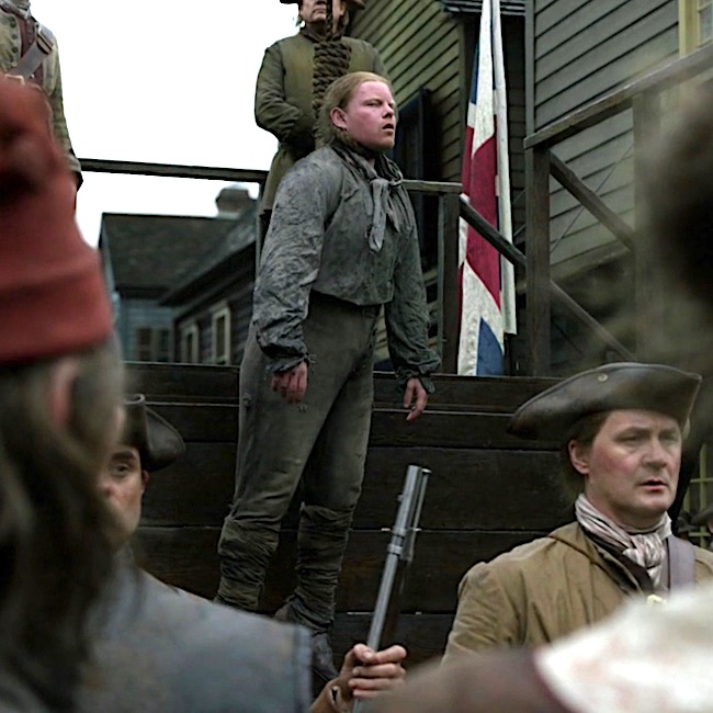
He could see; Hayes glared to and fro as they lifted him into the cart, twisting his neck, desperately looking. 

“Gabhainn! A charaid!” Jamie shouted suddenly. Hayes’ eyes found him at once, and he ceased struggling.

Jamie musters a wan smile for his auld friend and fellow Ardsmuir prisoner.

Drums of Autumn quote continues:

The captain of the guard stood poised, saber raised. 

Suddenly, the condemned man drew himself up straight. Eyes on Jamie, he opened his mouth, as though to speak. 

The saber flashed in the morning sun, and the drums stopped, with a final thunk! I looked at Jamie; he was white to the lips, eyes fixed wide. From the corner of my eye, I could see the twitching rope, and the faint, reflexive jerk of the dangling sack of clothes. A sharp stink of urine and feces struck…

The rope drops. Hayes death appears mercifully quick! Again from Drums of Autumn: 

The hangman had known his business; there had been no undignified struggle, no staring eyes, no protruding tongue; Gavin’s small round head tilted sharply to the side, neck grotesquely stretched but cleanly broken. 

OK, but, what about non-traditional hangings? Any of these in Outlander. But, of course!

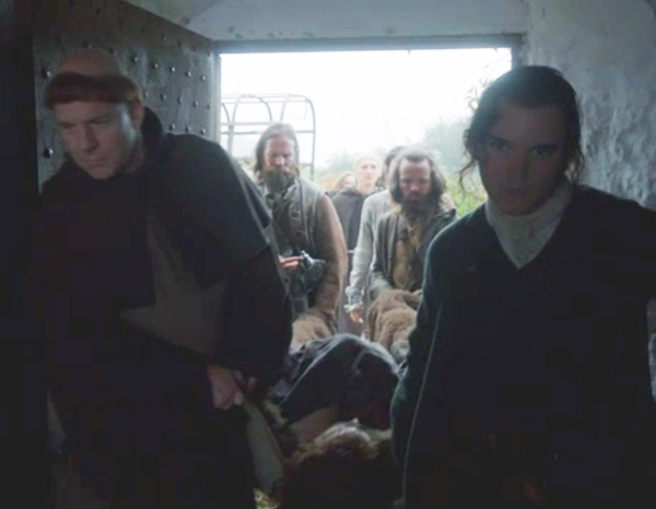
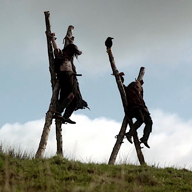
Two Jacobite traitors hang from crossed stakes courtesy of Red Coats in Outlander episode 105, Rent. Dougal and his party find the dead men with “T” for traitor carved into their flesh.  Although we don’t know how they died one can reasonably consider this a variation of a crucifixion hanging.

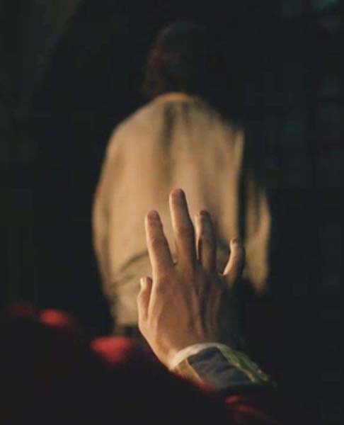
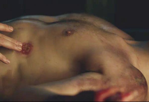
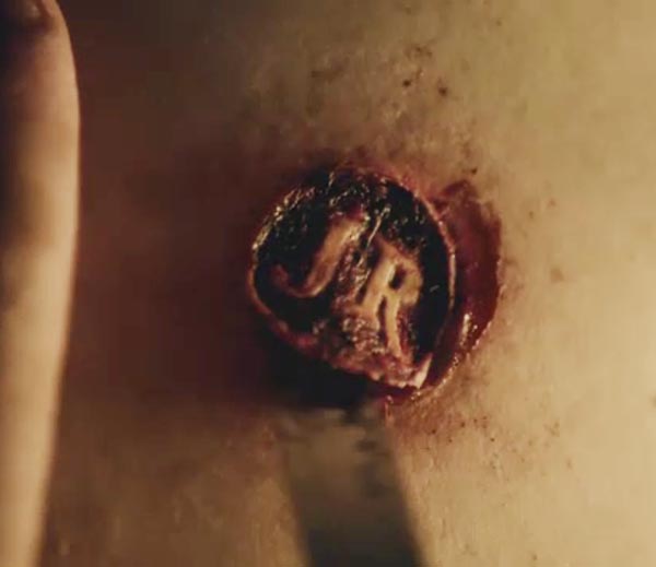
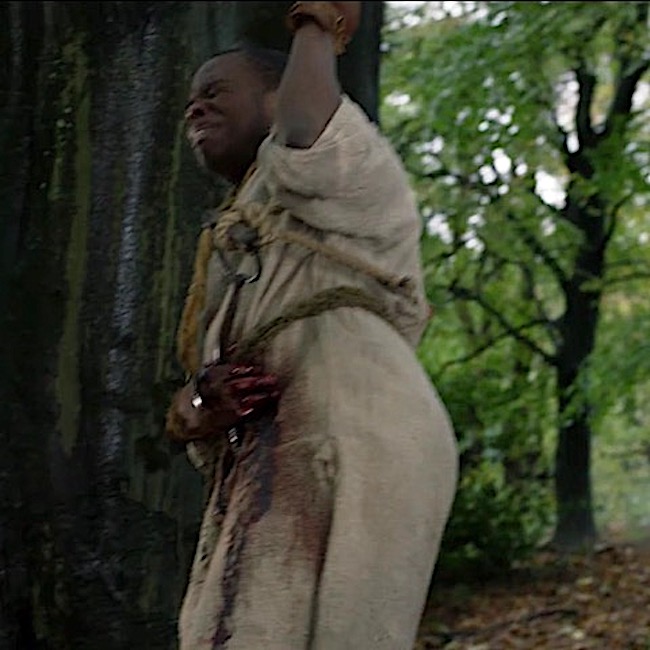
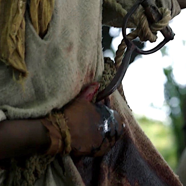
And, finally! In ep 402, Do No Harm, Jocasta’s slave, Rufus, dangles from a hook thrust under his ribs. This type of hanging is known as impalement.

From Drums of Autumn:

From everything I could sense and feel, I thought that the curve of the hook had gone upward through the liver. Likely the right kidney was damaged, and the jejunum or gallbladder might be nicked—but none of those would kill him immediately. 

So, one may reasonably conclude that Outlander has dutifully presented both traditional and non-traditional hangings!

Now, on to anatomy! Waaay back, Anatomy Lesson #12, Claire’s Neck or The Ivory Tower,  has a section on hanging but today we go deeper into the subject.

So, what does hanging have to do with human anatomy? Quite a lot actually, because hanging destroys normal anatomy. Let’s see how. 

Understanding traumatic events from hanging requires a bit of neck anatomy.  So….

Neck Anatomy: The neck is complex because it contains a mess of structures. These include:

    • pharynx for food and air passage
    • larynx for air passage and vocalization
    • trachea for air passage
    • esophagus for food passage
    • common carotid arteries supply blood to head
    • jugular veins drain blood from head
    • cervical vertebrae for support
    • hyoid bone for speech, swallowing, tongue movements
    • cervical spinal nerves, for neck and upper limb innervation
    • muscles for movement of head, neck, scapula and clavicle

Hangings typically traumatize one or more of these neck structures:

    • cervical vertebrae
    • hyoid bone
    • carotid arteries 
    • jugular veins
    • trachea and larynx

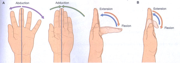
Cervical Vertebrae: The neck contains seven cervical vertebrae numbered from skull downward as C1-C7 (Image A). C1 is also known as the atlas; C2 is the axis.

Cervical vertebrae are smaller and more delicate than thoracic and lumbar vertebrae; this is especially true of C1 and C2. Collectively, all seven cervical vertebrae form the cervical skeleton and are bound together by strong ligaments.
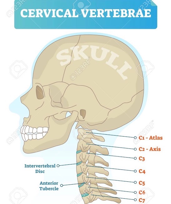
Vector illustration of cervical vertebrae. Medical scheme with close-up skull and isolated C1 atlas, C2 axis, C3, C4, C5, C6 and C7 vertebra. Diagram of Intervertebral disc and anterior tubercle.

Image A

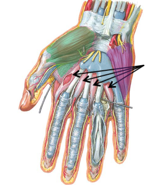
Hyoid Bone: The hyoid bone and cervical vertebrae are the only bones in the neck . The U-shaped hyoid (Image B, red) is unusual because it is the only bone of the body that does NOT articulate with other bones. Rather, it is suspended in the neck via muscles and ligaments connecting to neighboring bones and cartilages. More detail on hyoid and cervical vertebrae in Anatomy Lesson #12, Claire’s Neck – The Ivory tower.

Try This: You can palpate your own hyoid bone. Hold chin parallel to floor. Place thumb and forefinger of one hand on either side of your larynx (voice box). Move fingers upward until just under mandible. Squeeze gently, then swallow. You will feel a thin hard structure on each side, the R and L arms of the hyoid.

And, for book readers, Diana wrote about the hyoid bone in Written in My Own Heart’s Blood.
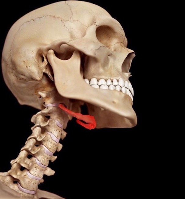
Image B

Common Carotid Arteries: The neck has a R and a L common carotid artery; each is a primary or secondary branch of the aorta arch (Image C).  Common carotids straddle trachea and larynx (Image C -blue/green structures) before splitting into internal and external carotid arteries which supply blood to neck and head.
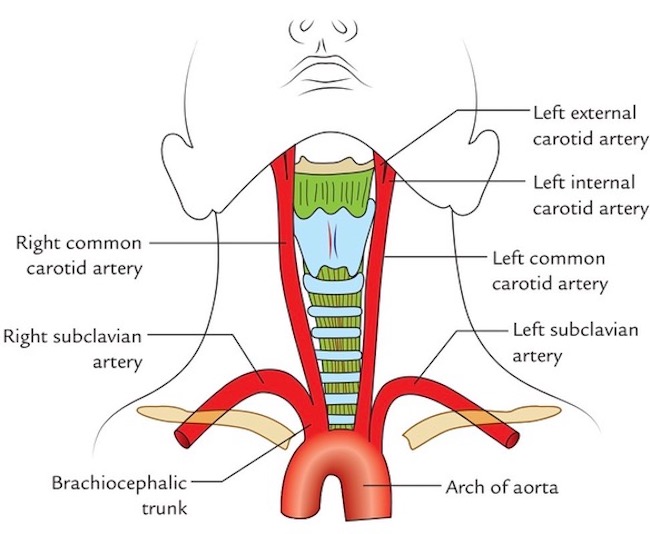
Image C

Jugular Veins: The neck has two jugular veins on each side; external and internal jugulars. These drain blood from head and neck structures toward the heart Image D).
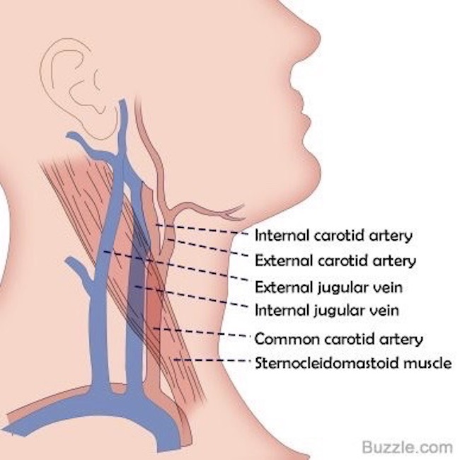
Image D

Trachea and Larynx: All of larynx plus upper trachea lie in the neck. Both are conduits for air to reach the lungs during inhalation and to leave the lungs during exhalation. In addition, the larynx produces vocalizations. Trachea and larynx provide the only normal route by which air can move to and from the lungs. Of course, a tracheotomy is an exception, but it is also not normal. (Psst…No S5 spoilers from  book readers, please!)

Image E shows trachea and larynx. Tracheal wall is stiff due to cartilage plates. The complex larynx consists of nine cartilages of various shapes and sizes. Learn more about the larynx in Anatomy Lesson #42, “The Voice – No, not that One!”
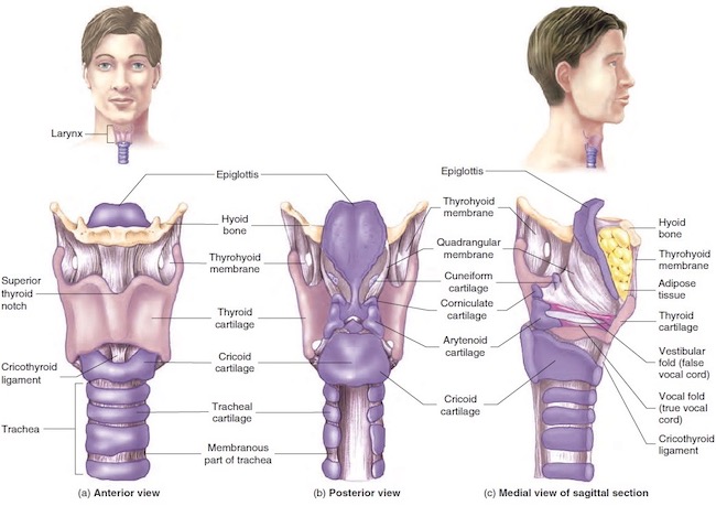
Image E

Now, for some info about hanging. 

Hanging: One might think hanging is a rather simple act, but it is not. There are many types of hanging although death is typically from either strangulation or neck fracture. Here are three common types of hanging.

*Short Drop: A condemned person is placed on a stool, ladder, cart, horse, etc. with noose around the neck. The object is removed leaving the person dangling from the rope. Although the person is unconscious within 15 seconds, death usually takes 10-20 minutes as the noose tightens causing slow strangulation.

*Standard Drop: A condemned person drops 4-6 feet (1.2-1.8m). This is considered more “humane” than short drop because it typically fractures the neck which traumatizes spinal cord and causes immediate unconsciousness and rapid brain death. 

*Long Drop: Drop varies. Height and weight of the condemned were used to calculate the drop needed to fracture the neck, preferably at C2, the axis. This is known as a hangman’s fracture.

Yes, I know! Horrifying to think about, but charts were developed to augment a swift death, the preferred outcome.

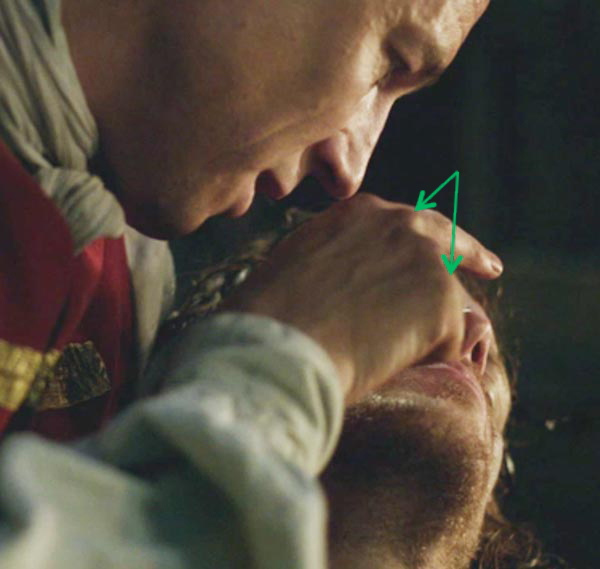
Submental Knot: Submental means “under the chin.”  Careful placement of the eye or knot of the noose under the chin jerks the head backward, contributing to neck fracture. Side and back knots tend to strangle rather than fracture the neck.

For more information about the chin, read Anatomy Lesson #26, Jamie’s Chin – Manly Mentus.

Back to Outlander: So, with knowledge about vulnerable neck structures and types of hangings plus their effect on anatomy, let’s review Outlander hangings. 

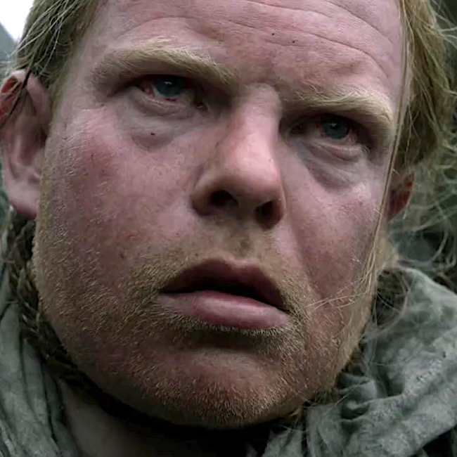
What about Taran from ep 115, Wentworth Prison? Taran was pushed off a not-very-tall scaffold – so in effect a short drop. Sadly, it didn’t break his neck and the Watchman kicked and struggled at the end of the noose! A member of the execution squad pulled down on his legs to break his neck, an act that was often required to end the misery. Of course, Jamie and other condemned prisoners witnessed this horror!

And, then there’s Hayes. Although his end was swift in Drums of Autumn book, not so in the episode. He did experience either a standard or long drop but afterward, his face is red and bloated and his eyes bloodshot. These findings are more consistent with strangulation than neck fracture, meaning his jugular veins were obstructed leading to death by brain edema and ischemia, wherein:

    • Face becomes engorged with blood
    • Facial skin turns red
    • Facial skin exhibits tiny petechiae/petechia (small blood marks)
    • Bleeding in conjunctiva overlying whites of eyeball (subconjunctival hemorrhage)

So the FX were very good but not well-matched for this type of hanging.

Just so we know, if the deeper lying common carotid arteries are occluded by strangulation, the face appears pale or white.

And, sphincters do relax spontaneously so urine and feces are evacuated. Thankfully, these weren’t part of the FX scheme! 😱

And poor Rufus! Impalement by hook is a very old punishment occurring in Babylonia! Later, it was used in 18th-century Ottoman-controlled Bosnia and by Dutch overlords in Suriname (South America) who impaled troublesome slaves under the ribs.

An 18th century observer recorded that an impaled person could hang as long as three days before expiring. The primary source of pain was extreme thirst.  Thankfully, this type of hanging has been pretty much abandoned.

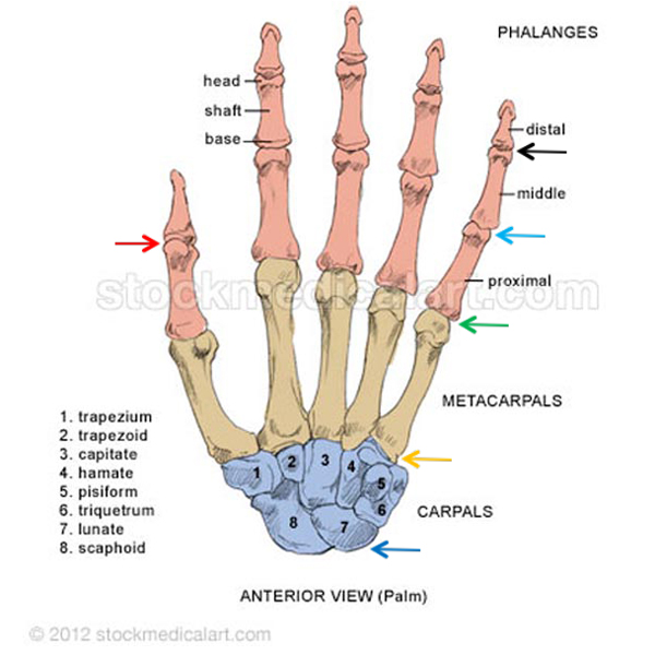
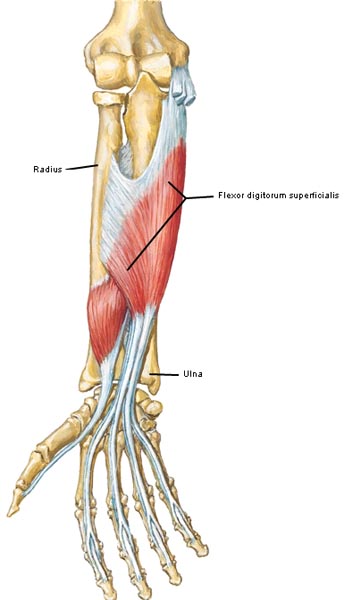
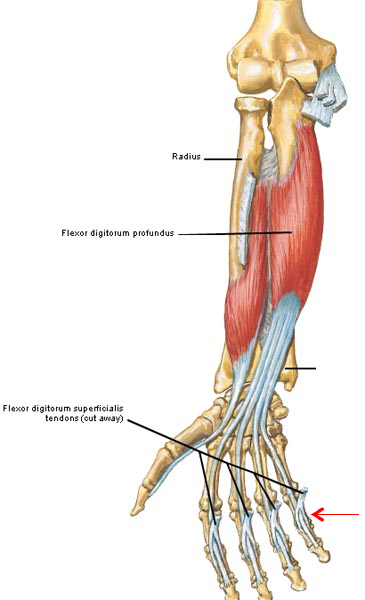
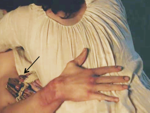
As for the hyoid bone…. It fractures in about a quarter of hangings but is not the cause of death. Forensic scientists look for a fractured hyoid in suspected homicides because about 50% of hyoids fracture with manual choking.

Fracturing the hyoid bone depends on applied force, age of victim, nature of instrument (ligature vs. hands) so even if the bone is not fractured, a murder is still possible (Image F –  red arrow).
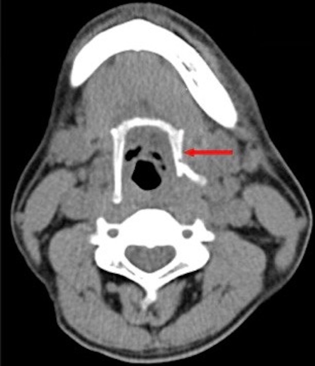
Image F

Today, we can be grateful that many countries have abandoned hanging as a means of punishment. An excellent film is available on this topic: Pierrepoint: The Last Hangman. Bonus! Tobias Menzies plays Lieutenant Llewelyn!

In Jamie’s time, hanging was an ignoble death. Why, we might ask? Here is one reasonable thought:

“The sword connoted an honourable way of dying, and an honourable return to the earth, but the rope left the body hanging between heaven and earth and was therefore an unseemly death.” 

Ron Brown, The Art of Suicide

Until next time….Hang in there!

The deeply grateful 

Outlander Anatomist

Follow me on:

  • Twitter @OutLandAnatomy
  • Join my Facebook Group: OutlandishAnatomy
  • Instagram: @outlanderanatomy
  • Tumblr: @outlanderanatomy
  • Youtube: Outlander Anatomy 

Photo Credits:


Fun Fact: phalanges

ep 202 phalanges

sing. noun: phalange [fuh-lan-jee]; pl. noun: phalanges [fuh-lan-jeez].

From the Greek, phálanx, meaning military formation; later used for the bones of fingers and toes

Anatomy def: 14 bones in the digits of each hand or in the toes of each foot; 56 phalange bones in total. Each thumb and great toe bears two phalanges and remaining digits each contain three phalanges.

Outlander def: Smashed but healing finger bones of Jamie’s puir left hand (right hand in Diana’s books). Claire’s own loving hands diligently labor to mended them!

Learn about phalanges in Anatomy Lesson #22, “Jamie’s Hand – Symbol of Sacrifice.”

Read about Jamie’s smashed bones in Dragonfly in Amber:

Jamie glanced dispassionately at his right hand. It really wasn’t too bad; a couple of fingers set slightly askew, a thick scar down the length of the middle finger. The only major damage had been to the fourth finger, which stuck out stiffly, its second joint so badly crushed that the healing had fused two finger bones together. The hand had been broken in Wentworth Prison, less than four months ago, by Jack Randall.

See Murtagh Fitzgibbons Fraser tighten the laces of Jamie’s amazing hand brace, in Starz episode 202, Not in Scotland Anymore. Designed to stabilize his healing phalanges, Clever Cunning Claire likely devised this awesome medical appliance!

A deeply grateful,

Outlander Anatomist

Anatomy Lesson #23: Harming Hands – Helping Hands – Healing Hands

Hallo Outlander Anatomy students! Welcome to Anatomy Lesson #23: The Hand – Part 2. Anatomy Lesson #22 introduced the hand but there is still much more structure and function to learn.

Today’s task is three-fold: to enrich our understanding of hand anatomy, to apply that anatomy using images from Starz episodes and to consider how hands are used. For most of us, our hands are with us throughout life so the way we use them helps define how we work, play and live (Photo A).

So, how do we use our hands? What tasks do we apply them to? Are the tasks helpful, harmful or neither? Outlander books and the Starz series provide abundant examples of hurtful hands, helpful hands and healing hands which are considered in this lesson. Just so we are all on the same page, all Starz images in this lesson are from episode 116, To Ransom a Man’s Soul, unless otherwise stated.

hands of love KLS edited

Photo A

Let’s begin the lesson with a brief review of digit and thumb movements and of the hand skeleton.

Our four fingers (U.S. definition or digits #2-#5) carry out four movements: abduction, adduction, flexion and extension (Photo B). Try all four movements with your own fingers.

finger movements KLS edited

Photo B

Because thumb anatomy differs from that of the fingers, it can perform seven independent movements: abduction, adduction, extension, flexion, opposition, reposition (Photo C) and circumduction (thumb moves in a circle – not shown in Photo C). Try all seven thumb movements.

Reposition (Photo C – right image) is the hand with all digits at rest or repose, a benign anatomical position maintained with little muscular effort.

thumb movements KLS edited

Photo C

In the image below, Claire’s healing hand is held in repose/reposition as she offers comfort to an angry, distraught guilt-ridden Jamie:

“Dinna touch me!” … “You canna save a man that doesna want saving!”

epi 116 good hand in repose KLS edited

Next, consider the crazy creepy crawler’s hand reaching for Jamie, the “object” of his obsession (Starz episode 115, Wentworth Prison). The palm is turned from our view but consider his hand. Is it at rest? Is it neutral? Anatomically, BJR’s hand yearning for Jamie is very nearly in reposition and although it may seem benign, we know it portends great harm. There’s a keen difference between anatomical positions of the hand and the intent behind such positions.

ep 115 BJR hand KLS edited

Next, let’s review bones of the hand (Anatomy Lesson #22). The complex skeleton of the hand contains 27 bones (Photo D – left hand, palm up): 8 carpals in the wrist, 5 metacarpals in the mid-hand and 14 phalanges in the digits. Each finger (digits #2-#5) contains three phalanges but the thumb (digit #1) has only two.

The bones of the hand create many joints (Photo D):

    • blue arrow shows wrist joints (between forearm and carpals)
    • orange arrow shows carpometacarpal joints (CM)
    • green arrow shows metacarpophalangeal joints (MP)
    • turquoise arrow shows proximal interphalangeal joints (PIP)
    • black arrow shows distal interphalangeal joints (DIP)
    • red arrow shows a single interphalangeal joint (IP) of thumb.

hand-bones-KLS-edited

Photo D: ©Stockmedicalart.com 2012

This creative rif on “The Scream” by Edvard Munch (Photo E) warns us that up next is toxic/nuclear waste in the form of handiwork by the bloody black-hearted bastard. In this lesson, I do resort to name calling – I canna help it when it comes to that wicked wretch!

hazard-symbol-munch-scream KLS edited

Photo E

Now for applied anatomy, first of the hand bones: the villainous Jack-Hammer mangled the bones of Jamie’s left hand (right hand in the book) such that Claire is tasked with treating nine fractures. From herself’s own words (Outlander book):

…”A single broken finger is enough to sink a strong man to his knees with nauseated pain…“I carefully picked up the smashed right hand, bringing it into the candlelight for examination. It would have to be set, and as soon as possible. The injured muscles were already clawing the fingers inward.

“It was a long, horrible, nerve-wracking job….. some parts, such as the splinting of the two fingers with simple fractures, went quite easily. Others did not…. I set his middle finger, exerting considerable force necessary to draw the ends of the splintered bone back through the skin.

The next image shows a compound or open fracture of the proximal phalange of Jamie’s middle finger. Compound fractures are characterized by broken bone(s) protruding through the skin and indeed the distal broken fragment is clearly visible (aqua arrow) as Claire rinses it with clean (sterile) water.

ep-116-compound-fracture-KLS-edited

Claire retracts the tip of Jamie’s finger pulling the bony fragment back into the finger. She then approximates the broken ends of the phalange. Retraction could require some effort because following a fracture, attached muscles undergo spasm to form a “natural splint” designed to keep the broken ends of a bone together as much as possible. This explains the quote from Outlander book:

“The injured muscles were already clawing the fingers inward.”

ep 116 compound fracture 01 KLS edited

And a final quote from Outlander book:

“All five fingers eventually lay straight as new pins, stiff as sticks in their bandaged splints… By good luck, only the one joint had been badly damaged…there was nothing I could do about the cracked metacarpal bones or the puncture wound.”

As Claire applies her skills to Jamie’s battered hand, she stitches the lacerations and places his hand on a small wooden pallet. She binds the badly damaged ring finger to the little finger using the latter as a split. All is then secured with a linen strip wound around a wooden peg that is then inserted into a hole in the wood pallet. Wow! This is so impressive and resourceful as no knots are require; the peg can be removed, the strip unwound and the wound attended without messing with knots. Overall, this ingenious device is an external cage which stabilizes the hand in the proper anatomical position and immobilizes it during the healing process. Brilliant!

Finally, did you ken the red, purple, and dusky blue bruises forming on Jamie’s hand? There is more about bruising later in this lesson.

ep 116 hand cage KLS edited

Now for applied anatomy of joints of the hand: in this jarring scene, we see the MP joints (green arrows) of BJR’s right fingers. He tells a traumatized Jamie “I know you can do better than this,” tapping the tip of his nose as one might correct an errant child. The total absence of regard for Jamie’s suffering tells us that the cunning cruel Captain does dwell in darkness and in darkness he shall remain.

ep-116-BJR-I-know-you-can-do-better-KLS-edited

Next, the unholy-one eyes Jamie writhing in agony: “Am I close? Have you reached your limit yet?” Oozing an air of utter boredom and sure knowledge of outcome, the mad menacing maniac sits with flexed left fingers that clearly display his left PIP joints (aqua arrows).

ep-116-BJR-have-you-had-enough-KLS-edited

Soon, BJR instructs Jamie to brand himself with BJ’s glowing wax seal stamp – over the left breast and near the heart. “Are you mine? Show me … do it!” The red arrow shows the flexed IP joint and the green arrow indicates the flexed MP joint of Randall’s right thumb. Remember: the thumb has only two phalanges with one IP joint shared between them.

ep-116-branding-Jamie-02-KLS-edited

Wracked with pain, Jamie brands himself but stubbornly sears his left side; his heart belongs to Claire! The flexed DIP joints (red arrows) are visible on Jamie’s right fingers as he grips the stamp.

ep-116-branding-Jamie-KLS-edited

Remember the anatomical mantra: the more bones in a body part, the more possible joints between the bones; the more joints, the more possible movements at the joints? Bone movements occur when muscles that attach to them undergo contraction. So, to better understand hand movements, we must next consider muscles operating the hands.

In Anatomy Lesson #22 we learned that some forearm muscles move bones of the hand. To review, the forearm is divided into two compartments. The anterior compartment contains eight muscles six of which attach to bones of the palmar (front) hand. The posterior compartment contains 12 muscles nine of which attach to bones of the dorsal (back) hand. Covering all the forearm muscles that move hand bones isn’t feasible in a single lesson, so let’s consider a few.

Three important muscles originate from the anterior forearm and end on bones of the digits; these are the long flexor tendons.

The first muscle is flexor digitorum superficialis or FDS (Latin meaning superficial flexor of digits); this muscle arises from humerus and radius (Photo F). Near the wrist FDS splits into four tendons, one to the intermediate phalange of each finger. Contraction of FDS mainly flexes PIP joints (Anatomy Lesson #22) but continued contraction flexes MP and wrist joints.

Try this: strongly flex (curl) your fingers and wrist joint. You used FDS to accomplish this. Identify wrist, MP and PIP joints. Flexion of these joints is critical for the grip (Anatomy Lesson #22).

Figure0426A FDS KLS edited

Photo F

Please think, if the digits have a superficial flexor will they also have a deep flexor? If you answered yes, then you are getting the idea of anatomy! The next anterior forearm muscle is flexor digitorum profundus or FDP (Latin meaning deep flexor of digits). Deep to FDS, FDP (Photo G) arises from the ulna and breaks into four tendons near the wrist to end on the distal phalange of each finger.

Now consider this: if tendons of FDS insert on the intermediate phalanges how will tendons of the deeper-lying FDP reach the distal phalanges? The body’s answer is ingenious! FDP goes through FDS: all four tendons of FDS split to allow FDP tendons to reach the distal phalanges (Photo G – red arrow). Do you see the splits? The engineering is awesome!

FDP flexes the DIP joints but it also contributes to flexion of PIP, MP and wrist joints.

Try this: Attempt to flex only the DIP joints of your fingers. Can you do it? I know a few folks who can but it is unusual. DIP joints typically flex in conjunction with PIP joints.

Figure0426B-FDP-KLS-edited

Photo G

The third muscle of the anterior forearm is Flexor Pollicis Longus or FPL (Latin meaning long flexor of thumb). FPL originates from the radius and inserts on the distal phalange of digit #1, the thumb (Photo H). Its major action is to flex the IP joint of the thumb but continued contraction also helps flex the MP joint.

Try this: flex your thumb and find the IP joint. Flexion is caused by FPL.

Figure0426B FPL KLS edited

Photo H

Do you recall those eight carpal bones arranged in two rows of four bones each (Anatomy Lesson #22) – aye, the ones with the funny mnemonic? These bridge the gap between radius and ulna and five metacarpals. More than a dozen strong fibrous wrist ligaments bind together carpals, metacarpals, radius and ulna (Photo I –  grey strands).

Figure0437B carpal arch KLS edited

Photo I

Two-dimensional images imply that the carpals lay in a flat plane, but this is not so. Wrist ligaments secure them into a curved carpal arch that is convex on the dorsum and concave on the palmar surface (Photo J – curved red line). The carpal arch is spanned by a fibrous sheet, the flexor retinaculum thus forming a potential space, the carpal tunnel (Photo J). But, the tunnel is NOT empty because it contains four FDS tendons, four FDP tendons, one FPL tendon and the median nerve (Anatomy Lesson #22).

Now for an important clinical correlation: truly a tight squeeze, any swelling of structures within the carpal tunnel compresses the median nerve and affects hand areas it subserves, a condition known as carpal tunnel syndrome. Symptoms may include tingling, numbness and pain with reduced strength of thumb and index, middle and half the ring fingers. Repetitive motions that over-extend the wrist such as keyboard use, piano playing, or prolonged use of vibrating tools may be contributing factors.

Figure0444B-Carpal-tunnel-KLS-edited

Photo J

Next, we consider the muscles of the back of forearm and hand: known as long extensor tendons, several muscles reach from posterior forearm to bones of the hand. We won’t name these but to summarize: three long tendons extend the wrist (Photo K – blue arrows) and 5+ long tendons extend the finger joints (Photo K – red arrows). Three long tendons extend and/or abduct the thumb (Photo K – black arrows). Whew!

Figure0424A-long-extensor-tendons-KLS-edited

Photo K

A closer view at the dorsal hand shows long extensor tendons as they cross the wrist. These tendons are held in check by a fibrous band, the extensor retinaculum (Photo L – black arrow). Note bands of fibrous tissue between the long extensor tendons. Known as intertendinous connections they augment extension of the fingers as a unit; if one extensor muscle contracts, it helps activate its neighbors (Photo L).

Try this: Intertendinous connections are quite variable but let’s try to locate one. Place forearm and hand flat on a surface. Pronate the forearm to expose the back of your hand. Try to lift (extend) just the little finger. Can you do it? Next, lift ring, middle and index fingers one at a time. Are some fingers harder to lift independently? I have greatest difficulty lifting middle and ring fingers independently so I know they are bound by an intertendinous connection. Try to locate one of yours.

Figure0453A-Extensor-tendons-KLS-edited_1

Photo L

Now, let’s consider intrinsic muscles of the hand beginning with the palmar surface. Palmar skin is tightly bound to an underlying fibrous sheet, the palmar aponeurosis, a feature that aids grasping (Photo M – red arrow). Two superficial muscles are apparent: palmaris longus and palmaris brevis. A forearm muscle, the tendon of palmaris longus blends with the palmar aponeurosis and tenses it during contraction. Many anatomists regard palmaris longus to be a vestigial muscle because it is absent in about 14% of people and its absence doesn’t really affect our ability to grip. Palmaris brevis is a tiny postage stamp-sized muscle near the base of the little finger (Photo M); its contraction puckers the palm skin and deepens a cupped palm.

Figure0442A-palmaris-longus-&-brevis

Photo M

Try this: Spread your digits and tap your palm. Feel that taut springy tissue? That’s the palmar aponeurosis. Now, bring pads of thumb and little finger together and strongly flex the wrist. Does a tendon stand out in the midline (Photo N – black arrow)? If so, this is palmaris longus. Repeat with the opposite side. What is the pattern of your forearm? Do you have two palmaris longus muscles, one or neither? Turns out I have one on the left but none on the right.

Palmaris longus KLS edited

Photo N

I love watching Jamie balance and twirl a dirk on the palm of his hand (Starz episode 105, Rent). See the puckered flesh at the base of his palm (red arrow)? This is caused by contraction of palmaris brevis to help cup his palm and support the dirk. Yep, Jamie has one! Is Claire watching his clever performance? Aren’t we all?

ep-105-palmaris-brevis-KLS-edited

The hand contains 19 intrinsic muscles (including palmaris brevis), meaning they arise and insert within the hand itself. These small muscles initiate fine movements of the digits. It would be tedious to name them all so understand that the thumb has four intrinsic muscles concerned with flexion, opposition, adduction and abduction (Photo O – green overlay). The little finger has three muscles which flex, oppose, and abduct (Photo O- purple overlay). Between the long flexor tendons and metacarpal bones are 11 additional muscles (lumbricals and interossei) which help flex, abduct and adduct the fingers (Photo O – black arrows). All totaled a whopping 34 forearm and hand muscles are involved in hand movements!

Figure0443B-intrinsic-hand-muscles-KLS-edited

Photo O

Like most body parts, the hand needs a blood supply. The large ulnar and radial arteries descend through the forearm, cross the wrist to form superficial and deep palmar arches (Photo P). Branches from both arterial arches produce two small arteries (proper palmar digital arteries) for each finger. The radial artery supplies branches to the thumb. The dorsal hand (not shown) has a smaller arterial arch that produces two more small arteries to each finger. Ergo, each finger has four supplying arteries; the thumb has three.

Figure0449B-palmar-arterial-arches-KLS-edited

Photo P

For those who do not understand blood flow, a full lesson on this subject lies in the future. For now, please know that the digital arteries carry blood ladened with oxygen and nutrients to the digits. They divide into smaller arterioles which branch into microscopic capillaries forming capillary beds (Photo Q). In the capillary bed, oxygen and nutrients are exchanged for carbon dioxide and waste products. The capillary bed reforms into venules that unite to form veins draining blood from the tissues. This general plan is followed throughout the hand and in most body tissues.

capillary bed KLS edited

Photo Q

Time for a clinical correlation about blood supply: capillaries have very thin walls. When subjected to trauma, these vessels burst leaking blood into surrounding soft tissues. Extravasated blood presents as a contusion, a reddish-purple-dusky discoloration which doesn’t blanch when pressed: the classic “black and blue” bruise (Photo R).

Bruising KLS edited

Photo R

Several students have inquired about Jamie’s blackened hand and digits following his rescue from Wentworth Prison. They were concerned that his hand exhibited tissue necrosis or gangrene. But no, discoloration is due to the beating administered by that depraved, demented devil. This gut-wrenching scene gives evidence of early bruising (black arrow): a vivid color range from deep red to purple to dusky blue. Very convincing!

ep-116-Jamies-hand-on-Claires-back-KLS-edited

Later on the Cristabel, Jamie’s thumb and fingertips are fully black and blue. The pounding mallet did a number on his hand capillaries and the bruising is fierce!

ep 116 Jamies hand on ship KLS edited

We are done with the real and applied anatomy lesson so let’s finish with examples of how hands are used in Starz episode 116 To Ransom a Man’s Soul.

“Dear God, you are a magnificent creature” utters the crazy cunning cutthroat with Jamie spread across his lap. As I gazed at this imagery, it seemed vaguely familiar. Then, B-I-N-G-O!

ep 116 BJR holding Jamie KLS edited

Intentional or not the position, posture and attitude are very nearly a mirror image of the Pietà, a Renaissance sculpture by Michelangelo Buonarroti (1475-1564) of the Madonna cradling a crucified Christ. This riveting juxtaposition of images reveals universal opposites: dark versus light; evil versus good; cruelty versus mercy, all deeply rooted themes in Diana’s works. Randall even draws the analogy to Jamie’s suffering: “Ah, so that’s your plan; to submit like Christ on the cross?” Helping hands, healing hands, or hurting hands. No brainer, here!

Pieta KLS edited

Later, the foul, festering fiend sensuously rubs lavender oil into Jamie’s painful burn telling him “These are Claire’s hands. Think of your wife.” There are new bruises on Jamie’s left shoulder. We don’t know what caused these – mayhap a mallet blow went astray? We shouldna be deceived by this perceived gentleness because BJ is about to deliver extreme mental pain to Jamie. Hurting hands, helping hands, healing hands?

ep 116 BJR these are Claire's hands KLS edited

After suffering hours of agonizing pain, BJ tells Jamie to:

“Think of Claire. Think of your wife. Wait for me. Claire’s here.”

Sadly, the soulless one said he would have Jamie’s surrender before he left this world and he does. Then BJ delivers his cruel, mocking coup de grâce: “I understand. How could she ever forgive you?” His hands are made for hurting and they are remarkably skilled at their work!

ep 116 how can she forgive you 01 KLS edited

Ugh! Enough of hurting hands…How about some healing hands? We already witnessed Claire’s healing hands as she cleanses and sets Jamie’s battered hand. Here she tidily stitches his skin lacerations. Her competency as a 20th century WWII combat nurse, her ingenuity at using 18th century tools and her brilliant, fertile mind are testimonies of her love and devotion to Jamie. Her hands have come to his rescue over and over throughout the Starz series; a true symbol of healing.

ep 116 stitching KLS edited

We’ve seen Claire reset Jamie’s broken bones and mend his wounds. Picking lavender, grinding buds in a mortar and pestle to make oil of lavender is yet another example of her healing hands as she prepares to enter the darkness with Jamie.

ep 116 lavendar oil KLS edited

As she rests her healing hand on her husband, he struggles with shame and self-loathing. “You belong to no one else but me…Well, Randall had your body but I’ll be damned if he has your soul as well.” Go Claire! Ye make us proud!

ep 116 Claire helping hands KLS edited

Finally, Godfather Murtagh’s healing hands are at work as he cuts JR from Jamie’s skin and casts the scrap into the fiery furnace! Mercifully, healing hands are plentiful in Starz episode 116.

ep 116 removing brand KLS edited

Let’s finish this lesson with examples of helping hands. First is a fantastic image of the three Highland musketeers rescuing Jamie from the “thug under the door.” Murtagh stolidly carries his precious cargo from Wentworth Prison to freedom. Angus furtively peers backward with dirk in hand while Rupert rides shotgun! Yeah, helping hands! Ye made me weep with relief!

ep 116 helping hands 02 KLS edited

So many helping hands at the monastery reach out to the party of Highlanders offering refuge, supplies and care for Jamie, wounded in body and soul.

ep 116 helping hands 03 KLS edited

More helping hands as the goodly Father comforts Claire after her confession: a far cry from Father Bain, no?

ep 116 priest helping hands KLS edited

Finally, we catch Angus’ au revoir kiss but did ye ken that the wee laddie’s helping hand helps itself to some of Claire’s personal real estate? Naughty Angus Mhor!

ep 116 Angus & Claire KLS edited

Helping hands, healing hands, hurting hands… how shall we use our own? Mine will continue to write lessons throughout the summer although most will be shorter and less complex. Thanks to all who love anatomy and Outlander!

Oh! Last week, I was invited by Jennifer Barnes, Ryan Hooks and Keetin Marchi (they are such great fun!) to give a mini anatomy lesson about the hand on the Outlander Aftershow at TheStream.TV. You can see it below around 16:30. Now that we face another bout of withoutlander, you can join our friendly hosts for the Outlander Book Club on Monday nights where they compare and contrast the book and the show: http://bit.ly/1dqHXxQ. Happy Summer!

The deeply grateful,

Outlander Anatomist

Follow me on:

Photo creds: Sony/Starz, Netter’s Atlas of Human Anatomy, 4th ed Gray’s Atlas of Anatomy for Students, 2005, ClinicallyOriented Anatomy 5th ed, www.artsology.com, www.imgbuddy.com, www.medicalartlibrary.com, www.parlmreadingperspectives.wordpress.com, www.wikipedia.org, www.wikiradiography.com, www.shutterstock.com