“Tremendous Tube – GI System, Part 2”

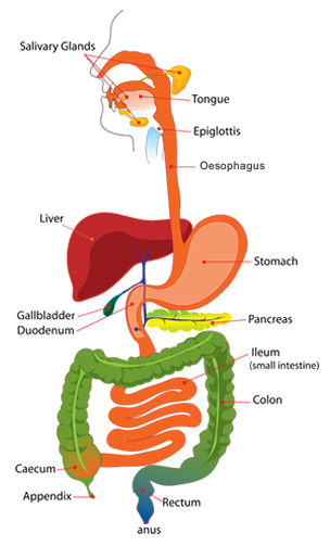
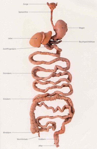
Greetings anatomy students! Time for another installment of our “slip and slide” down the gastrointestinal tract. Last time, Anatomy Lesson #44, “Terrific Tunnel – GI System, Part 1,” covered oral cavity, including tongue and teeth. There, we learned the major functions of the GI tract are ingestion, digestion, absorption, and excretion of food stuffs or their residues. As these activities are essential for survival (excepting parenteral feeding via intravenous route), every part of the GI tract is adapted for one or more of these functions. Please bear this in mind as we continue our guided tour down the Tremendous Tube!

Oh, before I forget, this lesson contains a book spoiler, so watch for the fleeing weiner dogs (a.k.a. Canis lupus familiaris); they will alert you!

img_3654

Fauces: Last lesson ended with the oral cavity. So picking up the thread, the fauces is the next part, a passageway connecting oral cavity with pharynx. The word derives from the Latin fauces meaning throat, but originally referenced the vestibule of an ancient Roman home.

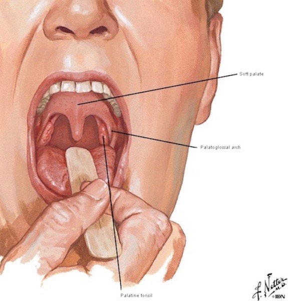
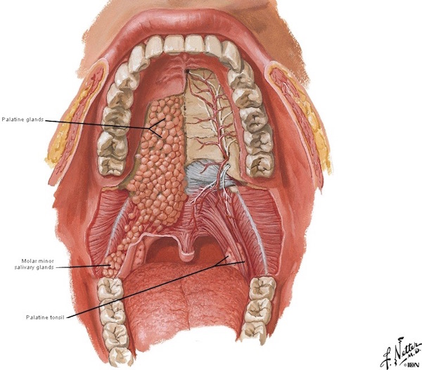
Fauces are formed by two pairs of arching pillars (Image A), one pair in front (anterior) and one pair in back (posterior).  Palatoglossal arches are anterior folds, reaching from soft palate to each side of tongue; these appeared in Anatomy Lesson #44, “Terrific Tunnel – GI System, Part 1.”  Paired palatopharyngeal arches are posterior folds, reaching from soft palate to sides of pharynx. All four folds contain small muscles used in chewing, swallowing, speaking, and so forth. Like the oral cavity, fauces are lined with mucosa, a layer containing numerous small salivary glands which add fluids to keep mucosal surfaces moist.
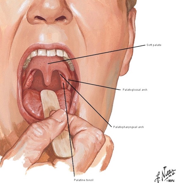
a-figure0047a
Image A

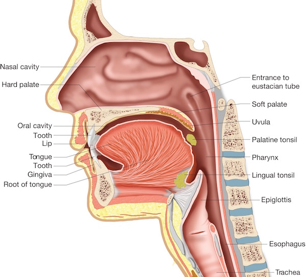
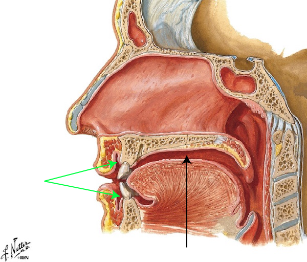
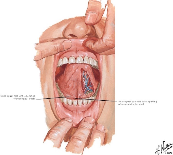
Faucial Arches: Together with the tongue, the palatoglossal arch and palatopharyngeal arch of one side form a triangular-shaped depression. This is readily apparent in Image B, a parasagittal section, meaning a vertical plane dividing the body into right and left sides (the plane passes slightly to the right of mid-line). Here, we view the inside of the right half of head. An oval, bumpy structure sits in this triangular depression; keep reading to learn about it.

Try This: Go to a mirror with a flashlight. Shine the light into the mirror and open your mouth; the reflected light will illuminate your fauces. Find the anterior folds, the palatoglossal arches. If you open very wide and depress your tongue strongly against the floor of mouth, you may be able to see the posterior palatopharyngeal arches. Try it!
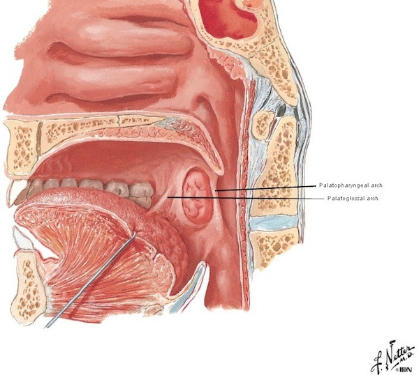
b-figure0060a-1
Image B

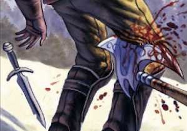
Pharynx: You may recall, Anatomy Lesson #42, “The Voice – No, Not That One!,” explained that pharynx is pronounced fare-inks, not far-nix. As noted in that lesson, one of my anatomy profs used to threaten: “if you say lar-nix one more time, I will rip out your far-nix!”

The pharynx is a muscular tube lined with mucosa and divided into three regions, named by their association with adjacent regions. Nasopharynx lies posterior to the nasal cavities (Image C – green). Oropharynx sits posterior to the oral cavity (Image C – yellow). Laryngopharynx (some physicians prefer the term hypopharynx) is posterior to the larynx (Image C – blue).

Like the oral cavity, pharynx is lined with mucosa (Anatomy Lesson 44: “Terrific Tunnel – GI System, Part 1”). A layer of muscle underlying the mucosa contracts to aid in swallowing, coughing, etc. Many tiny mucous (adj.) glands embedded in the pharynx release mucus (n; a.k.a. phlegm), a glycoprotein making the mucosal surface slimy. Talk about a slippery slope!
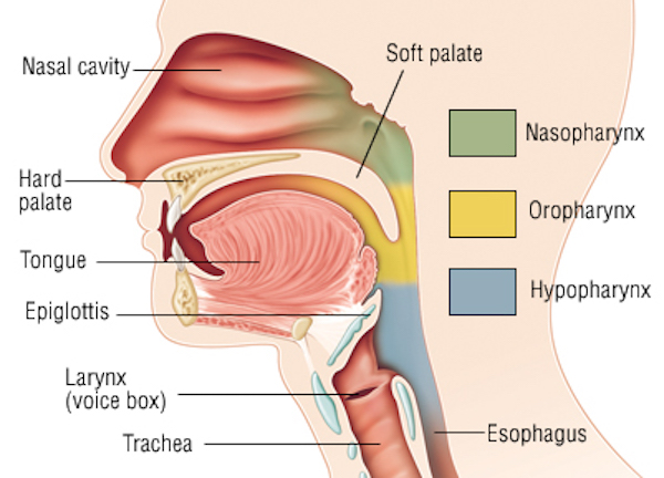
c-pharynx
Image C

Try This: Return to the mirror with flashlight in hand. Open mouth wide and look passed the oral cavity and fauces. Do you see the red wall at the back of throat? This is the posterior wall of the oropharynx. Without proper lighting and equipment, you cannot see naso- or laryngopharynges (pl.). Good work, all!

Pharyngeal Muscle:  Image D illustrates a coronal section wherein a vertical plane divides the body into front and back segments. The deep plane passed through the skull revealing back of pharynx and it’s muscles.

Muscles of the pharynx are complexly arrangement as they are telescoped inside one another. The right pharyngeal side of Image D is opened to reveal the back of larynx and uvula of soft palate. The left side shows intact pharyngeal musculature. Paired superior pharyngeal constrictor muscles are suspended from the skull. These fit inside the paired middle pharyngeal constrictor muscles which fit inside the paired inferior pharyngeal constritor muscles. This muscular arrangement allows the pharynx to usher food boluses from oral cavity and fauces into the esophagus (Image D).
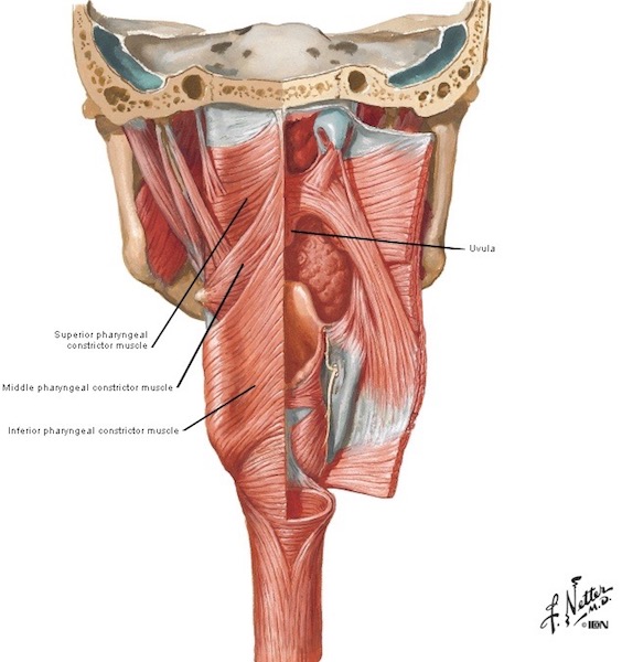
d-figure0063
Image D

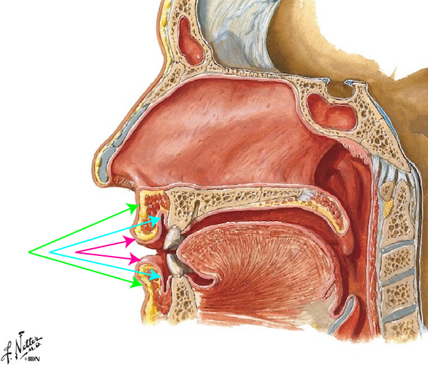
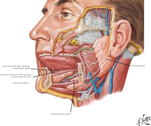
Tonsils: Oral cavity, fauces, and nasopharynx house the tonsils. Returning to the parasagittal image shown earlier, the tonsils are now labelled (Image E). Anatomists typically teach that humans have three sets of tonsils. But, to be precise, humans actually have four sets of tonsils: palatine, lingual, pharyngeal, and tubal.

By tradition, tonsils are written and named using the pleural form. This can be a bit confusing as two sets of tonsils are truly paired, but two are not. Her’s how this works. Lingual tonsils are not paired – rather, this is a large, single mass of tonsilar tissue under the mucosa at back of tongue. Pharyngeal tonsils, better known as adenoids, is a second singular mass of tonsillar tissue embedded high in the nasopharynx. Paired palatine tonsils are seated between the faucial arches; these structures are what most people mean when they say tonsils! Lastly, the oft ignored tubal tonsils (Image E – black arrow – they aren’t even labelled in the original figure) are paired masses near pharyngeal openings of each auditory (Eustachian) tube. Remember, the Eustachian tube leads to its respective middle ear (Anatomy Lesson #25, “If a Tree Falls – The Ear”).
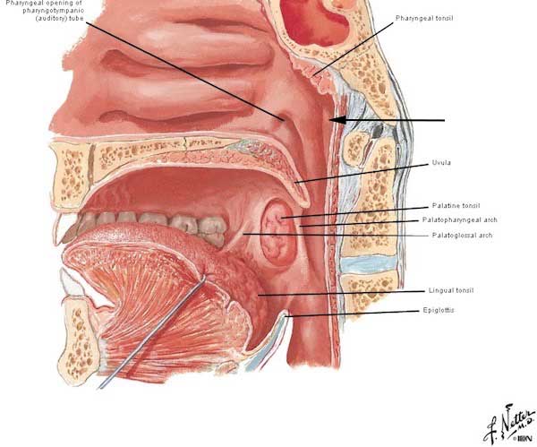
e-figure0060a-2

Image E

Waldeyer’s Ring: If one considers all four sets of tonsils from a frontal view (Image F –  right side), they form a “ring-around-the” nasopharynx and oropharynx. This so-called Waldeyer’s ring is named after a famous nineteenth century German anatomist, Heinrich Wilhelm Gottfried von Waldeyer-Hartz (Whew, glad I don’t have to sign that name on my credit card receipts!). This anatomical configuration is not accidental as tonsils are strategically placed to encounter pathogens and unwanted materials in food we ingest and in air we breathe.

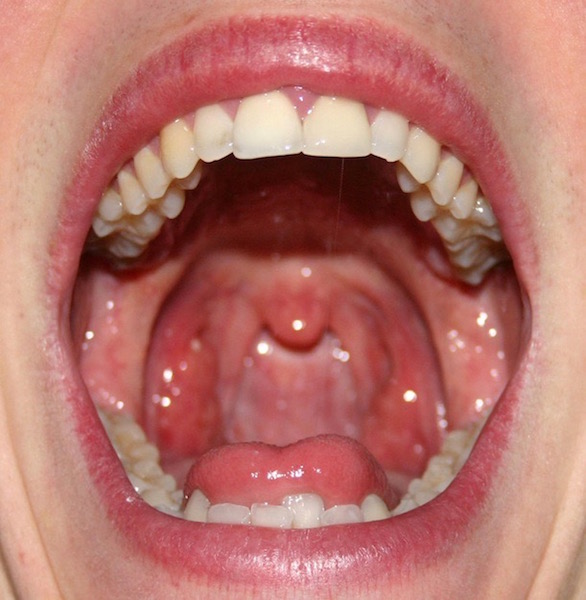
Try This: Unless a surgeon has already messed with your fauces, return to the mirror to find your own palatine tonsils situated between the faucial arches. Remember the surgeon’s chant: a chance to cut is a chance to cure! <G> Just so you know, palatine tonsils typically shrink with age, so if you are a senior citizen, don’t be alarmed if yours are way smaller than in your teens!
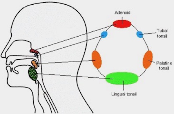
f-waldeyers-ring

Image F

Lymphoid Tissue: What the heck are tonsils? Tonsils are concentrated communities of lymphocytes, a particular class of leukocyte (white blood cell). Anatomy Lesson #37 “Outlander Owies Part 3 – Mars and Scars” stated that our blood contains five classes of leukocytes, one of which is the lymphocyte. There are also several types of lymphocytes, all of which work to provide us with immunity. Just so you know, lymphocytes are present both in circulating blood and in tissues outside of blood vessels.

Now, don’t run away screaming, but tonsils exhibit a classic morphology (appearance) by light microscopy (Anatomy Lesson # 34, “The Amazing Saga of Human Anatomy”). Image G shows a microscopic section through a bit of lingual tonsil.  The darker masses labelled lymphatic tissue (I prefer lymphoid tissue) are characteristic. Such clusters typically stain blue-purple (H&E staining for those with histology backgrounds). Notice that each cluster labelled “lymphatic tissue” contains smaller circles exhibiting a thin dark rim around pale centers – they kind of look like beads. Yay (I do beadwork)! This classical appearance of lymphatic/lymphoid tissue is present in various forms throughout the GI tract. Stay tuned. We will likely encounter them again during our TTT (tremendous-tube-tour)!
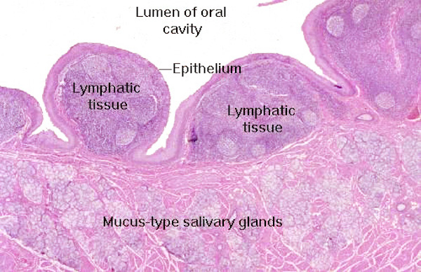
g-tonsils
Image G

Tonsillitis: Because tonsils are continuously exposed to pathogens (bacteria, viruses, and other microorganisms), they may become enlarged and inflamed, a condition known as tonsillitis. In fact, the suffix, “itis” attached to any word, means inflammation. Tonsillitis is invoked by various pathogens including viruses causing influenza or the common cold, or bacteria causing group A streptococcus (GAS), gonorrhea, and diphtheria. Image H shows the exudate common to a positive strep throat. BTW, exudate is the scientific term for the whitish gunk that may be exuded by inflamed tissues.
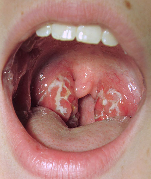
h-tonsillitis
Image H

If tonsils become chronically enlarged (hypertrophy) and/or repeatedly infected, they may be surgically removed by tonsillectomy (Image I).

WARNING: I try to avoid quotes from Diana’ books that have yet to be filmed, but sometimes naught else will do. Run free and flee with the hot dogs if you don’t want to skip the next three paragraphs!

img_3654

BOOK SPOILER: Later book readers know that Diana wrote about tonsillectomyAn Echo in the Bone has surgeon Claire performing a tonsillectomy of both palatine and pharyngeal (adenoids) tonsils on a youngster:

“Owg-owg-owg,” he said, grinning widely, but obligingly opened up. A faint putrid smell wafted out of his wide-open mouth, and even lacking a lighted scope, I could see that the swollen tonsils nearly obstructed his throat altogether. “Goodness gracious,” I said, turning his head to and fro to get a better view.

… Marsali had a small mirror with which to direct light, and that would perhaps help with the tonsils—the adenoids would have to be done by touch. I could feel the soft, spongy edge of one adenoid, just behind the soft palate; it took shape in my mind as I carefully fitted the wire loop around it, handling it with great delicacy so as not to let the edge cut either my fingertips or the body of the swollen adenoid…

Claire’s extraction likely produced specimens similar to these shown in Image I.
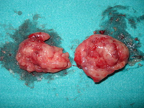
i-tonsillectomy
Image I

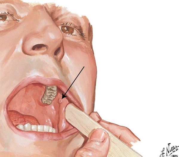
Tonsilloliths: Tonsils typically have surface creases known as tonsillar crypts. It is not uncommon for people to develop whitish globs in these crevasses. Such bodies are tonsilloliths, also known as tonsil stones, or sulfur granules. Tonsilloliths (-lith from the Greek lithos meaning stone) are stinky globs of mucus, dead cells, debris, and bacteria (Image J). They may be cheese-like or they may calcify into hard “stones” and are often the culprits in chronic bad breath due to the type of bacteria they harbor!

Most tonsilloliths are small but they can become huge, large enough to inhibit swallowing! Image J shows a tonsillolith embedded in a right palatine tonsil.  “Health-care-fingers” wield a tool to ease and tease the tonsillar stone out of its nest.

Social media carries a lot of advice about self-removal, including use of water picks, wet Q-tips, fingers, yadda, yadda, yadda. Warning! Palatine tonsils, the only pair you can readily see and access, has a very rich blood supply. Messing with deeply embedded or large tonsilloliths can cause bleeding and may result in subsequent infection. I cannot verify the efficacy of home-grown techniques but if you have doubts, please consult a professional!
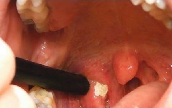
j-tonsilloliths
Image J

Not-So-Fun-Fact: Did you know, from 1926 through the 1960’s, children and adults suffering chronic middle ear infections were treated with radium to destroy the tonsils? Radioactive radium was inserted through the nasal cavities. Gah! And, during World War II, US, Canadian, and European military personnel also received NRI therapy (Nasopharyngeal Radium Irradiation – think Dana Scully and The X-Files!). Selected for treatment were soldiers with chronic middle ear dysfunction and who experienced job-related pressure differences.  Pilots, divers, and submarine trainees (Image K – submariners) were among the unlucky folks receiving such ghastly (my opinion) “health care.”  Consider the adage: “Yesterday’s heresy is today’s orthodoxy, is tomorrow’s fallacy.” Such is the checkered path of science and medicine! In its defense, science does maintain a check-and-balance system which helps it move onward and upward, even though the path may be uneven.
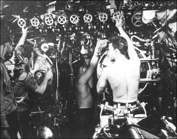
k-submariners-01
Image K

OK, moving on. Buh-bye tonsils!

Swallowing: So, food has entered the oral cavity, has been chewed (hopefully), and now it is time to swallow. What happens with swallowing? Well, swallowing involves oropharyngeal and esophageal stages.

Oropharyngeal Stage of Swallowing: Quite a few events occur almost simultaneously during swallowing and the list for the oropharyngeal stage is pretty astonishing:

  • Food is chewed
  • Saliva and food mix to form a wet blob known as a food bolus
  • Tongue lifts to press food bolus against the roof of mouth
  • Swallowing center (medulla of brain) initiates a reflex muscular closure of the laryngeal inlet to exclude food.
  • Epiglottis closes over the laryngeal inlet to block food bolus from entering larynx
  • Uvula constricts blocking food bolus from entering nasopharynx
  • Laryngeal muscles contract and respiration stops

You fully ken that things sometimes go wrong during swallowing; drink accidentally enters the nose or food gets sucked into the larynx. This is what happens if we try to breathe and swallow at the same time (please don’t try it as an experiment!). Such events stop us dead in the water until we cough out or blow out the offending materials. Hack! Honk!

Next is a video showing fluoroscopy of the oral cavity and pharynx during swallowing. Can you identify the oropharyngeal events in this video? Watch movements of the tongue, uvula, and epiglottis. Amazing! The esophageal phase appears in this video but it is discussed below.

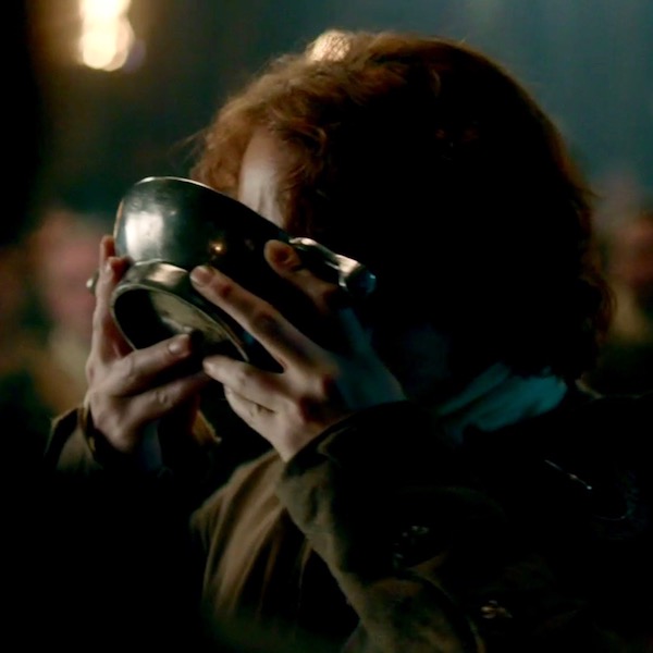
Does Herself write about swallowing? But, of course, Diana’s words are the “I Ching” for these Anatomy Lessons! Here, from Outlander book:

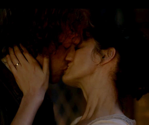
There was a lessening of the tension over the hall, and almost an audible sigh of relief in the gallery as Colum drank from the quaich and offered it to Jamie. The young man accepted it with a smile. Instead of the customary ceremonial sip, however, he carefully raised the nearly full vessel, tilted it and drank. And kept on drinking. There was a gasp of mingled respect and amusement from the spectators, as the powerful throat muscles kept moving. Surely he’d have to breathe soon, I thought, but no. He drained the heavy cup to the last drop, lowered it with an explosive gasp for air, and handed it back to Colum.

The dramatic scene comes to life in Starz episode 104, The Gathering. Better than Colum, Claire thinks (Outlander book), the lad is a natural born showman!
ep-104-swallowing

Muscle Detour: Before we study the esophagus, we must take a brief detour to glean basic info about muscle. This topic may seem misplaced, but bear with me, as the reason soon becomes apparent.  Thus far, muscles of oral cavity and pharynx have been skeletal muscles, the most familiar type. Calling all anatomy students: you should know our bodies contain three different types of muscle tissue: cardiac, skeletal, and smooth (Image L).
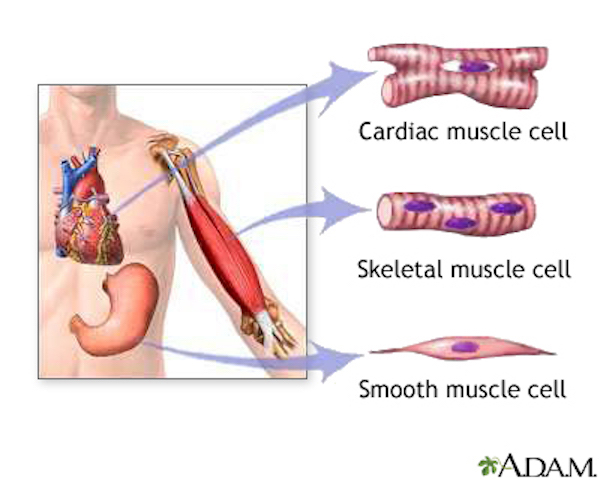
l-muscle-cells
Image L

Cardiac Muscle: Cardiac muscle is so named because it is found only in the heart (Image M). Psst … this isn’t really true, as it is also present in first part of the pulmonary veins (vessels carrying blood from lungs back to heart), a by-product of embryology.  But dinna write this on your biology quiz or try to correct your teacher. He or she willna appreciate it! <G>
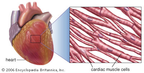
m-cardiac-muscle-01
Image M

Cardiac muscle is described as striated, involuntary in type. Striated because cardiac muscle cells bear microscopically-visible cross stripes (see them?): alternating dark and light bands across the cells (Image N – dark oval is a nucleus). Involuntary because we cannot voluntarily contract them; their contractions are regulated by involuntary mechanisms.
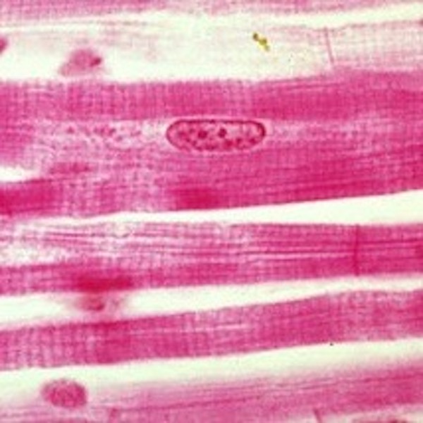
n-cardiac-muscle-02
Image N

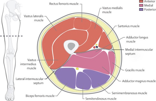
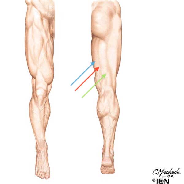
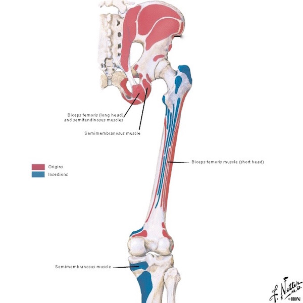
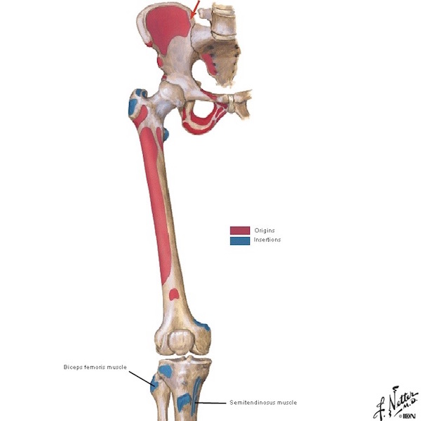
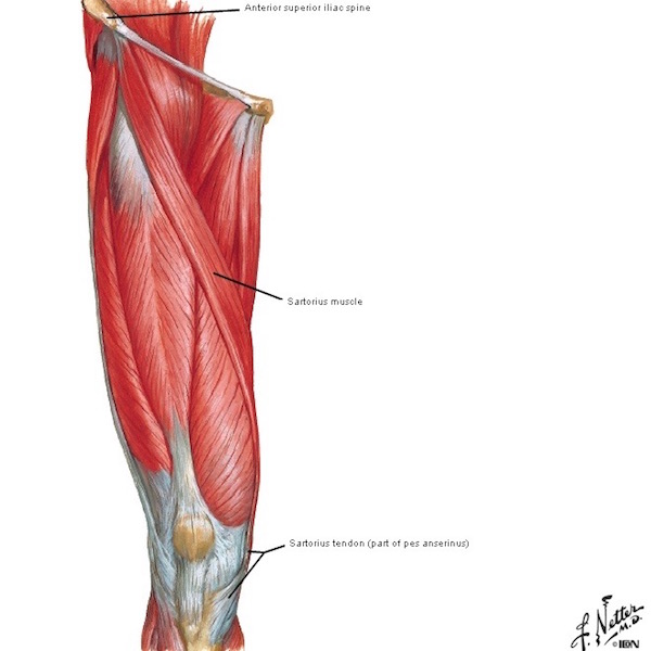
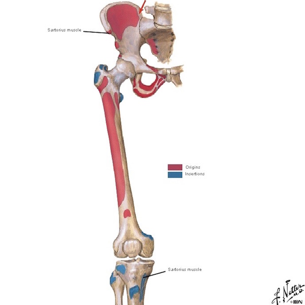
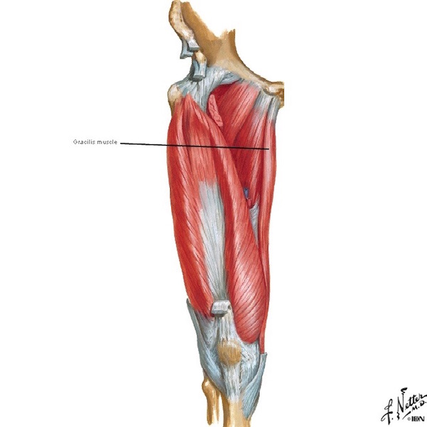
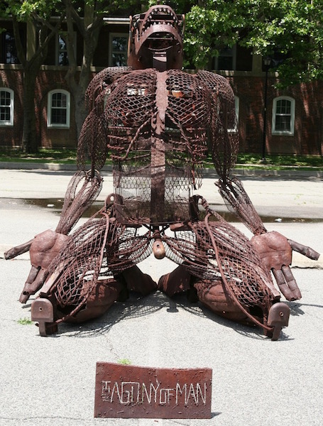
Skeletal Muscle: Skeletal muscle is so-named because it attaches to and moves bones of the skeleton (Anatomy Lesson #39 “Dem Bones – Human Skeleton”).  This type appeared in most of our anatomy lessons to date: pectoralis major, gastrocnemius, deltoids, quadriceps femoris, biceps femoris, blah, blah, blah, are examples.  Skeletal muscle contributes substantially to our over-all body weight: 38-54% in males and 28-39% in females! Many of the named skeletal muscles can be seen in this striking Body World’s display (Image O). BTW, the lifting partner wears ice skates but, the lifted partner wears none! Mayhap, a new Olympic sport?
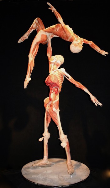
o-skeletal-muscle-01

Image O

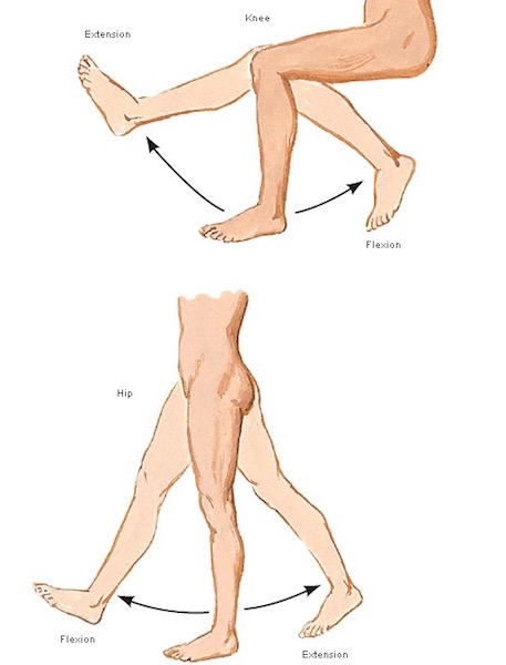
Skeletal muscle is further described as striated, voluntary muscle.  Striated, because like cardiac muscle cells, skeletal muscle cells bear cross stripes. Voluntary, because, with the exception of reflex contraction (think a knee hammer), we can contract skeletal muscle at will. As these muscles contract, they move the bones they insert into. Note the bold cross striations (stripes) along the length of skeletal muscle cells (Image P). These are typically more obvious in skeletal than in cardiac cells (dark ovals are nuclei).
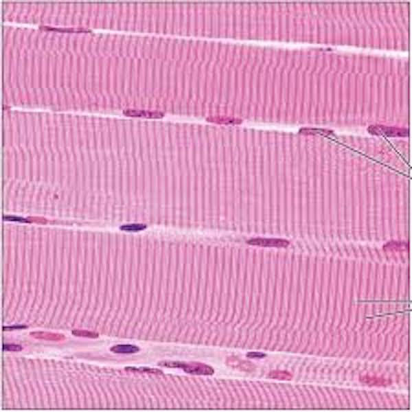
p-skeletal-muscle-02
Image P

Smooth Muscle: The final type is smooth muscle. Smooth muscle is not new to long-time anatomy students, as it appeared in two prior lessons. Anatomy Lesson #6, “Claire’s Hair – Jamie’s Mane” or “Jesus H. Roosevelt Christ!” introduced arrector pili muscles, made of smooth muscle cells whose contraction give us goose flesh. And, in Anatomy Lesson #31, “An Aye for an Eye – The Eye, Part 3,” we learned that the pupillary sphincter muscle is also made of smooth muscle. Smooth muscle appears in this lesson because hollow organs of the GI tract, such as esophagus, stomach, colon, and small intestine, contain layers of smooth muscle (Image Q) .
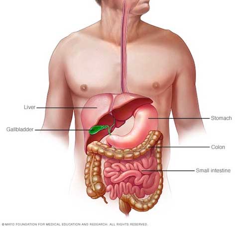
q-smooth-muscle-jpg-1
Image Q

Smooth muscle is classified as non-striated, involuntary. Non-striated, because its cells lack cross striations, hence the term, smooth (Image R – elongate purple bodies are cell nuclei). Involuntary because we cannot contract smooth muscle at will. Like cardiac muscle, it operates without our commands.

It is abundant in the walls of hollow organs of the GI tract, errector pili muscles, blood vessels, gallbladder, urinary bladder, reproductive tract, and in various other ducts and tubes. If you consider smooth muscle the wimp of muscles, think again: the uterus, endowed with thick layers of smooth muscle, can generate enough contractile force to push a 10 lb. babe out the bony pelvis!
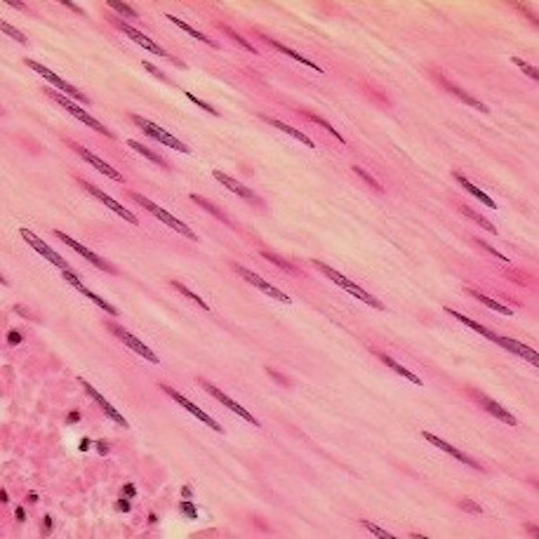
r-smooth-muscle-02
Image R

Esophagus: Next organ is the esophagus, a muscular tube connecting oropharynx with stomach. This tube goes by a mess of names, including food tube (ha ha!), gullet, gorge, oesophagus (British), throat, abyss, thrapple, gizzard, passage, maw and pa. Oops, should read, maw and craw! Hee, hee.

Esophagus begins at the laryngeal cricoid cartilage (Anatomy Lesson #42, “The Voice – No, not that One!”) and ends at gastroesophageal junction where it meets the stomach (Image S – black arrows). In adults, it measures roughly 20 – 24 cm (8″ – 9.5”) in length.
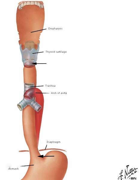
s-figure0229a
Image S

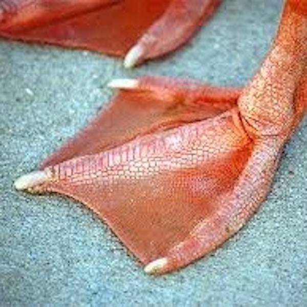
Esophageal Relationships: This tube pursues a long pathway as it “travels” through neck and thorax, ending in the abdominal cavity (Image T).  As the esophagus descends, it passes behind trachea (Anatomy Lesson #42, “The Voice – No, not that One!”), heart, and lungs, but in front of the spine.
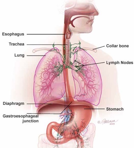
t-esophageal-relationships
Image T

Esophageal Hiatus: Long time anatomy students may recall that thorax and abdomen are separated by a muscular dome, the respiratory diaphragm (Anatomy Lesson #8, “Jamie Takes a Beating” and “Claire’s Healing Touch”). How does the esophagus bypass this structure?  Well, it doesn’t have to because there’s a hole through this skeletal muscle layer. Actually, three holes pierce the diaphragm: one for passage by esophagus (Image U – black arrow), one for the body’s largest vein (vena cava, Image U – turquoise arrow) and one for its largest artery (aorta, Image U – red arrow). If the hole (hiatus) for the esophagus is too large, a hiatal hernia may result wherein part of the stomach pushes up through the hiatus and into the thoracic cavity. Such hernias may cause heartburn, difficult swallowing, belching, or even chest or abdominal pain. But, for many folks, these are asymptomatic.
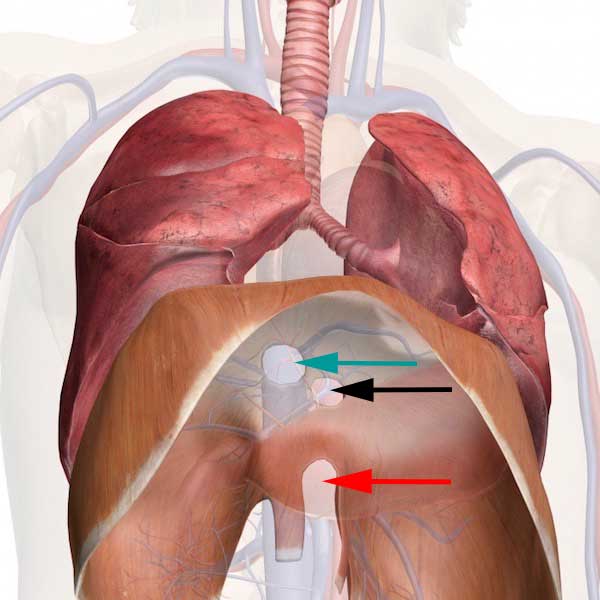
u-diaphragm
Image U

Esophageal Wall: An esophageal (adj.) “slice-of-life” reveals the organization of its wall (Image V).  The organ has a central lumen (cavity) which runs vertically.  Mucosa lines the lumen. Further out, two thick layers of muscle are present. Smooth muscle cells of the inner layer encircle the lumen forming an inner circular layer of smooth muscle. External to this, smooth muscle fibers run vertically forming the outer longitudinal layer of smooth muscle. Let’s take a closer look at these parts.
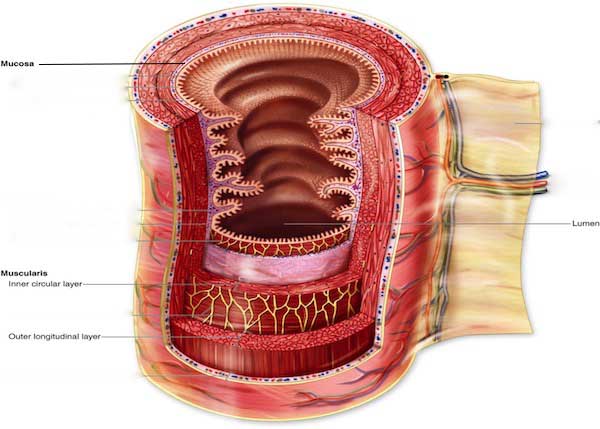
v-esophagus-01
Image V

Esophageal Mucosa: Like oral cavity and pharynx, the esophagus is lined with mucosa, including a thick layer of non-keratinized (living) surface cells (Anatomy Lesson #5, “Claire’s Skin – Opals, Ivory and White Velvet”). In animals such as the rat, mucosal cells of the esophagus and upper stomach are keratinized (dead) for protection because they eat a coarse diet highly fibrous foods, such as seeds. Mucous glands release their mucus onto the surface keeping it slipper, as apparent by endoscopy (Image W). Talk about disappearing into the depths; quite a tube!
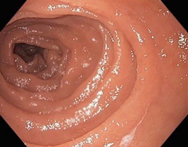
w-esophagus-02
Image W

Esophageal Muscle: Esophageal muscle changes type along its length, hence, the above muscle detour. Its upper 1/3 is skeletal muscle, the lower 1/3 is smooth, and the middle 1/3 is mixed skeletal and smooth. Ergo, we have voluntary control over the upper esophagus which activates during breathing, belching, swallowing, etc. But, as smooth muscle arrives, contraction becomes involuntary; bye-bye voluntary control. At this level, we also lose somatic sensation typical of skin (light touch, pressure, heat, cold, pain, vibration), replaced by visceral pain/discomfort. If a lighted match were to be placed against a visceral surface, the heat would not be detected. Although not as discriminating, visceral pain is fully capable of making us writhe (think passing a gallstone)!  Visceral pain is activated by ischemia (lack of oxygen), distension ( stretch), or inflammation (Anatomy Lesson #37, “Outlander Owies Part 3 – Mars and Scars”).

Try This:  Next time you swallow a hot cup o’ Joe, cocoa, or soup, notice when and where you no longer feel the heat as the food moves downward. If you are typical, heat is longer detected at about the top of the chest. This is where visceral sensation and involuntary muscle arrive on the scene!

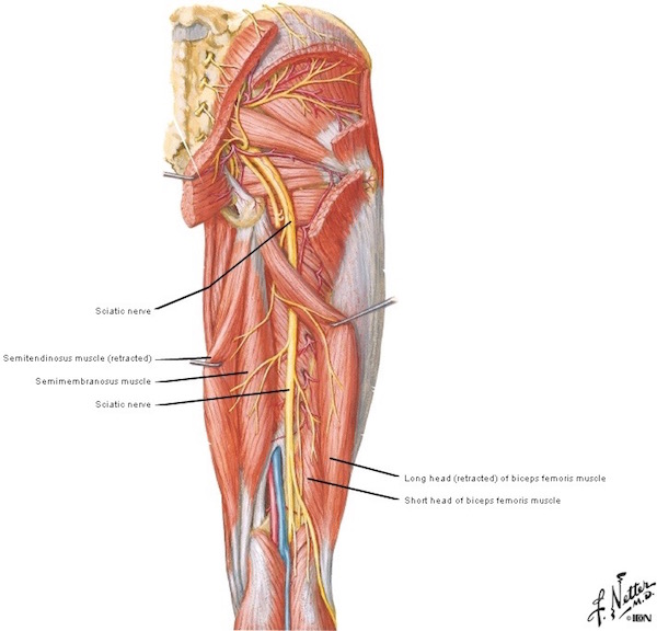
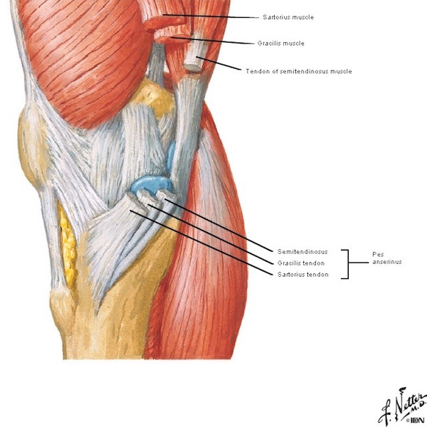
Esophageal Phase of Swallowing: As state above, esophageal muscle fibers are arranged in two layers: inner circular and outer longitudinal (Image X – back of esophagus). Contraction of the longitudinal muscle layer shortens the esophagus; contraction of the circular layer closes the lumen. This allows the organ to stretch and contract around a bolus of food, pushing it toward the stomach in coordinated waves known as peristalsis (review swallowing video above). Lastly, the esophagus has a functional lower esophageal sphincter (LES) which normally prevents reflux of gastric acids from the stomach back into the esophagus.

As noted in Anatomy Lesson #42 about the larynx, the back wall of trachea lacks cartilage (Image X). Filled with a fibrous connective tissue, this “defect” permits food to slide down the esophagus without rubbing against the tracheal rings. Remember, the secreted mucus keeps the food bolus and mucosal surfaces slippery (when wet <g>).
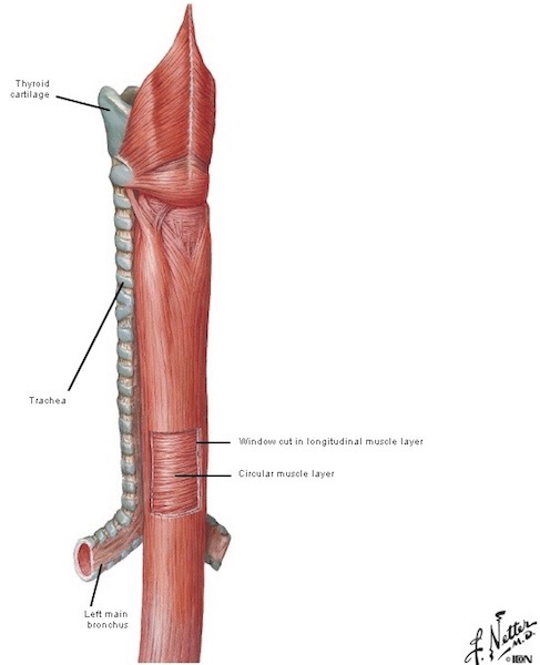
x-figure0230a
Image X

Because the esophagus delivers food and drink, and because our food choices are not always wise (think excessive distilled spirits), and because the entire gut is highly responsive to stress, the esophagus is subject to quite a few diseases and disorders. Here’s one familiar example, simply described.

GERD: Known by the longer term, Gastroesophageal Reflux Disease, folks with GERD experience chronic gastric reflux wherein highly acidic gastric juices regurgitate from stomach into the lower esophagus. Not just simple heartburn, in this instance, the LES fails to perform adequately. Over time, esophageal mucosal cells may be replaced with gastric lining cells, a change observable by endoscopy.  The pale pink area (Image Y – black arrow) is esophageal mucosa. The peachy area (Image Y – blue arrow) is gastric mucosa that has grown up and out of the stomach (dark area at the bottom of the pit) and into the esophagus. Why does does this happen? For defense: stomach surface cells are more resistant to acid than esophageal surface cells. However, as stomach cells do not belong in the esophagus, GERD is also accompanied by an increased risk of gastroesophageal cancer.
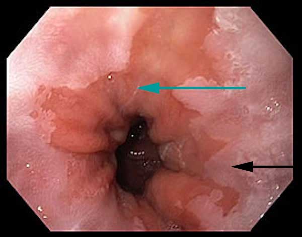
y-esophagus-barretts

Image Y

Fun Fact: Time for another obscure fact (I love these!): the first part of the esophagus is equipped with tastebuds! Yep. I know, hard to believe, but ‘tis true. Watch this fun video about taste buds and Bud beer (these are on the tongue; see the uvula hanging down?). The original ad starred John Belushi (RIP, John, ye were a funny man!). So clever, back in 1979, I called Anheuser-Busch begging for a copy of the film clip to use in my GI lectures. Sure enough, they kindly complied. But, that was before YouTube. So, enjoy!

OK, lesson done – time to play! Let’s visit Outlander’s outlandish examples of mouths, throats, gullets, thrapples, and gizzards!

This wonderful quote from Dragonfly in Amber book wasn’t filmed for S.2, so let’s use a substitute image. Just image King Louis of France popping wee birdies into his mouth and down his gullet instead of lusting after Claire in her (barely) scarlet gown (Starz episode 202, Not in Scotland Anymore).

After a triumphal tour of the table to show it off—to the accompaniment of murmurs of admiration all round—the dainty dish was set before the King, who turned from his conversation with Madame de La Tourelle long enough to pluck one of the nestlings from its place and pop it into his mouth. Crunch, crunch, crunch went Louis’s teeth. Mesmerized, I watched the muscles of his throat ripple, and felt the rubble of small bones slide down my own gullet. Brown fingers reached casually for another baby. At this point, I concluded that there were probably worse things than insulting His Majesty by leaving the table, and bolted. Rising from my knees amid the shrubbery a few minutes later….

Pregnant Claire feels the gorge rise in her gullet before bolting to upchuck in the Gardens of Versailles! Oops!
ep-202-gullet

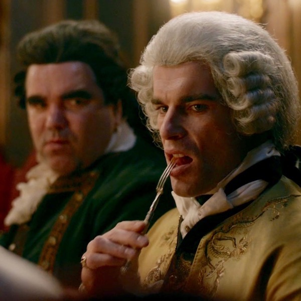
Dragonfly in Amber book again comes to our rescue as Comte St. Germaine forks fish (or something more sinister) into his mouth. His distain for hostess, Lady Broch Tuarach (Starz, episode 204, La Dame Blanche), eclipses the tasty trout! Canna appreciate good French cuisine if ye are intent on poisoning a bonny lass. Come on man, get a grip!  It was a ship, not your life. Oops, maybe it is your life! Who is your comely BFF? Why, Mary Hawkin’s intended, of course!

I caught Magnus’s eye as he served the Comte St. Germain, seated across from me, and beamed congratulations at him as well as I could with a mouthful of fish. Too well trained to smile in public, he inclined his head a respectful quarter-inch and went on with the service. My hand went to the crystal at my neck, and I stroked it ostentatiously as the Comte, with no sign of perturbation on his saturnine features, dug into the trout with almonds.

ep-204-eating

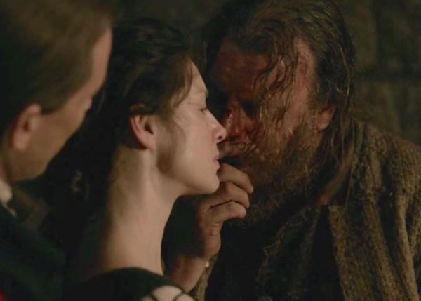
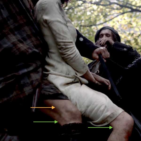
I love this scene between Dougal and Claire (Starz episode 106, The Garrison Commander) as they halt their flight from Captain Sicko’s grim grip. Ever the Heiland gentleman, Dougal invites Claire have a sip of a stinky water. “Oh, Aye, theres a stink to it. But, it’ll wet your thrapple, sure enough.” Thrapple? Yup, that is Scottish for throat, windpipe, or gullet. New word for our vocabularies. Yay!

St. Ninian’s Spring, the liar’s spring (Outlander book), purportedly burns throat and esophagus of any liar who drinks of it. Dougal does a dirk-draw to finish her off in case the burning starts:

The water had an odd dark color, and a worse smell—likely a sulfur spring, I thought. The day was hot and I was thirsty, though, so I followed Dougal’s example. The water was faintly bitter, but cold, and not unpalatable. I drank some, then splashed my face. The road had been dusty.

…“So at least you believe me when I say I’m not an English spy?” “I do now.” He spoke with some emphasis. “Why now and not before?” He nodded at the spring, and at the worn figure etched in the rock. It must be hundreds of years old, much older even than the giant rowan tree that shaded the spring and cast its white flowers into the black water. “St. Ninian’s spring. Ye drank the water before I asked ye.” I was thoroughly bewildered by this time. “What does that have to do with it?” He looked surprised, then his mouth twisted in a smile. “Ye didna know? They call it the liar’s spring, as well. The water smells o’ the fumes of hell. Anyone who drinks the water and then tells untruth will ha’ the gizzard burnt out of him.”

ep-106-liar-spring-01

No visible “fires of hell” in her thrapple, Dougal sheaths his deadly dirk! Claire, horrified that he would gut her gullet, asks why he spared her? His logic: weil, he wouldna have liked cutting her throat because she is a handsome woman and, because privately, he dreams of grinding her corn. Really, Dougal? This is your criterion for deciding whether to slice and dice a classy, sassy lassie? Geez, man, were you born in the 18th century?

“I see.” I spoke between my teeth. “Well, my gizzard is quite intact. So you can believe me when I say I’m not a spy, English or French. And you can believe something else, Dougal MacKenzie. I’m not marrying anyone!”

Ahhhh, weil, ye are wrong about that, Mistress Beauchamp. Jamie is waitin’ and rarin’ to go. Yay! Claire’s gonna get her corn ground, her corn ground, her corn ground….meal and multure free. Hee, hee!
ep-106-liar-spring-02

Let’s finish this Anatomy Lesson down the Tremendous Tube with a clever poem about Nessie’s own gullet. Composed by Keith Logan, it is entitled, Nessie’s Freedom. Yes, that Nessie!

Nessie’s Freedom

If you’re ever at Loch Ness
even in full highland dress,
put away your skean dhu,
order up a round or two.
Nessie really likes her bevvy
but drinks only Scottish heavy,
so don’t proffer Irish stout
or you may be carried out.
With a toast to the Black Watch
she might sip a little scotch,
though proportions of her gullet
spell a need for a deep wallet.
But if none of this concerns you
she may smile as she discerns you,
and there’ll ever be a welcome
as you drink to Nessie’s freedom.

Long live Nessie! BTW, Claire sees “her” in Outlander book. If you aren’t reading Diana’s books, you should start. Missing out on a mess of fantastic story!

Next Anatomy Lesson, the stomach and beyond!

A deeply grateful,

Outlander Anatomist

Photo Creds: Starz, Netter’s Atlas of Human Anatomy, 4th ed. (Images A, B, D, E, S, X), www.ddc.musc.edu (Images W, Y), www.doctors.net (Image G), www.en.wikipedia.org (Image I), www.flashblog2011.blogspot.com (Image O), www.health.harvard.edu (Image C), www.innerbody.com (Image U), www.jbbardot.com (Image T), www.kids.britannica.com (Image M), www.medcell.med.yale.edu (Image N), www.medline.plus.gov (Image L), www.mhhe.com (Image R), www.militaryhistoryonline.com (Image K), www.sites.google.com (Image F), www.mayoclinic.org (Image Q), www.studyblue.com (Images P, V), www.treatcurefast.com (Image J), www.wikipedia.org (Image H)

Anatomy Lesson #44: “Terrific Tunnel – GI System, Part 1”

Hello anatomy students! Time to launch a new but thrilling anatomic topic: the GI tract. What is the GI tract (Image A)? Well, it is a complex organ system that enjoys several monikers: alimentary canal, alimentary tract, digestive tube, GI tract, gastrointestinal tract, gastrointestinal system, and gut. Most anatomists prefer the term gastrointestinal tract or system, but since that term is a bit lengthy, this lesson will shorten it to GI tract.

meaning-of-gi1

Image A

Spoiler Warning: there are lots of Outlander images and book quotes within, including one waaaay forward from book eight. A scaredy cat warning appears before hand, so, watch for it!

scaredy-cat

You can easily scroll to the relaxing cat to skip the whole thing until you’re ready to read MOBY!

relaxed-cat

The GI tract is a tube extending from mouth through anus (yes, I wrote that and I warrant that team Angus and Rupert used that word, too!). It is divided into the following regions (Image B):

  • oral cavity
  • oropharynx
  • esophagus (oesophagus)
  • stomach
  • small intestine (duodenum, jejunum, ileum)
  • large intestine
  • rectum
  • anal canal and anus

The GI system includes several additional organs that are associated with the GI tract:

  • major salivary glands
  • liver
  • gallbladder
  • pancreas

gi-tract

Image B

GI Embryology: You might wonder why the last four organs belong to the GI system. This occurs because during embryogenesis, these organs develop as outgrowths of the developing GI tube. After birth, they retain connections with the parent GI tract via ducts (hollow tubes). Parental offspring!

Embryogenesis of the GI tract is extraordinarily complex but consider this simplified version (Image C – gold layer):

  • The human embryo starts as a flattened sheet of layered cells which folds and expands due to cell division (and death) directed by chemical growth factors.
  • By 18 days, the future gut expands at the head end as the foregut and a second hollow, the hindgut, develops at the rear. The open midgut lies between the two hollows.
  • By 22 days, the gut assumes a more tube-like configuration. The early liver appears.
  • By 30 days, the nascent pancreas appears.
  • Over the next days and weeks, the foregut differentiates into pharynx, esophagus, stomach, part of small intestine, liver, gallbladder, and pancreas.
  • Midgut develops into the rest of small intestine, and part of large intestine.
  • Hindgut gives rise to the remaining large intestine, rectum, anal canal, and part of anus.

embryology-of-gi1

Image C

The Tube (not the one in London!): Think of the mature GI tract as a long, hollow tube, albeit one that is variously folded and dilated. The tube shown in Image D has a wall made of zirconium but, consider that the space inside the tube is an extension of the outer world. Yes? Same is true with the GI tract: from mouth through anus, the lumen (space) inside the GI tract tube is an extension of the outer world. Even as ingested materials disappear after swallowing, anything in the lumen technically remains outside the body until it crosses the wall of the GI tract. Make sense? Yay!

tube

Image D

Length: The adult GI tract is also very long. Just how long is it? In adults, it measures 8.3 m (27’+) – almost the height of a three-story building! Much of its length is highly folded so it nicely tucks within the abdominal cavity. A Body Worlds exhibit (Image E) shows an expanded view of the GI tract and its associated organs. German labels reflect the language of the show’s creator, Gunther von Hagens. Lay the tube out straight, and it approaches 27’ in length.

The length isn’t a fluke of nature, rather, it is required for digestion of foodstuffs, absorption of nutrients, and preparation of residues for elimination. There are also many more functions provided by associated organs of the GI system. More about such functions soon.

bodyworld1

Image E

So, with those issues behind us (har har), the lesson begins and ends with the oral cavity or mouth. The mouth includes many components: inner lips, inner cheeks, gums, hard and soft palates, floor of mouth, tongue and teeth (Image F). Let’s start with the lips.

Anatomy Of Oral Cavity Structures Of The Oral Cavity, Pharynx, And Esophagus. Biology - Human Anatomy Library
Image F

Lips: Visible flaps at the mouth opening, the soft pliable lips are important additions to the GI tract because they augment food intake, chewing, and articulations as in speech (Image G). Hopefully, the lip-flash doesn’t freak you out!

People generally think of the lips as the part to which lipstick is applied. But, anatomists hold that the lips are more extensive.

lips

Image G

Anatomical lips include not only the lipstick canvas, known as the vermillion zone (Image H – pink arrows), but also the hair covered flaps above and below the vermillion zone (Image H – green arrows) as well as the moist and red inner surface of these flaps (Image H – blue arrows).

The hairy outer lip is part of the face which was presented in an earlier lesson (Anatomy Lesson #14, “Jamie and Claire” or “Anatomy of a Kiss”). The inner lips belong to the oral cavity and are lined with mucous membrane or mucosa (Anatomy Lesson #42, “The Voice – No, not that One!”). The vermillion zone is the transition area between these two surfaces.

image-h

Image H

Lips are equipped with orbicularis oris (Image I), a major muscle mass, rendering them highly mobile. For decades, orbicularis oris was described as a sphincter muscle encircling the opening of the oral cavity, a description that remains rampant on today’s Internet. Modern dissections have shown this description to be insufficient. Rather, orbicular oris is divided into four quadrants, each containing both circular and direct (diagonal) muscle fibers, lending an amazing ability to purse, pucker, and pout. For a more thorough consideration of orbicularis oris, visit Anatomy Lesson #14, “Jamie and Claire or Anatomy of a Kiss.”

Although not shown in Image I, lips are also provided with seven pairs (that’s 14 total!) of muscles that elevate (lift), depress (lower), and/or retract (pull back) the lips. Very mobile structures, the lips!

lip-muscles1

Image I

Jamie provides a fantastic example of pursed lips as he endures rambling threats and ignoble insults from BJR (Starz episode 109, The Reckoning). No choice – Cap. Mad Man holds a knife to his beloved Claire! Puckering requires simultaneous contraction of direct and circular orbicularis oris muscle fibers. ?“Nobody does it better … Nobody does it half as good as you. Baby, you’re the best!” ?

ep-109-lips1

Lastly, lips are anchored by webs of mucous membrane: a frenulum of the upper lip and a frenulum of the lower lip (Image J).

figure0047b2

Image J

Lips are highly tactile structures imbued with numerous sensory receptors which detect light touch, deep pressure, pain, vibratory sense, heat, and cold. These many tactile attributes help our lips close, grip objects, and engage in kissing and other acts of intimacy (Image K). Ha!

kiss1

Image K

Because lips are highly innervated and possess startling mobility, they enjoy a prehensile-like ability to grip things, even items they should avoid gripping (Starz episode 101, Sassenach). Small Saucy Sassynach!

ep-101-claire-cigarette1

Outlander kisses would be neither as passionate nor as (ahhhh!) satisfying sans nimble muscles and sharp tactile sensibilities of the lips. Smack down!

Jamie and Claire share many kisses, especially during Outlander Season 2 (Starz episode 210, Prestonpans). But a quote from Dragonfly in Amber book says it all – a bit of a love poem by Catullus:

Then let amorous kisses dwell

On our lips, begin and tell

A Thousand and a Hundred score

A Hundred, and a Thousand more.

ep-210-kiss1

I love writing about our fav couple’s lips, but we must move on. Sigh!

Oral Cavity: Before describing structures of the mouth, it is important to further define the oral cavity. The oral cavity is divided into two parts. The oral vestibule is a slit-like space between teeth and inner lips and between teeth and cheeks (Image L – green arrows). The oral cavity proper is the larger slit-like space behind the teeth (Image L – black arrow).

image-l

Image L

Oral Cavity Proper: However, with jaws open, the oral cavity dramatically expands into a rather large cavern (think foot-in-mouth). Gazing into the oral cavity proper, it starts at the vermillion zone and ends at the paired palatoglossal arches, two mucosal folds descending from the soft palate to the tongue (Image M). See the paired palatine tonsils (we have three pairs of tonsils)? These lie outside the oral cavity.

figure0047a

Image M

Wanna see Jamie’s proper oral cavity? Of course ye do! Jamie lets out a big-old howl after that bratty MacDonald lad slices and dices his left side (Starz, episode 110, By the Pricking of My Thumbs). Ouch, that hurts! Jamie is in for it now – his usually not-a-silent wifie willna appreciate such blatant butchery. Say ahhhhh, lad!

ep-110-oral-cavity

Palates: The roof of mouth is divided into two palates: an anterior hard palate, so named because its mucosa overlies bone, and a posterior soft palate which lacks bony underpinnings. Image N shows the palates as if you sat on the tongue and stared upward. The soft uvula is a midline structure suspended from the soft palate. The two palatoglossal arches drop right and left from the uvula.

Here is an interesting fact: The hard palate is characterized by permanent folds known as rugae. These ridges are stable and unique for each person suggesting they could be used for identification purposes. Their human function is not clear but is probably related to mastication (chewing).

Try This: With mouth open, use tip of tongue or finger to feel where hard palate and soft palate meet. Carry a flashlight/torch to a mirror. Open your mouth, but shine the light directly into the mirror; light reflects off the mirror and into your mouth. Identify your uvula, the dangly bit hanging from the roof of the soft palate. Several small muscles control it during swallowing, speech, etc. My fav muscle is the itsy bitsy teenie-weenie tensor veli palatini. <G>

palates

Image N

Mucous Membrane: Consider the mouth’s interior. Every surface, except teeth, is covered with mucous membrane, a red, shiny, wet, and sensitive surface (Image O). The membrane appears red because it has a rich blood supply. It is wet because saliva bathes its surfaces. It is shiny because saliva reflects light. It is sensitive because it is supplied with numerous sensory receptors.

With some exceptions, the mucous membrane of the entire oral cavity is non-keratinized (Anatomy Lesson #5, “Claire’s Skin” – “Ivory, Opal and White Velvet”), meaning its cells do not contain keratin. However, mucous membrane of gums near the teeth, top of tongue, and hard plate contain keratinized cells. Why? Because they are subject to abrasion during mastication. Keratin is an intracellular protein which hardens cells, making them resistant to abrasion. Make sense? Yay!

Remember when Angus Mhor administers a methodical beating to Jamie in Castle Leoch’s great hall (Rupert does the dirty deed in Starz episode 102, Castle Leoch)? Diane writes about Jamie’s oral mucosa in Outlander book. I’ve said it before and I’ll say it again, if you’re not reading Diana’s books, you are missing out on gobs of anatomy!

“Is your mouth cut inside too?” “Unh-huh.” He bent down and I pulled down his lower jaw, gently turning down the lip to examine the inside. There was a deep gash in the glistening cheek lining, and a couple of small punctures in the pinkness of the inner lip. Blood mixed with saliva welled up and overflowed.

Try this: Return to the mirror with a flashlight. Open mouth and inspect its lining. See that the mucous membrane surfaces are moist, shiny, and red. We all known how tender they are. With tip of tongue explore the surfaces of cheeks, floor of mouth, and inside of lips. All surfaces are smooth and moist excepting the rougher hard palate, gums near teeth and dorsum of tongue.

mouth

Image O

Minor Salivary Glands: Image P demonstrates a little known fact: deep to the mucous membrane, the entire mouth is riddled with thousands of minor salivary glands; these microscopic entities are much smaller than Image P implies. Tiny ducts (channels) lead from the glands, pierce the mucous membrane, and open into the oral cavity. Minor salivary glands produce and secrete saliva to moisten oral surfaces. Some of these glands specialize in producing mucus, a thick secretion rich in glycoproteins (protein molecules with carbohydrate side chains) which renders the saliva slippery.

Try This: Return to the mirror, grasp and evert one cheek (Ha, ha. No, not that one!). Can you see tiny surface humps and bumps? These are caused by aggregations of minor salivary glands.

figure0048a

Image P

Tongue: The human tongue is an organ. What is an organ? In anatomy, an organ isn’t a musical behemoth equipped with large keyboard and foot pedals; rather, it is a collection of tissues joined into a structural unit that serves a common function(s).

Ably demoed by Miley (Image Q), the tongue is rather large. We use it to help chew (as in mixing food with saliva), swallow, speak, and taste. And, to be bratty, of course!

miley-cyrus-sticking-tongue-out_112444

Image Q

The mature tongue is divided into (Image R):

  • Body – oral part (anterior 2/3)
  • Apex – the tip (part of the body)
  • Root – pharyngeal part (posterior 1/3)

The tongue is an extraordinary organ, arising from two different embryonic anlage (rudimentary parts) which fuse during development: one part gives rise to body and apex, and a second part produces the root. The two parts fuse leaving a remnant, the terminal sulcus (Latin meaning end groove), an inverted, V-shaped groove (Image R – dashed lines) on dorsum of tongue.

Take another look at Miley’s tongue (Image Q); the part you see is body and apex. The tongue root is not visible because it drops backward and downwards, out of view.

The tongue body has two different surfaces. A protected ventral (under) surface is covered with thin mucous membrane. The dorsum (top) of tongue is covered with mucous membrane but it is rough. Do you remember why?

image-r

Image R

Tongue Papillae: The dorsum of tongue is rough because the surface is covered with a mess of tiny bumps, the papillae (Latin meaning nipple). Image S is a vertical section through back of tongue showing surface papillae. Human tongues have four different types of papillae: fungiform, filiform, foliate, and circumvalate (the big guy marked papilla in Image S). Foliates are present on the sides of our tongues – they do not appear in the image

Papillae derive their names from their shape. Fungiform look like mushrooms, filiform resemble filaments, foliate are leaf-like, and the humongous circumvalate (valate) are bound by a deep circumferential moat.

BTW: See the blue ovals near the right edge of Image S (black arrow)? These are lingual tonsils . We have a large mass of tonsils embedded in each side of our tongue just posterior to the sulcus terminalis. Yes, we do!

Taste Buds: All papillae except the filiform type harbor taste buds, units embedded in the covering cells of the mucous membrane. The cells of filiform papillae are keratinized to help grip food particles and to protect the tongue dorsum during mastication. The human tongue has between 2,000 and 8,000 taste buds. But, here is an interesting Fascinating Fun Fact: taste buds are also found in the mucosa of soft palate, esophagus, inner cheeks and epiglottis (Anatomy Lesson #42, “The Voice – No, not that One!”)! Yep, ’tis true

Viewed vertically, taste buds resemble sections of a peeled orange (Image S – lower left), and are composed of supporting and sensory cells. A tiny taste pore opens into the oral cavity. Food molecules, delivered as a salivary cocktail, reach the taste pore, sensory cells detect the molecules and depolarize (change in electrical charge). this stimulates nearby nerve fibers which carry the impulse to the brain where it is interpreted as flavor. Whew!

Try this: Return to the mirror with a flashlight. Open your mouth and peer closely at the tiny papillae covering the tongue dorsum. Look carefully, you will likely see larger fungiform papillae scattered among the smaller, more numerous filiform type. You will not see the foliate because they are far back at the sides of tongue. If you are truly adventurous, place forefinger on the tongue dorsum and push it backwards as far as possible. Feel the rough bumps on either side? These are the circumvalate papillae. Rah!

image-s

Image S

Flavors: Since 1901, students have been taught that human taste buds detect four different flavors: salty, sour, bitter, and sweet. Recent lobbying by researchers have added a fifth flavor, umami (savory). And, for over a century, taste bud distributions were mapped out on the tongue dorsum to show where these flavors are detected (Image T). This myth has been debunked as research shows we can detect all five flavors anywhere on the dorsal tongue albeit more intensely in some regions than others. And, the detection of flavors is far more complex than the five types provided by taste buds, as we shall consider at lesson’s end.

taste-map

Image T

Ventral Tongue: The ventral (under) surface of tongue isn’t exactly beautiful, but it is covered with a thin mucous membrane similar to inside of cheeks and lips. This surface exhibits several folds and a midline lingual frenulum, a fold of mucous membrane which anchors the tongue to limit its movements (Image U). Bluish longitudinal squiggly ridges are created by large, underlying lingual veins.

figure0047b-2-1

Image U

BOOK SPOILER WARNING!

Skip the next paragraphs and scroll down to the relaxed cat if you don’t want to read a quote from Diana’s eighth book…

scaredy-cat

During embryogenesis, the lingual frenulum is quite long. Programmed cell death within the frenulum causes it to shorten (Fact: embryonic development involves gobs of programmed cell death). Occasionally, the programming goes awry and the frenulum fails to shorten leaving its owner tongue-tied (Image V – left side).

Known as ankyloglossia (Greek for fused tongue or fixed tongue), a simple case requires a clip through part of the lingual frenulum (Image V) to release the tongue from its tether (Image V – right side). This procedure is known as a frenotomy.

Diana provides! Claire performs this surgery on a character (no name!) from book eight, Written in My Own Heart’s Blood, known by faithful readers as MOBY:

I’m going to do just a frenotomy, at least for now. That is a very simple operation; it will literally take five seconds…. I had a tiny cautery iron, its handle wrapped in twisted wool… I had a fine suture needle, threaded with black silk, too, just in case. The frenulum is a very thin band of elastic tissue that tethers the tongue to the floor of the mouth, and in most people it is exactly as long as it needs to be to allow the tongue to make all the complex motions required for speaking and eating, without letting it stray between the moving teeth, where it could be badly damaged … the frenulum was too long and, by fastening most of the length of the tongue to the floor of her mouth, prevented easy manipulation of that organ.

Go Diana!

Try this: Return to “mirror-mirror-on the wall”. Open mouth and inspect undersurface of tongue and its anchoring lingual frenulum.

tongue-tied

Image V

relaxed-cat

We’ll leave the tongue with a timely quote from Dragonfly in Amber, Diana’s second book:

He didn’t break away from the kiss, but held himself motionless, gently exploring my lips, the tip of his tongue caressing, barely stroking. I touched his tongue with my own, and held his face between my hands.

Yep! a verra useful organ, the tongue (Starz, episode 107, The Wedding). Snort!

ep-107-tongue

Teeth: Moving on… Human teeth are designed to grip, cut, tear, and crush food in preparation for swallowing and digestion. Chewing grinds food into smaller bits so we can swallow a food bolus more easily and to expose more food surface to digestive enzymes. Ever tried to swallow a big chunk of poorly-chewed food? Even a spoon-full of sugar won’t help it go down. Stuuuuck!

Permanent (adult) teeth fall into four different groups (Image W): incisors, canines, premolars and molars. Incisors cut, canines tear, and premolars and molars crush food.

Anatomy Lesson #26 “Jamie’s Chin – Manly Mentus,” discussed teeth in detail, but to briefly review, an adult human is potentially equipped with 32 teeth (things do go awry). The maxillae (upper jaw) contain eight teeth per side. Ditto for the mandible (lower jaw).

Baby Teeth: As you know, youngsters have deciduous (baby or milk) teeth, meaning they are shed with age. There are 20 deciduous teeth, five per side in the maxillae and five per side in the mandible. Baby teeth have no premolars and only two molars per side, top and bottom, hence, a different overall count.

Claire gives Jamie an anatomy lesson about good teeth in Dragonfly in Amber book:

Eat those first, though; they’re good for you.” He shared the Highlanders’ innate suspicion of fresh fruit and vegetables, though his great appetite made him willing to eat almost anything in extremity. “Mm,” he said, taking a bite of one apple. “If ye say so, Sassenach.” “I do say so. Look.” I pulled my lips back, baring my teeth. “How many women of my age do you know who still have all their teeth?” A grin bared his own excellent teeth. “Well, I’ll admit you’re verra well preserved, Sassenach, for such an auld crone.”

figure0052b

Image W

Major Salivary glands: Because we have minor salivary glands, there must be “The Majors.” Actually, we have three pairs of major salivary glands but all reside external to the mouth. Then, why discuss them with the oral cavity? Because, as stated earlier, major salivary glands began existence as outgrowths of the embryonic oral cavity. As such, they are equipped with ducts that drain their secretions into the mouth.

What are those secretions? Well, saliva, of course. But, you should know that saliva isn’t just a slimy mixture of mucus, water, and ions, it also contains an enzyme that begins digesting starches before they leave the oral cavity. Known as salivary amylase and earlier as ptyalin (from Greek ptualon meaning spittle), this enzyme cleaves carbohydrates into smaller molecules. Once again, mom’s adage to chew your food slowly applies not only to breaking it down for easier swallowing but to mixing it with ptyalin to begin carbohydrate digestion. Smart lass, your mam.

The paired major salivary glands include parotids, submandibulars, and sublinguals (Image X). Parotid glands wrap around the ramus of each mandible (lower jaw bone), just anterior to the ear. Did you know, during a mumps infection, the offending virus takes up residence in the parotid glands? Submandibular glands lie in floor of mouth near the angle (back corner) of the mandible. Sublingual glands are located in the floor of the mouth but under the tongue. In case you forgot parts of the mandible, you can read about them in Anatomy Lesson #26, “Jamie’s Chin – Manly Mentus.

figure0057a-copy

Image X

Parotid ducts pierce the cheek mucosa and empty into the oral cavity adjacent to the upper second molars (Image Y – black arrow).

image-y

Image Y

The floor of mouth is equipped with a pair of sublingual folds, pleats of mucous membrane (Image Z). Each submandibular duct opens onto a sublingual caruncle, an anterior knob on each sublingual fold. Multiple, small sublingual ducts open onto each sublingual fold.

Try This: Place tip of tongue against cheek mucosa near the upper second molar tooth. Can you feel a small blip in the mucous membrane? This is the opening of a parotid duct, also known as Stensen’s duct. Place tip of tongue in floor of mouth and wiggle. Do you feel a long ridge of mucosa on each side? This is the sublingual fold. Return to the mirror, open mouth and lift tongue. Look for the sublingual folds on each side of the floor of mouth. At the front of each fold is a sublingual caruncle. Opening onto each caruncle is the submandibular gland duct, also known as Wharton’s duct. You probably won’t see openings of the sublingual ducts because they are small.  Just so you know, stones can form in salivary gland ducts just as they do in kidney and gallbladder.

figure0047b-3-1

Image Z

Saliva serves purposes other than moistening mucous membranes of the mouth and providing enzymes. These new uses are a bit more, social or antisocial as the case may be.

Gleeking: Ever hear of gleeking? Gleeking is the ability to propel a stream of saliva out through the submandibular duct orifices. Saliva accumulates in the submandibular glands and is forcefully expelled as the tongue compresses the glands. Some folks can gleek on both sides, others on just one. Take a peek at this amazing YouTube example! Talented gleeker!

Enticing Thoughts: As you know from Pavlov’s dog studies, eating stimulates salivary glands, but enticing thoughts may also flood the entire mouth with saliva.

Consider Malignant Marley, BJR’s own personal Icky-Igor, in Outlander book and Starz episode 115, Wentworth Prison:

Marley, who had begun to pant rather heavily during the search, stopped and wiped a thread of saliva from the side of his mouth. I moved as far away as I could manage, disgusted.

Eeeewww, poor Claire!

ep-115-marley


Great Expectorations or Spitting #1: Spitting is another great use of saliva! Again, not your typical biological function, but it has its purposes! Starz episodes provide three excellent examples of such mouth showers. Claire is the first spitter of the series, giving that redcoat bastard a wet blast right between the eyes (Starz, episode 101, Sassenach)! Well, he did have her between a rock and a hard place! Hee hee!

ep-101-spitting

Spitting #2: Next, Murtagh hawks an awesome, audacious loogie at Dougal (Starz episode 109, The Reckoning). He has no use for this MacKenzie war chief and does not ken why it takes Jamie until another 20 episodes to get rid of this Revolting Relative!

ep-109-spitting

Spitting #3: Black-Jack gets a well-aimed juicy one from Jamie (Starz, episode 116, To Ransom a Man’s Soul). And, why not? Jamie has little to lose because Claire is safe and BJR will proceed to extract his measure of agony no matter what. Just look at Jamie’s puir hand! Gah!

ep-116-spitting

Drawing this lesson to a close, here is a final tidbit. Taste buds detect the five basic tastes and their combinations, but more flavor detection comes from hundreds of nasal receptors that activate as food chemicals are breathed out through the nose (Anatomy Lesson #28, “The Savvy Sniffer – Claire’s Nose Knows!”). Detection of flavors is actually a complex combination of memory, movement, sight, smell, sound, touch and taste (Image AA). Amazing!

scan

So, remember this take-home message: the entire oral cavity and its associate elements are designed for the intake, mastication, taste, and breakdown of foodstuffs. These essential steps await further processing by the terrific tube! Very important, the oral cavity. I am grateful for mine every time I take a bite of something yummy. Mr. Bean’s complex poem says it all:

Food – by Mr. Bean

Food is good.

Hee, hee! Next Anatomy Lesson will continue with the GI Tract. TTYL!

A deeply grateful,

Outlander Anatomist

Photo creds:  Starz, Gray’s Anatomy, 39th ed. (Image I), National Geographic, Dec. 2015 (Image AA), Netter’s Atlas of Human Anatomy, 4th ed. (Images H, J, L, M, O, P, R, S, T, W, X, Y, Z), www.anatomy-bodychart.us (Image F), www.beyondthedish.wordpress.com (Image C), www.bodyworlds.com (image E), www.brettelliot.com (Image B), www.channelingerrik.com (Image of scaredy cat), www.histology.leeds.ac.uk (Image T), www.keyword-suggestions.com (Image N), www.mayoclinic.org (Image V), www.onedio.co (Image G), www.singtheidol.com (Image Q), www.smithsonianmag.com (Image O), www.smt.sandvik.com (Image D), www.superteachertools.net (Image K), www.youtube.com (Image A),

Anatomy Lesson #43 – “Hamstring – You Make My Heart Sing!”

Hi, anatomy students! So great to meet with you again. Today’s Anatomy Lesson #43 uncovers (ha) the posterior (back) thigh. We already met the anterior thigh waaaay back in Anatomy Lesson #7, “Jamie’s Thighs or Ode to Joy!” where we studied the quadraceps. Thus, we have much more to learn about the thigh.

Our object is to study muscles of the posterior thigh with two other thigh muscles thrown in for good measure. Some folks don’t find muscles all that compelling (unless they are Jamie’s, of course!); mayhap this lesson will change their minds. Anatomical tidbits are added to hold your attention.

As always, Outlander books and Starz episodes are blatantly scattered throughout. Let’s start with a great view of Jamie’s posterior thighs (Starz episode 112, Lallybroch). Och! Lad, best tuck in those flapping shirt tails! Oops, nothing to tuck into. Where’s a kilt when you need one?

ep-112-thigh-03

Ah, there’s that plaid (Starz episode 109, The Reckoning). Gird up your loins, Jamie! Hee, hee.

ep-109-kilt

Lower Extremity: This lesson starts with essential definitions. Anatomists define the lower limb or lower extremity as the appendage from hip joint through toes. Folks (including me, at times) refer to the lower extremity as the upper leg and lower leg but, in anatomy, this is not so. The thigh is that part of the lower limb between hip and knee joints (Image A – between dashed lines). The leg lies between knee and ankle joints and the foot is distal to (beyond) the ankle joint.

figure0467-thigh-01

Image A

Thigh Overview: Today’s lesson focuses on posterior thigh so let’s define this area. The human thigh is divided into compartments based on connective tissue septa (partitions) and actions; these are best visualized in cross-section (Image B). By convention, cross-sections are interpreted is as if you stand at a person’s feet and look toward the head.

A plane crossing the femur, reveals three compartments, each bearing different muscle groups: anterior (Image B – red), medial (Image B – pink; OK, OK – it’s dusty rose!) and posterior (Image B – lavender). Anterior means front; medial means toward the midline; posterior means back. We will study three muscles of the posterior compartment, one of the anterior and one of the medial.

thigh-x-section

Image B

Posterior Thigh Muscles: Three muscles make up the posterior thigh compartment; all are typically long and strong.

  • Semitendinosus
  • Semimembranosus
  • Biceps femoris

Try This: Open the space between thumb and forefinger and grip your posterior thigh about mid-femur. Your entire hand should grasp a sturdy muscle mass containing the three posterior compartment muscles. Appreciate the same anatomy with your other thigh or someone else’s. <g>

Topographical Anatomy: Looking at topography or surface anatomy of an intact posterior thigh, is it possible to see grooves and ridges created by its muscles? Yes, if the subcutaneous fat layer is not too thick and if the thigh is well-muscled. Posterior thigh (Image C – right lower extremity) bears three muscles in succession: Blue arrow marks semimembranosus, red arrow marks semitendinosus and green arrow shows biceps femoris.

figure0467-thigh-01-c

Image C

Not easy to see? OK, let’s look again using a male body builder (Image D). He is not mooning us, rather, he demonstrates posterior muscles of the lower extremity. Same color coding as Image C and not a challenge to recognize the long columns of posterior thigh muscles. Impressive!

Understand that semitendinosus (red arrow) and semimembranosus (blue arrow) fill the medial (inner) side of posterior thigh and biceps femoris (green arrow) occupies the lateral (outer) side.

hamstrings-01

Image D

Book Quote: Speaking of long columns of thigh muscles, Diana obligingly (yay!) provides a description of Jamie’s own fine muscular columns, here from Dragonfly in Amber. In case you don’t recognize the scene, these are Claire’s poignant thoughts the eve before she passes through the stones (sob!), back to Frank (gah!). Dinna ken if  “seasoned oakwood in the columns of his thighs” refers to anterior or posterior thigh muscles but, knowing Claire, it was very likely both. Snort!

I touched each soft hollow, the hidden places of his body. Felt the grace and the strength of each curving bone, the marvel of his firm-knit muscles, drawn lean and flexible across the span of his shoulders, smooth and solid down the length of his back, hard as seasoned oakwood in the columns of his thighs.

Book quote and photo (Starz episode 204, La Dame Blanche) aren’t a match, but will do in a pinch…the pinch being a love bite on Jamie’s thigh. Man, you are in BIG trubble! Yep, seasoned oakwood it is. Ha, ha!

ep-204-thigh-01

Remove the skin and, voila, three posterior thigh muscles pop into view (Image E – posterior right thigh).

Semitendinosus: Superficial semitendinosus courses down the medial side of posterior thigh. The muscular part ends roughly midway down the femur, transforming into a long, round tendon and curving behind the knee joint to end in the tibia. Did you know, part of semitendinosus tendon can be harvested during knee reconstructions to replace the ACL or anterior cruciate ligament (Anatomy Lesson #7, “Jamie’s Thighs” or “Ode to Joy!”)? Yep, a very useful muscle.

Semimembranosus: Semimembranosus (Image E) is a broad, flat muscle so named because it has an unusually membranous tendon of origin. Although it lies deep to semitendinosus, it is the most medial of the posterior thigh muscles. Semimembranous also drops across the knee joint to end in tibia. Semimembranosus is variable; it may be reduced, enlarged, duplicated or absent.

Biceps Femoris: Longtime students will ken that we studied biceps brachii back in Anatomy Lesson #20, “Arms! Arms! Arms! – Redux.” Well, like the arm, posterior thigh also has a biceps muscle, biceps femoris. Biceps derives from Latin, meaning two heads and femoris reflects its close association with femur. Indeed, biceps femoris (Image E) does have two heads of origin: the long head courses the length of the thigh and the short head is roughly half as long. Both muscle parts unite as a single tendon ending in the fibula. The short head of b. femoris may be absent but otherwise, the muscle demonstrates few variations.

Clinical Correlation: Recently, a professional soccer player suffered major posterior thigh pain. Imaging revealed a large, anomalous (duplicated?) right semimembranosus which was crossed over and compressed by the tendon of semitendinosus. This unusual anatomy created what is known as entrapment syndrome of the posterior thigh, which compresses the popliteal artery (see below) restricting blood flow. This is especially acute during exercise and hence, pain.  Muscles object when deprived of oxygen….. “blood of my blood.”  We know the drill.

figure0477a-thigh-03

Image E

Remove gluteus maximus and posterior thigh muscles spring into view (Image F). Remove semitendinosus and long lead of b. femoris and, ta da,  semimembranosus and short head of b. femoris say hello! Image F reveals the long, flat membranous tendon of origin for semimembranosus. Short head of b. femoris arises from lower half of femur.

figure0477b-thigh-04

Image F

Moment of Silence, please!: In Image E, the tops of posterior thigh muscles are not visible because gluteus maximus covers them. G. maximus is a large, flat quadrangular-shaped muscle honored in Anatomy Lesson #1, “Jamie’s Tush” and was the original inspiration for Outlander Anatomy lessons! Thus, it is only fitting that we interrupt this lesson to pay homage to lovely body curves created by these, the body’s most massive muscles (Starz, episode 107, The Wedding). Gasp, thud!

ep-107-thigh-06

Muscle Attachments: Next, let’s suss out bony attachments of the posterior thigh muscles – know these and muscle actions make sense. Image G of the posterior hip bone and femur is busy but nicely shows muscle origins (red) and insertions (blue). Origin means the body site (almost always a bone) giving rise to a muscle. Insertion means the site where the muscle ends by attaching to a different bone. Muscles usually cross one or more joints (there are exceptions) – a site where bones meet to allow for movement. Make sense? Dandy! Succinctly put:

  • Origin:
    • Proximal (nearer the body center)
    • Fixed point (least moveable site)
  • Insertion:
    • Distal (further from the body center)
    • Movable (moves with muscle contraction)

Origins and Insertions: Long head of biceps femoris, semitendinosus, and semimembranosus, all take origin from the ischial tuberosity (Image G) of the respective hip bone, cross the hip joint and pass down the posterior thigh. As noted, the short head of b. femoris takes origin from the lower half of femur (Image G). BTW, the sturdy ischial tuberosities are known at gyms as “sits bones” and, indeed, we do sit on them!

After coursing down the posterior thigh, each muscle claims a different insertion site.

Semimembranosus crosses knee joint to end in medial tibia (Image G – inner leg bone).

figure0473-thigh-02

Image G

The tendon of semitendinosus swings behind the knee joint and inserts into anteromedial (front/inner) surface of tibia (Image H).

Long and shorts heads of biceps femoris unite as a single tendon which crosses the knee joint to end in the head of fibula (outer leg bone) Image H.

figure0472-use

Image H

Actions: Now for the nitty-gritty: What actions do these muscles perform? Well, they are critical for successful lower limb ambulation: walking, running, climbing, kicking, squatting, jumping, etc. Semitendinosus, semimembranosus and long head (not short head) of b. femoris cross the hip joint so as they contract, the hip joint extends or straightens (Image I – bottom right). All three muscles (including short head) cross the knee joint so as they contract, the knee flexes or bends (Image I – top right). All three muscles counteract forward bending at the hips to keep us from doing a face plant. Whew, busy muscles!

figure0525b

Image I

Hamstrings: Now, here’s an interesting tidbit: Anatomists call semitendinosus, semimembranosus and long head of b. femoris true hamstrings; “ham,” from Old English hom, meaning the hollow or bend of the knee and “string” referring to tendons located there. To receive this “anatomical blessing,” a posterior thigh muscle must meet the following criteria:

  • Take origin from the ischial tuberosity (ergo, short head of b. femoris doesn’t quality)
  • Cross the hip joint causing extension (short head of b. femoris doesn’t quality)
  • Cross the knee joint causing flexion
  • Same innervation (short head of b. femoris has a different innervation)

Hamstringing is an dreadful event wherein a victim is incapacitated by severing hamstring tendons at the back of the lower thigh (Image J – in cartoon form, thank goodness!). This laceration was crippling, painful and often caused death by exsanguination from severed vessels at back of the knee (see below). Hamstringing was a time-honored (eek!) method of permanently crippling animals and humans so they could not reengage in future warfare.

Hamstringing is a very old practice. Identified as houghing in the King James Version of the Bible, both Joshua and David ordered hamstringing of chariot horses. Carthaginians hamstrung their Roman enemies as did Germanic tribes. Romans hamstrung elephants and the practice has been revived most recently in Zimbabwe!

hamstringtcg

Image J

After he is stabbed in the torso by the mouthy MacDonald, Jamie doesn’t fail us! Pulling a dirk with his left hand, he prepares to strike his attacker (Starz episode 110, By the Pricking of My Thumbs). Who’s the cowering coward behind the tree? The Duke, of course. Off with his head!

ep-110-thigh-04

And, strike he does! Down goes MacDonald as Jamie’s blade slices into the unguarded right biceps femoris tendon, hamstringing his opponent. Given the state of 18th century medicine, this MacDonald willna be farming his croft. Ei, Ei, Ohhhhh….. Well, Jamie is a warrior – even if he is cute!

Try This: Want to feel your own hamstrings? Sit in a chair or on a sofa with knees bent and right foot turned outward. Place right fingers beneath outer right thigh (near knee). Pull right heel back against chair rung or couch base. Feel the tendon? This is biceps femoris. Relax and place fingers of left hand behind inner lower thigh, this time with turn foot inward. Pull heel back again. Feel two tendons? The superficial one belongs to semitendinsus and the deep is semimembranosus! A+ for you!

ep-110-thigh-05

“Down by the old mill stream,” the tendon of Jamie’s right biceps femoris muscle is clearly visible as he shifts weight to the mill wheel. See the taut tendon behind his right knee? Oops, lost some of you! Please focus on the hamstring. Don’t see it? Bummer! <G>

ep-112-thigh-04

Let’s try again. How about both Jamie and Claire this time (Starz episode 101, Sassenach)? “Here’s to you lass, for tipping us to the villains in the rocks, and gieing us a wee bit of fun,” boasts and toasts Rupert. Green arrows mark tendons of biceps femoris muscles. Can you spy a second band of taut tissue, just forward of Jamie’s biceps tendon? This is the iliotibial tract or IT band (orange arrow – Anatomy Lesson #7, “Jamie’s Thighs or Ode to Joy!”). Yep, keep your eyes peeled for all sorts of Outlander anatomy!

BOOK QUOTE: Both Claire and Jamie have something to say about rubbing thighs during that intimate ride together, although Claire’s is slightly more high-brow. From Outlander book:

My companion seemed to be having little trouble, in spite of being unable to use this right hand. I could feel his thighs behind mine, shifting and pressing occasionally to guide the horse. I clutched the edge of the short saddle in order to stay seated; I had been on horses before, but was by no means the horseman this Jamie was….

but then that ride through the dark together – with that lovely broad arse wedged between my thighs…with a bum like that… What does it matter if she’s a f-face like a ah-ah- sheep?”

Baa, baa, bleats Claire! 🙂

ep-101-thigh-01

It took more than a bit of sleuthing to find an Outlander image showing inner hamstring tendons. Probably the best comes from The Wedding (Starz episode 107). Squee! Shift eyes, ahem, to Jamie’s left inner thigh; green arrow marks a ridge created by tendons of semitendinosus and semimembranosus. Flexed knee brought to you by power of the mighty hamstrings!

Although a bit later in their relationship, this quote from Dragonfly in Amber book says it all!

I scooped out a good bit of the salve and spread it down the long muscle of the thigh, pushing Jamie’s kilt above his hip to keep out of the way. The flesh of his leg was warm; not the heat of infection, only the normal heat of a young male body, flushed with exercise and the glowing pulse of health. I massaged the cream gently into the skin, feeling the swell of the hard muscle, probing the divisions of quadriceps and hamstring. Jamie made a small grunting sound as I rubbed harder. “Hurt?” I asked. “Aye, a bit, but don’t stop,” he answered.

ep-107-thigh-05

Hamstring Injury: Moving on… anyone who has suffered a hamstring pull understands just how much we rely on these muscles. You can scarcely engage in a single lower-limb activity without feeling the burn. Yeow! This is a short list of things which contribute to hamstring injury:

  • Sitting for long periods of time
  • Sitting on a hard surfaces, putting pressure on the hamstrings
  • Kicking any ball (football, soccer)
  • Hurdles
  • Splits
  • Poorly conditioned folks suddenly trying things they once did as teenagers!

Hamstring Stretch: Because we are so dependent on hamstrings, it is important to keep them supple by stretching. This can be done sitting, standing or reclining (Image K). Anatomically, the reclining stretch is safest because shoulders, head, spine and hips are supported by the floor; sitting and standing stretches can result in hunched backs, arched necks and hips out of alignment. Here are some pointers:

  • Keep hips and shoulders flat and aligned
  • Contract abdominal muscles to support the lower back
  • Flex both ankles
  • Grip hands behind the thigh (NOT behind the knee)
  • Gently lift one limb until resistance is met – then stop! Don’t force the lifted limb
  • Don’t arch the neck or back
  • Alternatively, flex the down knee to release tension on the raised hamstrings
  • Alternatively, pass a towel behind the up thigh and gently pull on towel to lift

hamstring-stretch

Image K

Videos can be helpful, so the next one demonstrates a good hamstring stretch. As with the above photo, hands go behind the thigh with the interesting addition of flexing alternating with not flexing the ankle joint. Some yoga practitioners grip the great (peace) toe, which works wonderfully if one is a seasoned hamstring stretcher, otherwise avoid!

Sciatic Nerve: A note about the sciatic nerve, the largest of the body (Image L). This behemoth (in adults, it rivals the size of my thumb!) courses down the back of the thigh hidden by gluteus maximus and hamstrings. It gives off twigs to innervate all the hamstring muscles, crosses the knee joint and supplies most of the leg and foot. It is a very busy nerve, hence its size. Sciatic nerve pain is a common affliction but must wait for another lesson. So sorry!

Popliteal Vessels: Behind the knee lies the popliteal fossa, a potential space filled with fat and blood vessels. The large popliteal artery and vein descend through this space to supply knee, leg and foot. These are at risk during hamstringing. If severed, the victim will bleed out, very quickly!

figure0484-sciatic-nerve

Image L

Switching gears, do you recall this terrified wee fellow? He served as Colum’s tailor-of-the-day (Starz episode 103, The Way Out). His wife was a MacKenzie, but kinship won’t protect him from Colum’s dirk! The MacKenzie chieftain was mightily p.o.ed at the tailor because his newly minted frock coat was too long. Did the tailor mean to hide Colum’s legs (Anatomy Lesson #27, “Colum’s Legs and Other Things too!”)? Well, erm, aye, he did, but he hadn’t planed to lose his larynx over a Heiland fashion statement (Anatomy Lesson #42, “The Voice – No, not that One!”).

What in the world does a tailor have to do with the posterior thigh? Read on!

ep-103-tailor-01

Perhaps you recall at lesson start, I mentioned we would cover two other muscles, one from anterior and one from medial thigh compartments (review Image B). Here we go!

Sartorius: The sartorius muscle spirals down the anterior thigh. Its name derives from the Latin sartor meaning, tailor. Why? Because, back when garments were hand sewn, many tailors assumed a cross-legged posture so the knees could support a garment under construction (Image M). Sartorii (pl.) are engaged in adopting the cross-legged position, hence the name.

tailor

Image M

The striking sartorius muscles are very apparent in this female body builder (Image N). Thin, flat and superficial, we rarely see sartorii except in folks who are heavily muscled and/or express little subcutaneous fat (Anatomy Lesson #5, “Claire’s Skin – Opal, Ivory and White Velvet”).

sartorius-01

Image N

Spiraling down the thigh but superficial to the quadriceps (Image O), sartorius is the body’s longest muscle (love superlatives)! Sartorii can be pulled during activities which require a forcefully push off, as with sprinting, jumping and running. Thus, sports, such as hockey, rugby, football or basketball, place one at higher risk of injuring this muscle, although improperly-performed squats can also do the trick. Pain can occur anywhere along the length of sartorius with groin and inner knee as common sites of complaint.

figure0474a-sartorius

Image O

Sartorius belongs to the anterior thigh compartment. It takes origin from a knob of hip bone, the anterior superior iliac spine or ASIS (Image P). Crossing both hip and knee joints, the muscle arcs across the front of the thigh to end in the anteromedial tibia.


figure0472

Image P

As each sartorius contracts, hip and knee joints flex and knee rotate laterally (to the side) as in yoga’s lotus pose (Image Q). Sartorii don’t appear in Image Q because the feet cover them.

lotus-pose

Image Q

However, this image of a yoga practitioner entering half-bound lotus pose nicely shows the left sartorius (Image R – double arrow); its tendon creates a skin groove just above the arrow.

half-bound-lotus

Image R

And, Jamie reveals a near perfect sartorius groove (of course) as he scolds new wifey: Lassie, I tell you the truth about Ned Gowan and that strumpet! Ned paid a high price for your splendid wedding dress; a dirty job against which he fought, valiantly. Ha!

See the thigh groove created by Jamie’s left sartorius (Starz episode 107, The Wedding)? Look closely, a red arrow marks the groove directly behind his vast vastus medialis, (Anatomy Lesson #7, “Jamie’s Thighs” or “Ode to Joy!”). Got it? Yay!

ep-107-thigh-03

Gracilis: Graceful gracilis belongs to the medial thigh compartment  (review Image B). Derived from the Latin gracilis meaning slender, this is the most superficial muscle of the medial thigh (Image S). It is thin and flat, broad above, narrow and tapering below. Like true hamstrings and sartorius, gracilis also crosses both hip and knee joints.

figure0474b-gracilis

Image S

Gracilis takes origin from the pubic bone and inserts on anteromedial tibia (Image T). It helps adduct the thigh (draw toward body midline) and is a weak flexor of both hip and knee joints. Interestingly, gracilis is widely used in reconstructive surgery as a replacement for facial muscles, hand muscles or the external anal sphincter. Loss of the gracilis is not disabling as larger, stronger muscles of the medial compartment are able to compensate.

Clinical Correlation: A few years back, surgeons teaching in my anatomy course presented a case wherein they replaced the external anal sphincter with gracilis muscle, performed on a young woman who had suffered a horrific injury in an auto accident. So shocking and graphic, one of our first year medical students fainted, undoubtedly from vasovagal syncopy wherein heart rate and blood pressure suddenly drop due to extreme emotional distress. Yes, that brought this memorable lecture to a halt! But, the student soon regained consciousness and composure and this gave the surgeon a teaching moment to explain what had happened. I am pleased to report that not only student recovery but the reconstructive surgery were both successful. Surgeons I worked with were very resourceful docs and imaginative thinkers. Their motto: A chance to cut is a chance to cure! Very confident folks, the surgeons.

figure0472-gracilis

Image T

Pes Anserinus: Now, perhaps you spy an insertion theme: tendons of sartorius, gracillis and semitendinosus all converge and insert as a unit into anteromedial tibia. At insertion, they form a webbed configuration known as the pes anserinus, Latin for goose foot. The name, “goose foot,” arises from a three-pronged appearance assumed by the three tendons, each from a different thigh compartment (Image U).  The order of insertion from front to back can be recalled using the acronym for sergeant: SGT for sartorius, gracilis, semitendinosus. Now, this seems especially appropriate because sergeant comes from the Latin servier meaning “one who serves”; an apt moniker for these three hardworking “guy ropes” as they are sometimes called!

All three pes anserinus muscles are primarily knee flexers but studies show that the peculiar three-pronged insertion provides rotatory stability to the knee, meaning together they stabilize external (outward/lateral) rotation of the knee. Hallelujah! Why? Remember, the knee joint is inherently unstable because its constituent bones do not enjoy a ball and socket type of interaction (Anatomy Lesson #7, “Jamie’s Thighs or Ode to Joy!”). Ergo, the joint is provided with numerous ligaments and tendons, including pes anserinus, to ramp-up stability. Go anatomy!

figure0488b

Image U

Goose Foot: Did you see a goose’s foot in the pes anserinus? No? OK, then, let’s look at a goose’s foot! How about now (Image V) – see the three pronged toes joined by webbing? Still not convinced? Understand that early anatomists didn’t have magazines, internet, social media, TV or movies for creative word imagery so they drew on the natural world to devise a new language describing body parts and passions (Lesson #34, “The Amazing Saga of Human Anatomy”). Many of their words may seem quaint but, some 500+ years ago, they were cutting edge!

pes-anserinus-01

Image V

That’s it for the hamstrings and its two closely allied pals. Remember, these muscles are important in virtually all types of ambulation. The next time you stride through a mall seeking new fashions, kick a soccer ball or chase down an errant child, please give a brief nod of thanks for these wonderful muscles!

Do you love art? I love art. Created by Steel Neal and installed at Cooper Square, NYC, “The Agony of Man” is a 1200 pound, 3 times life-size rendition of the human form (Image W). It is constructed entirely of salvaged scrap metal: I-beams, railroad track, rebar, boilers, New York City garbage cans. Teeth (Anatomy Lesson #26, “Jamie’s Chin – Manly Mentus”) were repurposed from road resurfacing equipment. Bony orbits (Anatomy Lesson #30, “Aye, Eye – The Eyes – Part 2!”) were salvaged from original park benches at Madison Square Park. Ribs (Anatomy Lesson #15,“Crouching Grants – Hidden Dagger”) are from the original concrete island of Worth Square.

Sartorii? Yes, there they are, spiraling across the front of thighs – these are made from an extinct type of rebar salvaged from the original foundation of the Union Square Subway Station. Indeed, an evocative rendering of human agony!

agony-of-man

Image W

How does human agony fit into a lesson about “hamstring – you make my heart sing?” Well, because we, the wretched, are suffering from drrrrrouuuuughtlander (Photo X)! Gasp! Ron.com, must have a glass of OUTLANDER soon! Dying here!

droughtlander

Image X

All together now:

Hamstring!

You make our hearts sing!

You make everything,

groovy!!! (shamelessly lifted from the Troggs)

A deeply grateful,

Outlander Anatomist

Photo Creds: Starz, Netter’s Atlas of Human Anatomy, 4th edition (Images A, C, E, F, G, H, I, L, O, R, S, T), Outlander Anatomy photo collection, www.christineshipjoint.blogspot.com (Image M), www.getbig.com (Image N), www.keyword-suggestions.com (Image U), www.mnn.com (Image W), www.popsugar.com (Image K), www.sartoriusmusclepainigi.wordpress.com (Image V), www.seannal.com (Image D), www.sensation-yoga-poses.com (Image Q), www.thelancetnorway.com (Image B), www.wowwiki.wikia.com (Image J), www.yogatrail.com (Image P)