Welcome to Anatomy Lesson #19: The Arm. Wonderful arms of all types inhabit the Outlander books and the Starz series including BJR’s “long arm of the law” (he has too many arms; the blackguard is a heartless squid), Jamie’s willingness to “give an arm or a leg,” anything to be free of that mad Captain of Dragoons, the highlanders who are frequently “up in arms” fetching Claire from her scrapes and Claire’s efforts “to keep at arm’s length” the teenage witch, Laoghaire (Lass, stay away from me and my hubby!). An anatomy lesson on this topic serves us well!
Let’s review…In Anatomy Lesson #4, you learned that anatomists divide the upper limb into shoulder, arm, forearm and hand (Photo A). The shoulder extends from base of neck to shoulder joint (Anatomy Lesson #2). The arm extends from shoulder joint to elbow joint. The forearm is between elbow and wrist joints. The hand lies distal to (away from) the wrist. The human upper limb enjoys a high degree of mobility with the ability to grip, strike and conduct fine motor skills. Although many of these abilities are rightfully credited to the hand, the shoulder joint is the most mobile joint of the entire human body and thus adds its credentials to upper limb versatility.
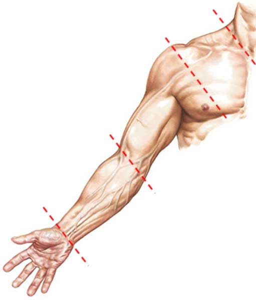
Photo A
We’ll begin the lesson with dem bones (shades of Joe Abernathy): The bony skeleton of the upper limb provides the foundation for its functions. Anatomy Lesson #2 and Anatomy Lesson #3 discussed three bones of the upper limb that are important again today: clavicle, scapula and humerus (Photo B). Each upper limb contains 32 bones but the remaining 29 bones will be postponed for later lessons.
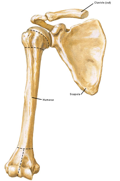
Photo B
First, the clavicle: As noted in previous lessons, human clavicles act as struts holding each upper limb away from the torso and allowing for increased range of motion. Viewed from the front (Photo B) the clavicles appear to be straight bones but this is not so. Viewed from above (Photo C – superior surface) each clavicle is S-shaped: The sternal half curves toward the viewer, the acromial half curves away from the viewer and the acromial end curves forward again to complete the S-shape.
Due to muscle pull, the clavicle curves more deeply in athletic individuals or manual laborers such as our hard-working farm-lad, JAMMF. The sternal end articulates with the manubrium at the sternoclavicular joint (Anatomy Lesson #15). The acromial end articulates with the acromion at the acromioclavicular joint (Anatomy Lesson #2). Both joints are typically visible in the lean and fit.
Try this: Demonstrate the S-shape by placing fingers on the sternum and following one clavicle towards the shoulder point. Near the sternum, the clavicle is subcutaneous, but nearer the side it becomes more difficult to palpate because it curves backward; at the acromioclavicular joint, it curves forward again.
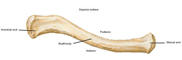
Photo C
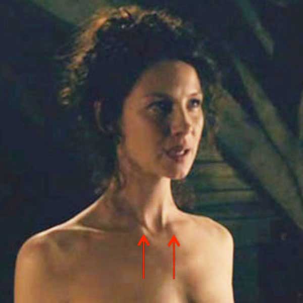
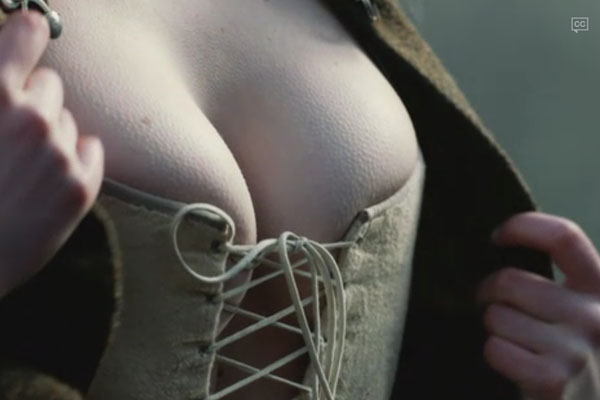
For our viewing pleasure, Claire presents us with a perfect pair of, erm, sternoclavicular joints (Starz episode 107, The Wedding)!
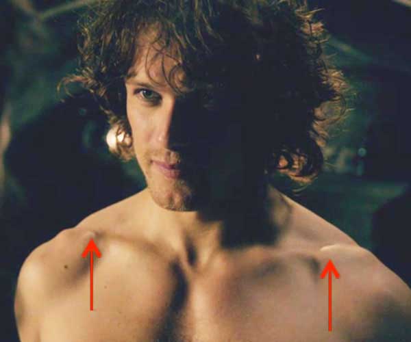
And, several students have asked if the knobs on Jamie upper chest are due to prior injuries (Starz episode 107, The Wedding). No, these are normal acromioclavicular joints.
The next bone is the weird-looking scapula which on side view resembles a ship’s propeller (Photo D). The red arrow marks the acromion or point-of-shoulder; the green arrow shows the pear-shaped glenoid cavity for articulation with the humeral head; the yellow arrow marks the inferior angle.
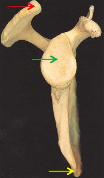
Photo D
Each scapula is large, thin and triangular in shape (Photo E – posterior view). The medial border lies parallel to the vertebrae. A large scapular spine arises near the medial border and ends as the acromion. The inferior angle lies at the 7th intercostal space and is used as a clinical landmark. The small glenoid cavity is at the side. Like puzzle pieces, acromion and clavicle meet at the acromioclavicular joint (green arrow) and the humeral head glides in the glenoid cavity at the glenohumeral joint (red arrow); strong ligaments and muscles stabilize these bony interactions.
Try this: Only a few scapular landmarks are readily palpable. Feel the subcutaneous acromion or point of shoulder. Cop a yoga pose or choose a partner and feel the long medial border and inferior angle. Follow the scapular spine which is mostly subcutaneous. You cannot palpate the glenoid cavity because it lies too deeply.
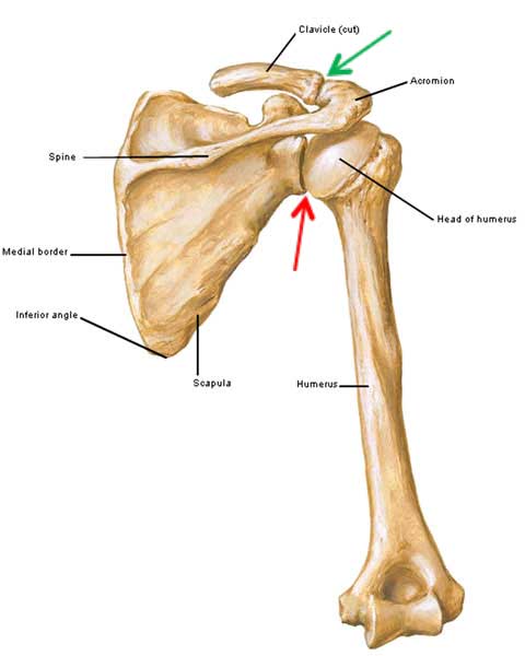
Photo E
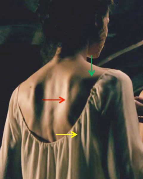
Clad only in her low-backed shift, trim Claire is our beautiful model for her right scapula (Starz episode 107, The wedding). The long medial border (red arrow) lies parallel to the vertebral spines, the scapular spine lies mostly horizontal (green arrow) and her shift covers the inferior angle (yellow arrows).
Together clavicles and scapulae form the pectoral girdle, an incomplete bony ring that supports both upper limbs. An aerial view (Photo F) shows the pectoral girdle sans skull and humeral bones (Can you identify the dens of C2 vertebra?). The sternum (yellow arrow) stabilizes both clavicles (blue arrows) at the sternoclavicular joints (green arrows). Scapular spines are marked by the red arrows. Clavicles articulate with acromia at the acromioclavicular joints (pink arrows).
Hopefully this visual clarifies how the clavicles serve as struts holding upper limbs away from the torso and how both upper limbs hang from the bony pectoral girdle. Lastly, force from a blow to either arm is transmitted along the clavicle to the sternum where it is absorbed by the bony thorax. It’s quite an ingenious and elegant design from which to hang our arms, ye ken?
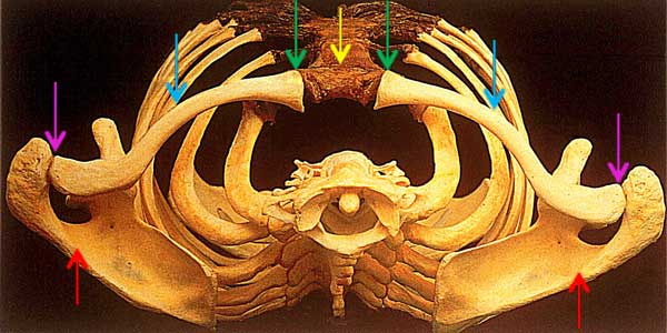
Photo F
Radiograms increase understanding so this x-ray (Photo G) shows a left pectoral girdle including clavicle (blue arrow) and scapular spine (red arrow). The yellow arrow marks the articulation between head of humerus and glenoid cavity of scapula at the glenohumeral joint. The humeral shaft is stretched to your right.
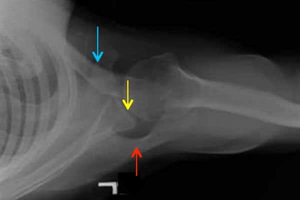
Photo G
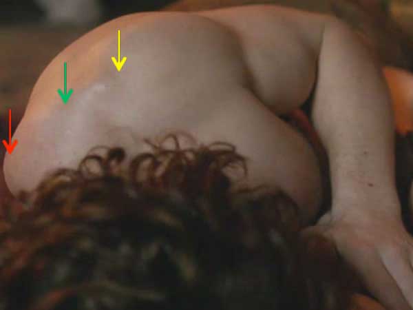
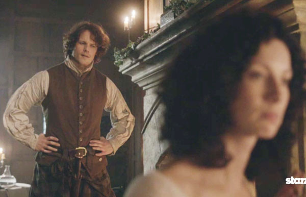
Despite his well-muscled physique, Jamie’s pectoral girdle (Starz episode 109, The Reckoning) can be appreciated from Claire’s perspective. The red arrow marks the medial upper border of scapula, yellow arrow points to acromioclavicular joint and green arrow marks scapular spine. We can be certain that while we might value this anatomical perspective we all know that Claire is most appreciative and well-deserving and all that…ahem and amen! Gah, what a sublime pectoral girdle and bonny hair!
Each arm contains only one bone, the humerus (Photo H); its large head glides within the small, shallow glenoid cavity at the glenohumeral joint (GHJ). A ball and socket joint, the GHJ is highly moveable but not very stable because of the size differential between head and socket although a cartilage labrum (lip) helps deepen the socket and ligaments and muscles help stabilize the joint. Just below the humeral head is an anatomical neck. The region marked surgical neck is noteworthy because it is the mostly likely site to sustain a fracture to the upper humerus. A bony elevation halfway down the humeral shaft is the deltoid tuberosity. The lower end of humerus helps form the elbow joint but it will be covered in a later lesson.
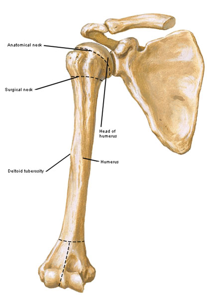
Photo H
Being a ball and socket joint, the glenohumeral joint is multi-axial meaning the humeral head moves with many degrees of freedom inside the glenoid cavity. Basic movements are shown in Photo I but other movements of the arm are combinations of these basics. Flexion draws the arm forward. Extension draws the arm backward. Abduction moves the arm away from the body midline. Adduction moves the arm toward the body midline. Lateral rotation turns the arm outward and away from the body center. Medial rotation turns the arm inward and toward the body center. Circumduction is a circular motion of the arm as in back stroke or baseball pitch, a movement that may be performed clockwise or counterclockwise. A bent elbow joint is not required to perform these motions.
Try this: execute each of these movements with one arm.
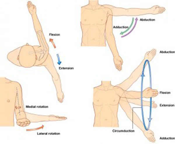
Photo I
Can we see these arm movements in the Starz Outlander episodes? Oh, to be sure!
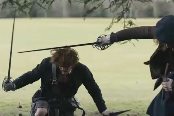
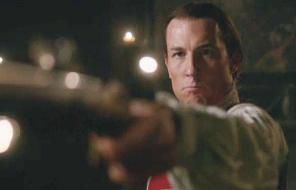
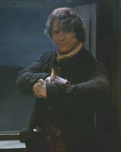
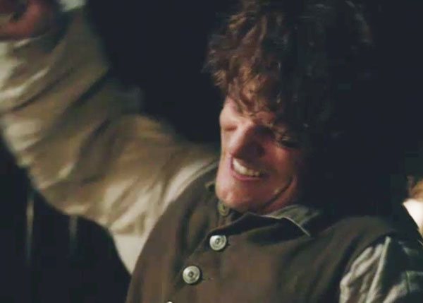
This is a wonderful example of a horrible man “up in arms” when things don’t go his way. BJR’s arm is in flexion as he fires an empty musket at Jamie. Surprise! (Starz episode 109, The Reckoning)!
And this is a great example of humeral extension at the start of the best make-up-sex-ever-filmed (Starz episode 109, The Reckoning)! Jamie canna wait to remove his sark. Both arms are draw backwards as he sheds that garment…off ye damn shirt!
Try this: extend both arms behind your back. Most people can extend the arms to a 45° angle. If ye are flexible, extend arms behind your back and interlock fingers. Gently lift your arms more than 45° but not if it hurts. Good job!
Handsome Dougal is next (Starz episode 109, The Reckoning). Yes! An army of leddies have been clamoring for more of our bold Highland war chief! Both arms are drawn against his sides in full humeral adduction as he awaits Colum’s tongue-lashing. I love this scene! With little more than a sideward glance, a twitch of the ‘stache and some brow crunch, Dougal conveys fierce frustration with his meddling Laird-bro.

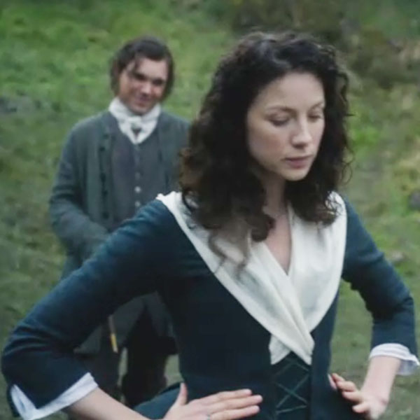
Next, Claire offers a fine example of humeral abduction with arms akimbo awaiting Professor Angus’ stabbing lesson (Starz episode 108, Both Sides Now). Two arms are drawn away from the body midline although bent elbows aren’t required for this action. Doesna that gown look fantastic on her trim torso?
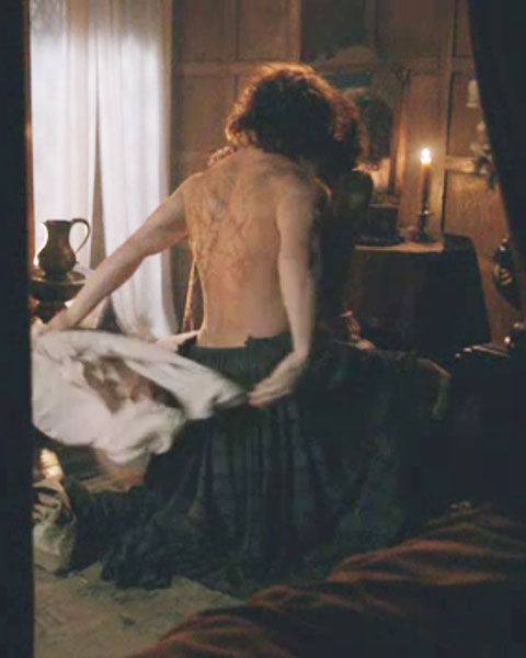
Now, I dinna want to rekindle a fire storm with this next image, but Laoghaire offers (weel, it’s not all she offers) an excellent example of lateral rotation of the arms (Starz episode 109, The Reckoning). Here she sheds her cape while aiming her 16 year-old charms at new hubby Jamie. Two arms are turned outward and away from the body center. She knows this position gives her better advantage to show off her, um, well to show off! Geez, where did she find that corset with the wee side vents? Mayhap she raided Granny Fitz’s hidden trunks where Claire’s awesome closet resides (kudos to Terry & Co on another unique costume that tells a story all its own)!

Here are contracted arrector pili muscles. Mmmphm, must be a tad chilly by the burn! Oops, wrong lesson! Goose flesh belongs wit’ the skin in Anatomy Lesson #6. But, if ye ken yer anatomy verses, ye can pretty much identify anything! Moving on….
I always laugh at this image of our elegant Claire caught in a not-so-elegant squat (Starz episode 105, Rent). As she hikes up her skirts to pee in the pot, her right arm is turned inward and towards the body center as in medial rotation. Geez, Angus you buamastair (oaf), knock next time! Who knew wild women waulked wool?
Finally, Dougal’s guilty rampage over the death of his not-so-bonny wife Maura presents several outstanding displays of humeral circumduction (Starz episode 110, By the Pricking of My Thumbs). The image below shows counterclockwise circumduction (as in pitching baseball). Watch the entire scene again to see his arm movements; they are fantastic! Dougal is a sad Shakespearean mess in this scene but his acting chops are top shelf. Good thing Nurse Claire ministers one of her Spanish potions before he slices and dices everyone and everything in the hall brawl!

Now that we ken arm movements, let’s discuss arm muscles: a whopping 17 muscles per side move the shoulder and arm. Eight of these muscles move the pectoral girdle which indirectly alters humeral position. Nine muscles directly move the humerus. Any wonder humans enjoy such versatility in shoulder and arm movement? There is no way 17 muscles can be covered in a single lesson so today we will review trapezius, latissimus dorsi and pectoralis major and learn a new muscle, the deltoid.
Trapezius was discussed in Anatomy Lesson #2 and Anatomy Lesson #3 (Photo J). This muscle acts directly on the shoulder joint and indirectly on the humerus. Arising from skull and vertebrae, it inserts on scapular spine, acromion and clavicle. Upper fibers lift the shoulder joint dragging the humerus along for the ride; middle fibers retract (pull back) shoulder joint and humerus and lower fibers depress (pull down) shoulder joint and humerus. The bottom line: more shoulder movements = more arm movements. Trapezius creates the web between base of skull and acromion and is most prominent in the muscular.
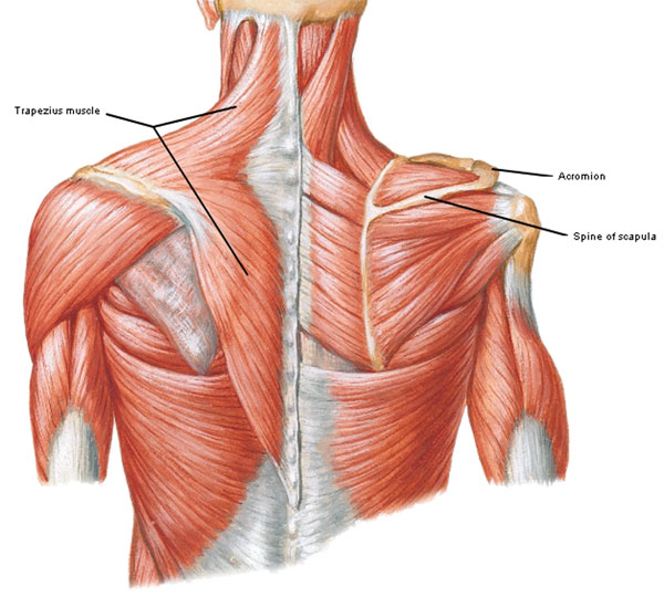
Photo J
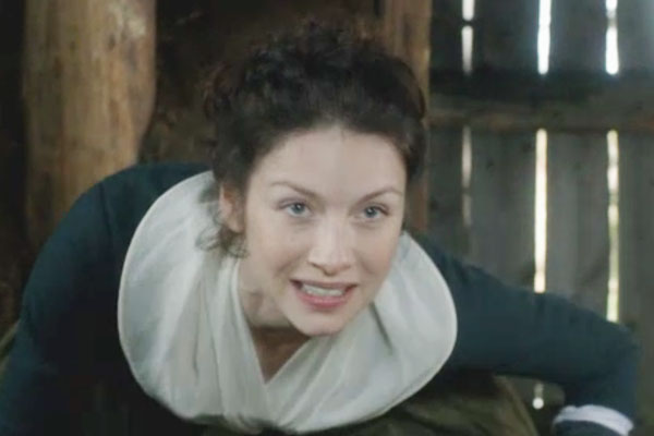
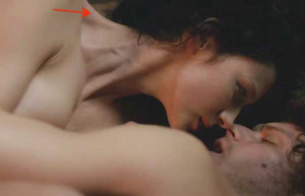
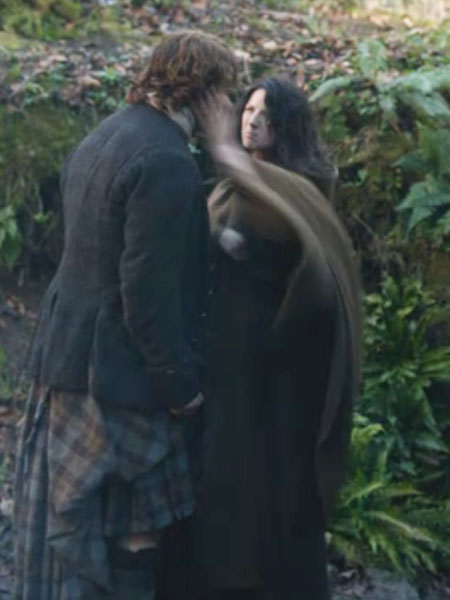
Here’s a delightful example of Claire’s beautiful trapezius as she reaches for Jamie’s mouth (Starz episode 109, The Reckoning). Jamie was out to master Claire but discovers there’s a price to pay. Sorry laddie, every coin has its flip side. Herself’s magic pen strikes again (Outlander book):
“Oh, aye, Sassenach,” he answered a bit ruefully. “I am your master … and you’re mine. Seems I canna possess your soul without losing my own.”
The next review muscle is latissimus dorsi (Anatomy Lesson #10). These large flat fan-shaped muscles arise from vertebrae, sacrum and hip bones via the thoracolumbar fascia. Fibers wrap around the body, converge and insert into each humerus (Photo K). Latissimus is the only human muscle connecting upper and lower limbs, making it a potential powerhouse for lifting body weight!
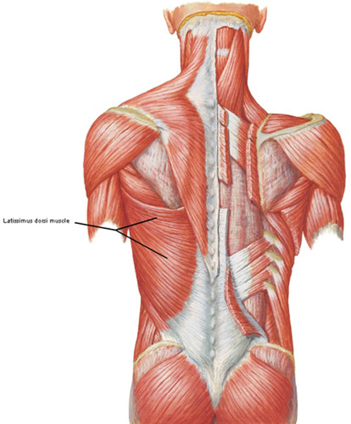
Photo K
As latissimus dorsi nears the armpit (axilla), it turns tendinous before inserting on the humerus (Photo L – front view). It extends, adducts and medially rotates the arm. Latissimi dorsi (pl.) are the pull up (palms face forward) and chin up (palms face backward) muscles.
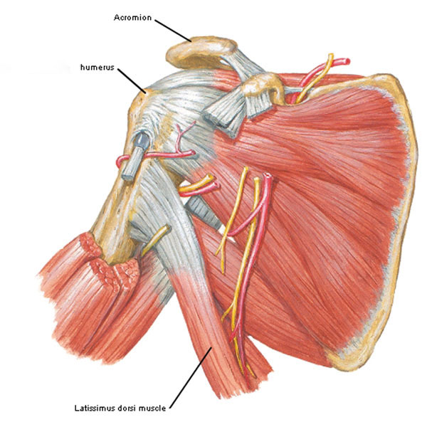
Photo L
Jamie is our poster boy for latissimus dorsi (Starz episode 106, The Garrison Commander). Here his left muscle clearly sweeps from the lumbar spine, around the body and upward to end on the humerus. Captain Randall is our poster boy for the maddest-baddest villain in literary and TV history!
The third review muscle is pectoralis major (Photo M). It has two heads: one from the clavicle and a second from sternum and ribs (Anatomy Lesson #4). A wee slip comes off abdominal structures but anatomists often ignore it. Muscle fibers converge as they approach the humerus where they insert near latissimus dorsi. Pec major flexes, adducts and medially rotates each arm.
Oh, and many students ask the name of the large vein traversing the arm and disappearing between deltoid and pectoralis major muscles. This is the cephalic vein, so named because it carries blood in a headward direction. And, the groove it runs in between deltoid and pectoralis major is the deltopectoral triangle.
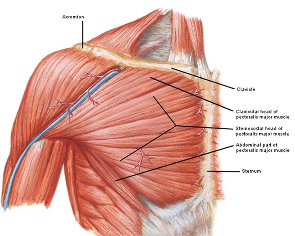
Photo M
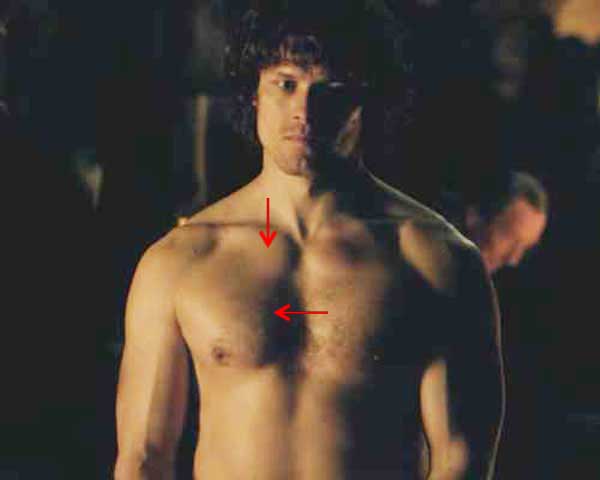
Half-clad Jamie is our model yet again (Starz, episode 105, Rent). Well-developed pec majors are clearly visible under the skin. Red arrows mark clavicular and sternocostal heads. Puir lad, he dinna get no Dougal R-E-S-P-E-C-T!
The fourth and last muscle is the deltoid, named because it resembles an inverted capital delta Δ, 4th letter of the Greek alphabet. Viewed from the side, the shape is clear: broad at the top narrowing to an apex at the bottom. Each deltoid arises from clavicle, acromion and scapular spine (Photo N) and inserts on the deltoid tuberosity of humerus (Photo H). Middle fibers contract to abduct the arm; front fibers flex and back fibers extend the arm.
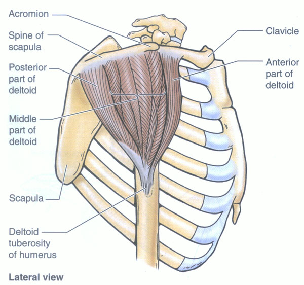
Photo N
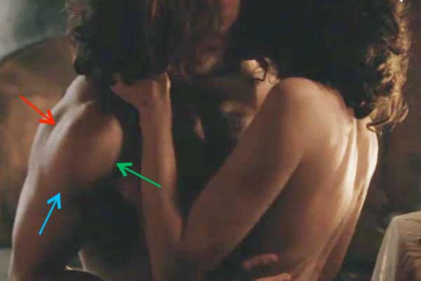
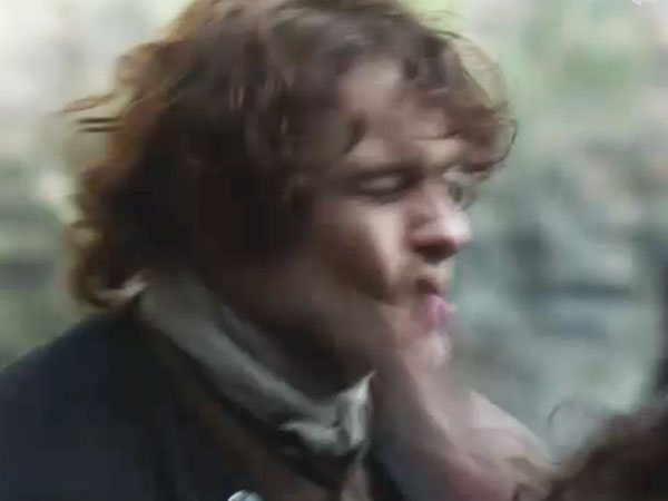
The next image shows Jamie’s fantastic right deltoid when he and Claire are still vertical (Starz episode 109, The Reckoning). The posterior border is marked by a red arrow, anterior border by a green arrow and insertion (hehe) on the deltoid tuberosity (hehe) by a blue arrow (canna help it, anatomists have a ribald sense of humor).
Okey-dokey, we are done with the arm muscles. For fun, let’s examine images from Starz episode 109, The Reckoning, for more arm movements. See if you can you identify the arm positions – some are combos.
1: Name the position of Jamie’s right arm and of his left arm.
Answer: Jamie’s right arm is flexed and his left arm is abducted. Get the idea? I knew ye would like it – this is gobs of fun!
2: Next, for our viewing pleasure, BJR has just taken an assisted face plant on the desktop and is out for the count: 10-9-8-7-6 …… name the position of his left arm.
Answer: flexion
Let’s take a keek-peek at our first couple’s first fight! Whew, as much passion here as on the mattress, um, floor! Claire’s aboot to knock some brains into that hard-as-an-iron-pot highland skull!
3: Name the movements of Claire’s left arm (there are two).
Answer: Abduction and flexion.
No question here, but I call this “the slap heard round the world!” Ouch Jamie, she wallops you in front of yer three best bros? Whew, they are going at it “hammer and tongs.”
In the next image, Jamie’s right arm is lifted up, up and away from the body midline (his button line). Aye, he is blistering Claire’s bum with his doubled belt. Weel, Claire, ye did put the entire party in peril and you promised Jamie you would be at the grove when he returned. Och, dinna make promises ye canna or willna keep. Herself explains Claire’s view of the spanking (Outlander book):
I felt deeply betrayed that the man I depended on as friend, protector, and lover intended to do such a thing to me. And my sense of self- preservation was quietly terrified at the thought of submitting myself to the mercies of someone who handled a fifteen- pound claymore as though it were a flywhisk.
4: What movement is performed by Jamie’s right arm?
Answer: vertical abduction (as in “Reach for the sky, partner!”). The downward smack is adduction.
Here, Jamie tap dances for his beloved (mayhap he enjoyed that abduction/adduction a bit too much?). Lad, if ye ever want to do the horizontal boogie with Claire again ye better buckle on those show-biz shoes and get on wi’ it! You’ve done considerable damage spanking yer new wife. Herself explains (Outlander book):
“Whatever the justice of the situation—and I had to admit that at least some of it lay on his side—my sense of amour-propre was deeply offended….”
Ohhhh, she is royally pissed – in such a cold fury she brushes that same curl at least 200 strokes and we all ken that 100 does the trick!
5: Name the position of Jamie’s arms.
Answer: the begging position…Hah. No, both arms are abducted.
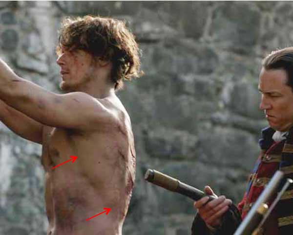
Good job with identifying arm movements! Before this lecture ends, I canna help but offer a few wee anatomical observations from Starz episode 109. We ken well that Jamie was shot through his right trapezius so I was happy-happy to see scars! First we see the exit wound (blue arrow)….
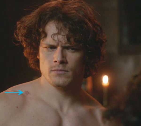
and we also see the entrance wound (red arrow)! Yessss, the special effects folks remembered that Jamie’s gunshot wound must have two scars!
Whaaat? – Entrance wound in back and exit wound in front? Well yes, Herself being the final arbitrator of such supreme-court issues (from Outlander book)!
No, when we were running from the English, I realized we were near the edge of the Fraser lands, and I thought I’d take my chances there. So I spurred up and cut to the left, around Dougal and the rest. There was a good deal of shooting goin’ on, mind ye, but the ball that hit me came from behind. Dougal, Rupert, and Murtagh were back of me then. And the English were all in front—.“
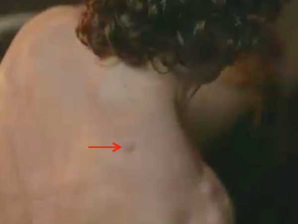
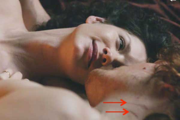
In the afterglow of bliss-land, our newly-wed redhead looks a bit worse for wear after Claire extracted her own measure of justice! We book readers expect cheek scratches but did ye ken his neck gouges (red arrows)? Herself writes (Outlander book):
It had been a most unpleasant night. My reluctant acquiescence had lasted precisely as far as the first searing crack of leather on flesh. This was followed by a short, violent struggle, which left Jamie with a bloody nose, three lovely gouges down one cheek, and a deeply bitten wrist.
Weel, if ye bed a vixen … Come here vixen and bite me again!
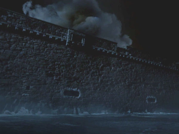
To arms! To arms! While not anatomy-related, did ye see the mushroom-shaped cloud over Fort William – proof that toxic waste in the form of BJR resides inside that stony edifice!
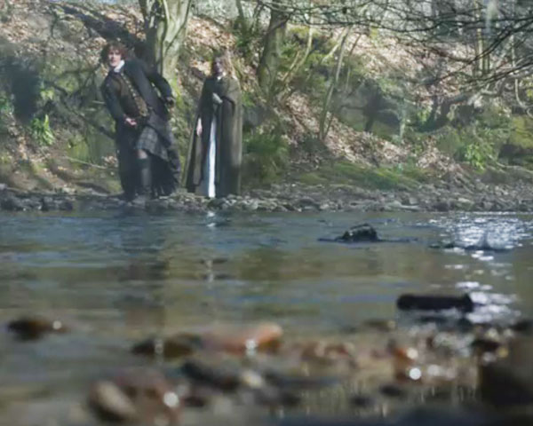
This last image is verra funny. Jamie’s right arm is in fine fettle as he sends many rocks artfully skipping across the burn. But as soon as Laoghaire sidles up, his verra next toss goes kerplunk! The stone drops like, well, like a rock. Let that rock-flop be a warning to ye laddie; she is NO a good girl … she’s armed, dangerous and on a stealth mission!
To Arms! Too Arms! Two Arms! That’s it for the bones, muscles and movements of the arms. Raise yours in grateful joy! Aren’t they grand?!
A deeply grateful,
Outlander Anatomist
Photo creds: Starz, Gray’s Anatomy, 39th ed., Netter’s Atlas of Human Anatomy, 4th ed., Clinically Oriented Anatomy, 5th ed., Hollingshead’s Textbook of Anatomy, 5th ed., www.bestperformancegroup.com, www.ceessentials.net/article31.html, www.wikipedia.com