Anatomy def: narrow band of muscle fibers arising from the fascia over each masseter muscle, inserting into tissues at the corners of the mouth, and acting to retract the angles of the mouth.
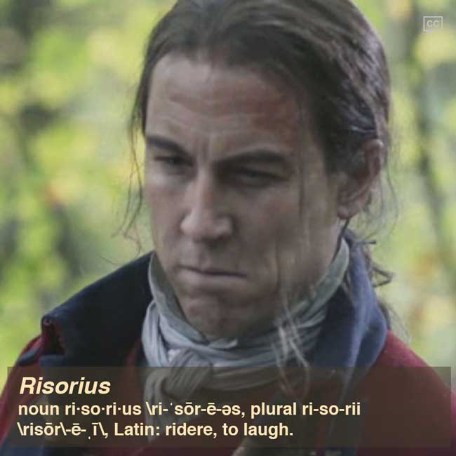
Outlander def: paired, strong muscles pulling back the corners of the mouth as in a grin, or a grimace to menace wary victims!
Learn about the risorius muscle in Anatomy Lesson #13: “Frank and BJR” or “Face Off.”
Read about BRJ’s face in Outlander book:
Whatever this man’s cousin looked like, the man himself might have been Frank’s brother. There was the same lithe, spare build and fine-drawn bones; the same chiseled lines of the face; the level brows and wide hazel eyes; and the same dark hair, curved smooth across the brow. But this man’s hair was long, tied back from his face with a leather thong. And the gypsy skin showed the deep-baked tan of months, no, years, of exposure to the weather, not the light golden color Frank’s had attained during our Scottish holiday.
See BJR’s risorii muscles in action as he glowers at Claire (Starz episode 101, Sassenach)!
Risorii muscles are likely the bases for the vertical skin creases commented upon in a recent interview with Tobias Menzies.
And, for the truly dedicated, the December 2015 issue of Smithsonian showcases an interesting article “Face to Face” (pp. 46-51) about software being developed to decode facial expressions and change the way we interact with our devices and each other. No more emoticons? Gasp!
A deeply grateful,
Outlander Anatomist