Anatomy Lesson #29: The Eyes Have It!

ep 109 Dougal eyebrows 01 KLS editedAn apocryphal story about Abraham Lincoln, 16th US President, claims he was taking a vote during a cabinet meeting on whether or not to sign the Emancipation Proclamation. All of his cabinet secretaries voted nay, whereupon Lincoln raised his right hand and declared: “The ayes have it!” 🤗

Today, the “eyes have it” as we begin a discussion which, due to complexity, will span more than one anatomy lesson! So, welcome all students to the first in a series of eye lessons: Anatomy Lesson #29, The Eyes Have It!

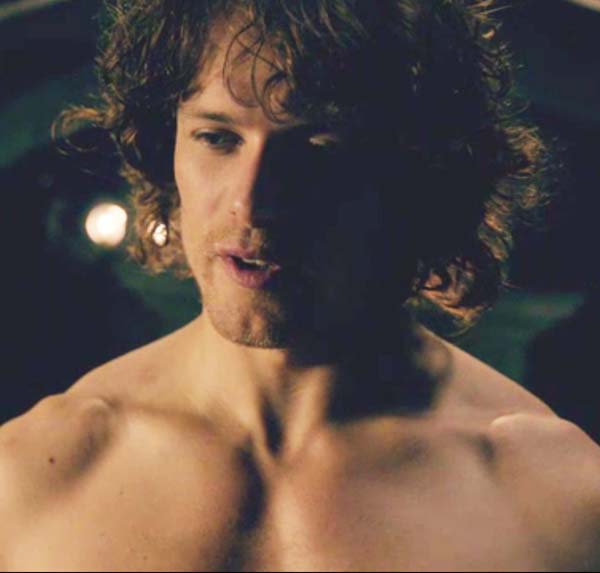
The eye is arguably the most elegant and complex sense organ of the human body. Its duty is to provide vision thereby increasing our perception of both inner and outer worlds (Photo A). John Ruskin (1819 – 1920), a leading English art critic of the Victorian era, put vision into clear perspective: “The greatest thing a human soul ever does in this world is to see… To see clearly is poetry, prophecy and religion, all in one.”

Eye KLS edited

photo A

The eyeball is marvelous but it doesn’t work in a vacuum. Surely, you’ve read about celebrities who are accompanied by a full entourage of assistants? Well, the eyeball has a similar assemblage. For the eyeball to work optimally, it requires help from a small host of accessory structures: eyebrows, eyelids, eyelashes, bony orbit, periorbital fat, lacrimal apparatus and extrinsic muscles. Today, we will learn how eyelids, eyebrows and eyelashes augment vision.

We’ll start today’s anatomy lesson with the eyebrows, a major human facial feature. In anatomy, the eyebrow is known as the supercilium (pl. supercilia). Eyebrows are linear growths of coarse hair that protect the eyeballs from the sun’s rays as well as sweat, rain, dandruff and other debris. The Greek physician Herophilos (335-280 B.C.) first suggested that the brows are “adorned with hair, so that if copious perspiration came, it would be contained by this ‘check-point’ of hair placed in its way until it is wiped off, so that it could not obstruct the eyes.” The eyebrow arch favors this explanation as it typically peaks over the eye and slants to each side ensuring that moisture wicks around the eyeball and towards temple or nose.

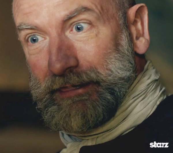
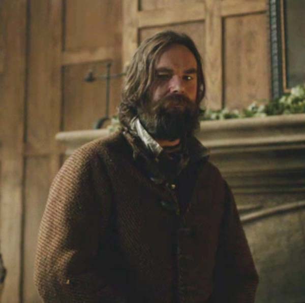
Delicious Dougal-Dude exhibits a fine example of manly arched brows; brow power to the max as he reminds Colum that he is dad-of-the-lad, Hamish. Colum should be grateful; after all, Dougal was showing fealty by lying wi’ his laird’s lovely Lady Letitia (Starz episode 109, The Reckoning).

ep 109 Dougal eyebrows 01 KLS edited

Besides protecting the eyeballs, eyebrows are vital in human communication by signaling mood changes such as anger, surprise, concern and even sexiness. Jamie’s eyebrows are furiously flapping in this Starz scene (episode 109, The Reckoning). Och! Weel, Claire’s dark dirk dare is agonizing for Jamie – runs counter to the man’s natural instincts. Ken the distress telegraphed by his eyebrows? At this point, yes, yes, lassie! Anything!

ep 109 Jamie brows KLS edited

Eyebrows also add major definition to the face. In a recent MIT study, subjects were asked to identify celebrities whose eyes or eyebrows had been digitally erased. Surprise! Subjects could correctly identify 60% of celebrities if eyes were blocked but only 46% if eyebrows were erased suggesting that eyebrows are more significant than eyes in identifying faces.

We humans like to mess with our anatomy and eyebrows are no exception: tattooing, waxing, plucking, brushing, coloring, trimming, stenciling, darkening, shaving, piercing, tweezing and threading (Photo B). And, of course, cosmetic surgery can lift eyebrows or Botox can be used to reduce skin lines near the brows to create a more youthful look.
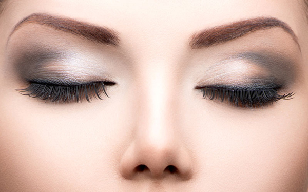
Beauty eyes makeup closeup. Long eyelashes, perfect skin

photo B

Time for an eyebrow quote from Outlander book:

“Because I want to look at you,” I said. He was beautifully made, with long graceful bones and flat muscles that flowed smoothly from the curves of chest and shoulder to the slight concavities of belly and thigh. He raised his eyebrows. “Well then, fair’s fair. Take off yours”…

Here ye go. Breathe! Panting is OK, but no fainting (Starz episode 107, The Wedding)!

ep 107 Jamie eyebrows KLS edited

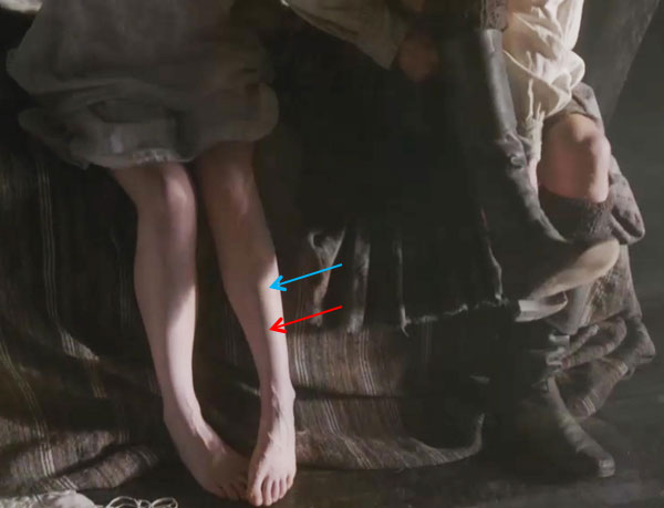
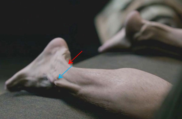
Next up, anatomy of the palpebrae (pl.) or eyelids. Eyelids (Anatomy Lesson #11) are moveable flaps of skin that overlie the eyeballs. Upper and lower lids join on the nasal side at the medial canthus (Photo C – red arrow); the lateral canthus marks their fusion at the temporal side (Photo C – blue arrow). The elliptical opening between the eyelids is the palpebral fissure.

The upper lid is larger and more mobile than the lower and each lid covers the eyeball differently. With the eyelids open, the upper lid normally overlaps the upper cornea (and deeper-lying iris) but the lower lid sits just below the cornea (Photo C). With the eyelids closed, the upper lid moves down to cover the entire cornea. Note: if the lids of an eye at rest are open widely such that the entire cornea (and iris) is exposed, then one should consider being evaluated for possible causes (Grave’s disease, orbital cellulitis, exophthalmos, to name a few).

Try this: Open your eyelids and gaze into a mirror. Find medial and lateral canthi (pl.). Where are your eyelids relative to the iris (the iris is the best landmark because the transparent cornea is difficult to identify in a forward gaze)? Close the eyelids and gently probe the corneal bulge and realize that in this position, the upper lid completely covers the cornea.
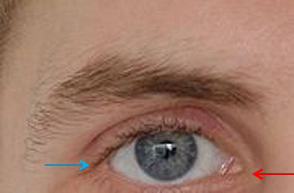
photo-C-edited

photo C

Except for parts of the external genitalia, eyelid skin is the thinnest of the entire body. Each lid contains a flexible connective tissue plate: the superior tarsus of the upper lid and inferior tarsus of the lower lid. Tarsi (pl.) add body and shape to the eyelids allowing them to cup the eyeball surface (Photo D – vertical section through eyelids and eyeball). Eyelids close to protect the eyeball from trauma, debris, and excessive light; they also help create the tear film and spread it across the eyeball surface.

Try this: Grip your upper lid between thumb and index fingers and gently squeeze. Feel the superior tarsal plate pop between your fingers? Now repeat with the lower lid and realize that the inferior tarsal plate is much smaller than the superior version.
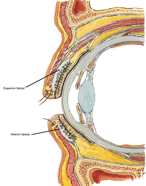
Figure0077B tarsus KLS edited

photo D

Eyelids are well equipped with sebaceous glands to keep the skin supple and sweat glands to cool the skin and eliminate wastes (Anatomy Lesson #5). But, the inner surfaces of superior and inferior tarsi house unique tarsal (meibomian) glands, specialized glands that release meibum, an oily substance that seals the eyelids when closed and also traps the tear film so it won’t spill over the eyelids (Photo E).

Try this: Look in a mirror and gently pull down your lower eyelid to evert and expose the inner surface (the upper lid is harder to evert). Locate the pale yellow lines arranged perpendicular to the lid margin – these are your tarsal glands!
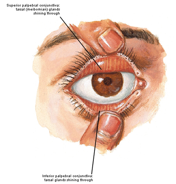
Figure0077A KLS edited

photo E

Each set of eyelids (Anatomy Lesson #13) is moved by three muscles: orbicularis oculi, levator palpebrae superioris and superior tarsal muscle of MĂźeller.

Orbicularis oculi muscle (OO) has two parts: an orbital part consists of large muscle loops that overlie bones of the eye socket (Photo F – labelled O); a palpebral part is confined to upper and lower eyelids (Photo F – labelled P). Both orbital and palpebral parts attach to the medial palpebral ligament, a tough band of connective tissue at the medial canthus of each eye (Photo F – red arrow).
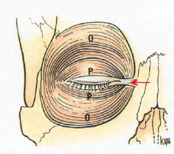
photo-F-edited

photo F

The orbital part of OO is under voluntary control meaning we can contract the muscle at will; this action squeezes the flesh surrounding the eyelids.

Jamie presents an agonizing example from Starz episode 116, To Ransom a Man’s Soul: his eyelids are slightly open but the skin around them is puckered because the orbital part of OO is contracted. He has confused his beloved Claire with the awful yuk-man! Pax, Jamie, Claire has ye in her healing hands!

ep 116 Jamie Orbicularis oculi KLS edited

The palpebral part of OO contracts voluntarily to close the eyelids in a conscious blink. Volition is especially evident when we wink – voluntarily contracting the palpebral part of one eyelid.

Do you know how often we blink? Although we can voluntarily contract the palpebral part of OO, it is routinely under unconscious control meaning the brain signals the muscles to contract about 15 times every waking minute. This automated blinking cleanses, renews and redistributes the tear film. Also, if an object flies toward the eyeball, the palpebral part reflexively contracts and closes the lid to protect the cornea. This unconscious response relieves our conscious brain of instructing the palpebral muscles to contract every four seconds while we are awake (see the gif below in slow-mo).

Try this: Open your eyelids. Now contract the flesh around the lids, an action accomplished by the orbital part of OO. Now, relax the flesh around the eyelids and then deliberately blink both eyes or wink one; contraction of the palpebral part of OO causes this action. Next, close the lids of one eye and place a fingertip in the medial canthus. Move the finger gently up and down. Feel the tough ridge of tissue? This is the medial palpebral ligament which provides an attachment site for OO.

220px-Eye_opening_and_closing

The second set of eyelid muscles is levator palpebrae superioris (LPS). Each of these strap-like muscles originates deep in the eye socket and passes forward to insert on the superior tarsal plate of the same side (Photo G). LPS is under voluntary control; its contraction lifts the upper eyelid so light can enter the eyeball. Lacking comparable muscles, the lower eyelids glide open by gravity.

Finally, the third muscle acting on each eyelid is the superior tarsal muscle of Müeller; this small muscle spans from the underbelly of LPS to the superior tarsus (Photo G). Made of a different type of muscle (the body has three types – the rule of three in anatomy!), it can never be moved voluntarily and thus is always under autonomic control. Surprise, shock, pain, loud noises or scary events cause the superior tarsal muscle to contract swiftly and suddenly and the eyelids fly open. This is considered a survival mechanism allowing more light to enter the eyeball so we can quickly identify potential threats.
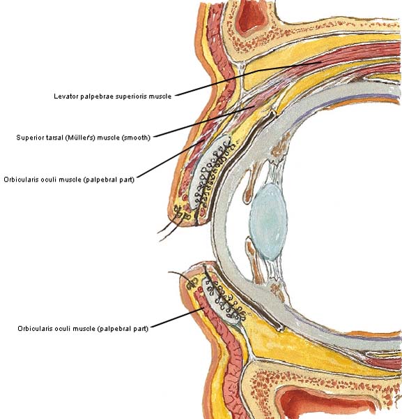
Figure0077B eyelid muscles KLS edited

photo G

Do we see Müeller’s muscle at work in Starz episodes? Oh, aye. Here are three good examples!

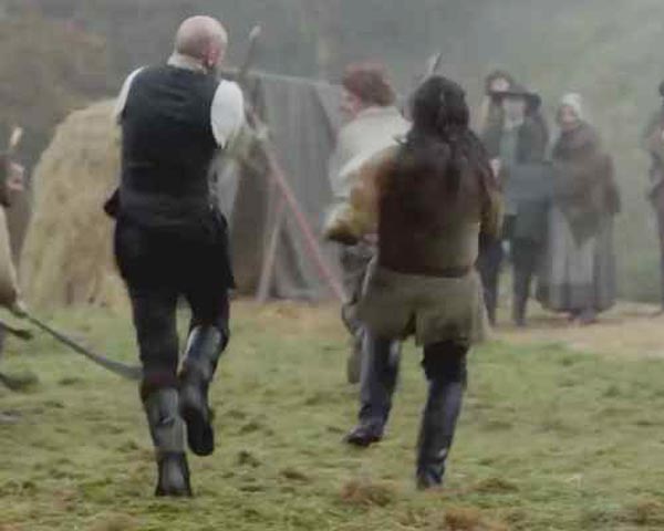
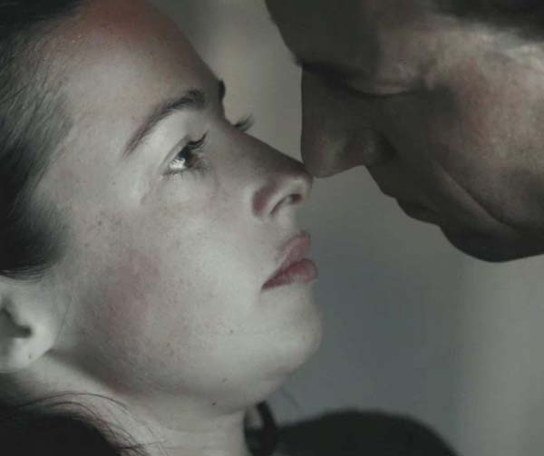
SURPRISE! Across a time barrier of two centuries, Jamie hears Frank shout “my wife is NOT with another man!” Sorry Frank, but she is! Naw… Jamie’s upper eyelids fly open because Hugh Munro’s arrow interrupted some serious hand sex (Starz episode 108, Both Sides Now). Bad timing Hugh – no more gaberlunzies for you!

ep 108 Jamie eyelids KLS edited

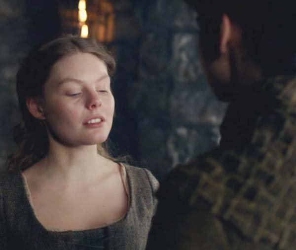
SHOCK! Jamie’s upper lids flash open; he is shocked, shocked I say, as he kens that bears aren’t the only creatures that poop in the woods! Wee Willie says he was busy taking a piss (yeah, right) when the red coats captured Claire. She was thrashing about and she’s gonna get thrashed again after Jamie gets her (Starz episode 109, The Reckoning)!
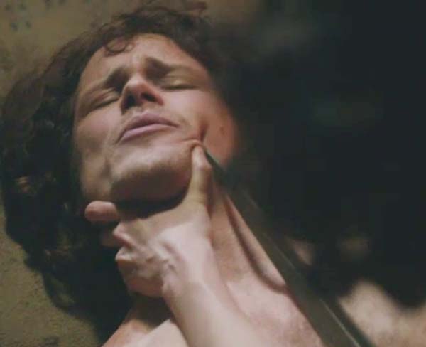
ep 109 Jamie Muller muscle KLS edited

PAIN! MĂźeller’s muscle quickly activates as Claire silently embroiders her initials into her handsome hubby’s side. Jamie, she warned ye to stay away from that damned double-dealing Duke! Och, aren’t nurses supposed to be gentle (Starz episode 110, By the Pricking of My Thumbs)?

ep 110 Jamie Muller muscle KLS edited

Next, let’s view some fun facts about eyelashes. Also known as cilia, eyelashes are short, thick hairs that grow in double or triple rows along the rims of our eyelids (Photo H). Eyelashes naturally curl: upper lashes curve upward and lower lashes curve downward; this design keeps the hairs from interlacing when the lids close. Also, please don’t pull out eyelashes; lashes take 7-8 weeks to regrow but constant pulling can lead to permanent loss. Natural eyelash color may differ from head hair although dark-haired individuals tend to have darker eyelashes than light-haired folks. Eyelashes help capture debris headed for the eyeball surface. Similar to cat whiskers, they are sensitive to touch and provide warning if an object approaches the eyeball so the lids can reflexively close.
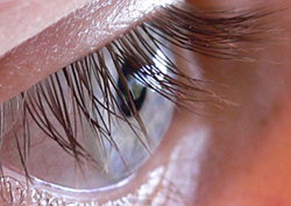
250px-N2_Human_eye KLS edited

photo H

Many cultures consider long eyelashes a mark of beauty although there is one (Hazda of Tanzania) wherein women trim their eyelashes! Eyelash length is enhanced with mascara, extensions or false eyelashes although enhancing is nothing new. As far back as the Bronze Age kohl was used as eyeliner and lash enhancer and remains the eye cosmetic of choice in some parts of the world. In the west, mascara, eyeliner, eyeshadow, eye putty and tints are used to color the lashes or their bases (Photo I). Also, a mess of tools are used to alter eyelashes such as curlers, applicators and shields. Eyelash transplants are available although it uses head hair so if you go that route,  plan on regular haircuts! Finally, there is also a glaucoma-treatment drug with a side effect of eyelash growth. Whew!

panda bears KLS edited

Photo I

And as if this isn’t enough fussing with our lashes, eyelash jewelry is now in vogue (Photo J)!
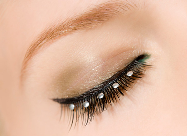
eyelash-jewelry_72423193 KLS edited

Photo J

Eyelashes are fraught with many problems: infection with parasitic crab lice; ingrown lashes; loss of lashes; abnormal grown of lashes on other parts of the eyelid; itching, redness and flakiness; and styes/stys. And, did you know that 98% of humans harbor a harmless commensal mite (Demodex folliculorum) in the follicles of our eyelashes and eyebrows? In the US, the National Center for Biotechnology Information (NCBI – PubMed/NIH) posts numerous concerns about dreadful reactions to glues used in eyelash extensions and other cosmetic eye issues. Wherever you live, please get informed before enhancing the eye or its accessory parts with anything new or weird.

Clinical Correlation: Two major types of sores plague the eyelashes. One type is the stye/sty, the blockage of a sweat gland at the base of the eyelashes that produces a hordeolum, a painful, red, swollen bump (Photo K). A chalazion develops if a tarsal gland becomes blocked. Initially both types of sores are red, swollen and painful. However, over time, the chalazion becomes hard and non-tender whereas the sty/hordeolum continues to sting like a skelping!
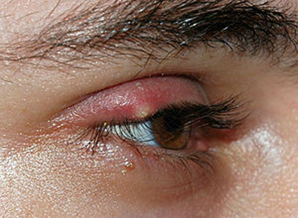
Stye02 KLSedited

Photo K

Let’s finish this lesson with Outlander images and book quotes about eyelashes and eyelids. Want to see a lovely lassie with long lush lashes? Yep, its wee Jenny staring into shark eyes after BJR sticks his bloodied finger into her mouth (Starz episode 112, Lallybroch). Jamie says his sis has great eyelashes (Outlander book):

—she’s got blue eyes, like mine, but prettier, wi’ black lashes all around.

Maybe so; maybe faux? They look real to me!

ep 112 Jenny eyelashes KLS edited

Shortly after their marriage, Claire muses that Jamie has unusual eyelashes (Outlander book).

… His lashes were long… Oddly colored, though; dark auburn at the tips, they were very light, almost blond at the roots.

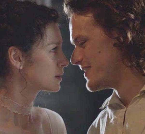
Claire gits up close and personal as she gazes at Jamie’s long, thick ginger lashes (Starz episode 107, The Wedding). She just interrupted his kiss and he can still smile? What a lad!

ep 107 Jamie eyelashes 01 KLS edited

Of course, Outlander book readers ken that our Hero Jamie canna wink (urging non-readers to get with the remedial reading program. Wink, wink!):

He blinked at me like a large red owl—some congenital tic made him incapable of closing one eye in a wink—and I laughed.

Now, Jamie’s not quite blinking like a big old red owl in the image below but the palpebral parts of OO are contracted (Starz episode 107, The Wedding). Seems he is totally bummed because he thinks that Claire didna like IT! What the hey is IT? Ha ha! Seems Murtagh, Rupert and Ned gave Jamie a load of shite about women: they told him women didna like IT! But, they were dead wrong about not-a-wet-nurse Claire. She does like IT!

ep 107 Jamie eyelids 02 KLS edited

Even cray-cray LegHaire, that lying little lass, contracts the palpebral part of OO as she lowers her upper lids. She is sooo sad for her puir Jamie – sob – trapped in a loveless marriage with a cold English bitch (Starz, episode 110, By the Pricking of My Thumbs)! Her lad must get swine drunk before he can stand to plow Claire’s field. SMACK! Seems LungHaire’s cheek just received a bit ‘o Claire’s “healing touch.” Oh, and she’s dead wrong about Mistress Claire; she does like IT! Shouting now!

ep 110 Laoghaires eyelids KLS edited

Murtagh’s Morning Mistake: he interrupts Jamie’s breakfast (Starz episode 110, By the Pricking of My Thumbs). Snort! Godfather, your eyelids are open now (levator palpebrae superioris is contracted) and ye look a wee bit sheepish. That’s right…baaaad timing! Baaaa!

ep 110 Murtagh KLS edited

Here’s proof of Murtagh’s uh, oh (Starz episode 110, By the Pricking of My Thumbs): with both eyelids closed (palpebral parts of OO contracted and covering her corneas), Claire is basking in the afterglow of “the gift that keeps on giving” – Ira’s words, not mine. Yep, she likes IT!

ep 110 Claire eyelids KLS edited

So students, Outlander proves that the eyes have it: lashes, lids and brows working together for the greater good!

All together now:

Sing me a song of gal who’s not gone,

Say could that lass be Claire?

Some are dead wrong as she loves her man strong,

Challenge her will if ye dare!

Jamie’s her man,

She belongs to his clan,

She’s part of his blood and bone!

Her eyes are for him; she’ll risk life and limb

‘till their life on earth shall be done!

Oh, sing me a song…


A deeply grateful,

Outlander Anatomist


photo creds: Netter’s Atlas of Human Anatomy, 4th ed. (Photos D, E, G); Clinically Oriented Anatomy, 5th ed. (image of orbicularis oculi); Starz; www.consumerreports.org (image of eyelash jewelry); www.giffy.com (blinking eyes); www.quotesgram.com (Panda bears); www.rakis.com.au (image of eyebrows); www.web.md.com (image of stye); www.en.wikipedia.org (images of man’s eyebrow; sty; eyelashes; gif of blink)

Colum’s Legs and Other Things, Too!

Thanks for running to class today, Outlander anatomy students! Today’s Anatomy Lesson #27 is on the Leg. Upper and lower limbs are both fascinating and complex puzzle pieces of our human anatomy (Photo A). Four Anatomy Lessons (#19, #20, #22, #23) have covered the upper limb but only one dealt with the lower limb (Anatomy Lesson #7). This leg-a-thon has been slooow in coming, but Colum’s legs are begging for some well-deserved attention!

Jim_Shaw_1202395255_1KLS edited

Photo A: Dream Object by Jim Shaw

We all know that Colum’s legs captured Claire’s attention from the get-go. Here, her glass face shows what she thinks “plain and clear” (Starz episode 102, Castle Leoch). She’s surprised when the laird catches her riffling through his personal letters and books and then she is startled by his unusual physique.

ep 102 Claires face KLS edited

Herself records Claire’s thoughts (Outlander book):

“At the moment, though, my discomfort arose from the fact that the beautifully modeled head and long torso ended in shockingly bowed and stumpy legs. The man who should have topped six feet came barely to my shoulder. … ‘I welcome ye, mistress,’ he said, with a slight bow. ‘My name is Colum ban Campbell MacKenzie, laird of this castle.’ ”

ep 102 Colums legs 03 KLS edited

Colum’s bowed and twisted legs burden him with two “gifts that keep on giving”: pain and discomfort. Scientific explanations for his disability are coming up soon but first we must learn normal anatomy of the leg. So let’s get a leg up and start trotting!

In Anatomy Lesson #7, “Jamie’s Thighs or Ode to Joy,” we learned that anatomists define the leg differently than is common. In anatomy, the lower limb is divided into thigh, the region between hip and knee joints; leg, the region between knee and ankle joints; and foot, the remainder of the limb (Photo B). Colum’s entire lower limbs are affected by his disability but as that entails too much anatomy for a single lesson, we will focus on the anatomical leg.

Figure0467-KLS-edited-02

Photo B

As always, we begin with the bony foundation. The lower limb contains 29 bones, one fewer than the upper limb: the thigh contains one bone, the leg contains two and the foot has 26.

The femur or thigh bone is the longest and by most measures the strongest bone of the human body (Photo C – right femur). Its bony details (and Jamie’s thighs) were covered in Anatomy Lesson #7 so we will skip them today.

Figure0471A femur KLS edited

Photo C

Each leg contains two bones, the tibia situated medially (closer to body midline) and the fibula positioned laterally (away from body midline). Although not shown in Photo D, strong ligaments bind tibia and fibula to each other and also to femur and a few foot bones.

Parts of the sturdy tibia are subcutaneous thus its sharp anterior border and medial surface are easily palpated. Its proximal (near) end forms a bony plateau for the knee joint and its distal (far) end forms the medial malleolus or inner ankle bone (Photo D).

The needle-shaped fibula (Latin meaning clasp – as the pin of a brooch) is difficult to palpate except at its ends: the head forms a bony knob at the outside of each knee and the lateral malleolus is the outer ankle bone (Photo D). Fibulae are useful because surgeons can harvest most of their lengths for bone grafts (e.g. mandibular reconstruction) with little deficit as long as both fibular ends are left intact.

Try this: Place fingers on the front of your leg. Palpate the sharp anterior border of tibia or shin bone, the part that gets barked on projecting surfaces. Ouch! Move fingers toward the inside of your leg and feel the hard, flat medial surface of tibia. Move fingers to the inner ankle and tap the bony medial malleolus of tibia.

And try this: Next, move fingers to the muscle mass at the side of your leg; it covers most of fibula except at each end. The bony knob you feel near the knee is the head of fibula. Find the bony outer ankle or lateral malleolus of fibula. Together, medial and lateral malleoli (pl.) form a strong box-like frame for a foot bone which will be covered in a later lesson (Photo D- green arrow).

Figure0495A-tibia-and-fibula-KLS-edited

Photo D

The leg contains 12 muscles; we’ll cover four. The first muscle is tibialis anterior (Anatomy Lesson #9), the muscle savaged by a boar’s tusk at the tynchal (Starz episode 104, The Gathering). Tibialis anterior is a strong fleshy muscle that hugs the outer surface of the tibia. It arises from the tibia and inserts into a foot bone (Photo E – right tibialis anterior). Contraction dorsiflexes the foot meaning it lifts the toe-end of the foot toward the sky, an action critical for walking and running. It also helps invert the foot, meaning to turn the sole of foot toward the midline.

Try this: Plant a shoeless foot on the floor. Leave the heel planted but lift the toe-end of your foot skyward. This is dorsiflexion. Replant the foot and turn sole inward to face the midline; this is inversion (some practitioners call this supination). Return the foot to dorsiflexion and palpate the tense fleshy muscle just lateral to your tibia; this is tibialis anterior.

Figure0501 tibialis anterior KLS edited

Photo E

Want to see a really cute tibia (red arrow) and tibialis anterior muscle (blue arrow)? Of course ye do! Here is Claire the morning after (Starz episode 107, The Wedding). Yep…her curvaceous legs are beautiful and her crossed toes make her look soooo innocent but we all ken what those toes did last night! Jamie, weel, he’s hungry. How about a bite, Claire?

ep-107-Claires-legs-01-KLS-edited-1

At the side of the leg is fibularis longus (aka peroneus longus). A long spindle-shaped muscle that arises from head of fibula, its tendon passes under the foot and inserts on a bone near the instep (Photo F). Fibularis longus contracts to evert the foot meaning it turns the sole away from the body midline. Think of a novice ice skater: ankles knock together and soles turn outward – this is eversion of the feet. Fibularis longus also helps plantar flex (point) the foot.

Try this: Plant a shoeless foot on the floor. If you can, turn your foot so the sole faces to the side and your inner ankle moves closer to the ground. This is eversion (some practitioners call this pronation) of the foot. With the foot in this position, feel the tense muscle running down the outside of your fibula; this is fibularis longus. Now point the foot, this is plantar flexion also aided by fibularis longus.

Figure0503 fibularis longus KLS edited

Photo F

The back of the leg contains seven muscles but we will cover only two. First is gastrocnemius (Latin and Greek meaning stomach of the leg); this odd name reflects the bulging shape of the calf created by gastrocnemius. This muscle spans two joints: knee and ankle. It also has two heads, one arising from each side of the femur (Photo G). The heads unite high in the leg but about midway down the calf the muscle gives way to the calcaneal or Achilles tendon.  The longest and strongest tendon of the body, it inserts into the calcaneus or heel bone (Photo G, green arrow).

Figure0498-gastrocnemius-KLS-edited

Photo G

Contraction of the gastrocnemii (pl.) flexes the legs at the knee joints and plantar flexes (points) the feet. These are powerful muscles and each is active during fast movements such as running, jumping and dancing.

Gastrocnemii are verra active during a shinty game (Starz episode 104, The Gathering). Run ruaidh Jamie! Big bad Uncle Dougal is hot on your handsome Highland heels! Run, run as fast as you can; you can’t catch the ginger-haired man!

 ep 104-Dougal shinty KLS edited

Covered by boots, we canna see the gastrocnemii but they are also busy as Murtagh, um, “executes” his version of the Highland sword dance (Starz episode 114, The Search).

ep 114 Murtagh dancing KLS edited

The soleus (Latin meaning sandal) is a large, flat muscle deep to each gastrocnemius. Soleus arises from the back of tibia and fibula and joins gastrocnemius to form the Achilles tendon (Photo H).  Because gastrocnemius and soleus share the Achilles tendon, some anatomists consider them a single muscle, the triceps surae (Latin meaning three-headed calf muscle). Soleus crosses only the ankle joint so it helps plantar flex the foot. But equally important, soleus is a postural muscle that helps us stand aright; if not for its constant backward pull, we would do a face-plant!

Now, here’s an interesting factoid: besides plantar flexion and posture maintenance, soleus helps prevent stasis of venous blood. A large leg vein (posterior tibial) passes deep to soleus so when the muscle contracts, venous blood is moved towards the heart against gravity. For this contribution, the soleus is also called the sural pump. This is so important that some health providers recommend soleus exercises for inactive patients to help prevent deep venous thrombosis (DVT or blood clots). And, smart passengers on long air flights should flex and point their feet every couple of hours (activates the soleus pump), walk around, wear loose-fitting clothes, etc., to help prevent blood clots.

Figure0499 soleus KLSedited

Photo H

A comment or two about the calcaneal/Achilles tendon, the most commonly injured tendon of the human body!  Explosive actions such as jumping or a sudden pushing off can tear the tendon or avulse it from its insertion on the heel bone. Achilles tendon injury is much more common in males than females and accompanies increased athletic activity (especially after years of sedentary lifestyle), the effects of aging or the use of some antibiotics.

And, just so we understand its origins, the Achilles tendon is named for the mythical Greek hero Achilles who was slain during the Trojan War when Paris’ poisoned arrow hit the heel, his only vulnerable body part (Photo I). As a wee lad, his mom held him by the heel and dipped him in the River Styx to grant him indestructibility but, sadly, that heel didna get baptized!

achilles1 KLS edited

Photo I

Want to see a manly example of an Achilles tendon (Starz episode 112, Lallybroch)? Weel…Jamie has one! First off, the lad has long tibiae and fibulae (pl.) which lend his calves a lean, lengthy leg-look. Red arrows mark the junctions between Jamie’s gastrocnemius muscles and his Achilles’ tendons which continue down the legs to end at the heel bones (blue arrow). Nice runner’s legs, laddie! Getting into that mill pond? Take good care; Claire willna like it if ye freeze off her fav body part!

ep-112-Jamies-legs-01-KLS-edited

Now, we are done with normal anatomy, so let’s apply it to Outlander! Returning to Colum MacKenzie (Starz episode 104, The Gathering), Claire’s considers Colum and his brother, Dougal (Outlander book):

“Now there was a strange man. A cultured man, courteous to a fault, and thoughtful as well, with a reserve that all but hid the steely core within. The steel was much more evident in his brother Dougal. A warrior born, that one. And yet, to see them together, it was clear which was the stronger. Colum was a chieftain, twisted legs and all. Toulouse-Lautrec syndrome. I had never seen a case before, but I had heard it described. Named for its most famous sufferer (who did not yet exist, I reminded myself), it was a degenerative disease of bone and connective tissue.”

ep 104 Colums legs 03 KLS edited

Question #1: Does Claire correctly diagnose Colum as suffering from Toulouse-Lautrec syndrome? Let’s take a closer look.

Henri de Toulouse-Lautrec (aka Henri Marie Raymond de Toulouse-Lautrec-Monfa, 1864-1901) was a 19th century artist who garnered fame as a painter, printmaker, illustrator and draughtsman. A contemporary of Gauguin and van Gogh, his work embraced the colorful, daring and glaring life of Bohemian Paris and his subjects were often members of that social stratum. His works are highly valued. Ten years ago, his painting of a young laundress (La blanchisseuse, 1889) sold for a record US $22+ million (Photo J)!

200px-Henri_de_Toulouse-Lautrec_018 KLS edited

Photo J

Toulouse-Lautrec was the first born child of an aristocratic French family whose parents were first cousins. By eight years of age, his talent for drawing and painting had surfaced, but problems with growth and development soon followed. At 13, Henri fractured his right femur and a year later, his left, but the breaks never healed properly. His legs ceased to grow such that as an adult, he stood 4’ 8” (1.42 m). His trunk was adult-sized but his legs remained those of a child (Photo K).

Fast forward to 1962; a pair of French physicians (Maroteaux and Lamy) described a rare clinical entity (1.7/1 million births) dubbed pycnodysostosis (Greek meaning dense, defective condition of bone) or Toulouse-Lautrec syndrome. Pycnodysostosis is inherited if a child receives a recessive gene for the disease from each parent, an unlikely outcome unless parents are closely related. However, because genetic testing was unknown during Henri’s lifetime, it is surmised that he suffered from this disease (there are other possibilities).

Photolautrec KLS edited

Photo K

Now, lace up your shoon, gobble down your haggis and bear with me for a bit of microscopic anatomy. We humans think of our skeletons as static structures but this is so not so! Each bone is a living organ that is restructured and remodeled throughout life by action of two bone cells: osteoblasts (bone-forming cells) that make bony matrix and osteoclasts (Greek meaning bone + broken) that breakdown bone. Osteoclasts (Photo L – blue dashed line) are multinucleated giant cells many times larger than our red blood cells (Photo L – red dashed line). A third bone cell, the osteocyte, also has bone activity but we will not cover this cell today. Our bones are remodeled as we grow, age, gain or lose weight, experience pregnancy, change exercise regimes, or mend broken bones; all possible because of osteoblasts and osteoclasts.

It turns out that pycnodysostosis/Toulouse-Lautrec sufferers cannot make an enzyme (Cathepsin K) essential for protein breakdown. Because osteoclasts lack this enzyme these cells cannot break down bone; thus, bones neither remodel nor mend properly. Make sense?

osteoclast

Photo L

Lacking just ONE enzyme, Toulouse-Lautrec sufferers experience these painful and debilitating symptoms:

  • Stature: adult males stand less than 150 cm (4’ 11’); adult females are shorter.
  • Bones: bones are dense because osteoclasts cannot resorb them. Bones are brittle and fracture easily especially weight-bearing bones of the lower limbs, mandible (Anatomy Lesson #26) and clavicles (Anatomy Lesson #3).
  • Skull: bones of forehead (frontal bone), back of head (occipital bone), nose and mandible as well as the teeth do not form properly. The anterior fontanel (‘soft spot” on top of the head) remains widely open, possibly why Toulouse-Lautrec consistently wore a hat.
  • Hands: skin over the back of fingers is deeply wrinkled; nails are flat and grooved. Distal phalanges are short (Anatomy Lesson #22 & Anatomy Lesson #23).
  • Spine: defective vertebrae cause the spine (Anatomy Lesson #15) to  curve laterally (scoliosis).
  • Impotency and/or sterility: Toulouse-Lautrec purportedly contracted syphilis from a prostitute who served as his model – his history suggests he was not impotent but he is not known to have sired children. Alcoholism (absinthe for pain), tertiary syphilis and his weakened constitution took his life at 37. Colum was apparently sterile because Dougal ensured his bloodline by begetting Hamish with Letitia (Starz episode 109, The Reckoning). Dougal also assured Colum “that she was tender and sweet as a ripe peach and all that a man could want in a woman” (Outlander book) implying that Colum did not know his wife sexually (impotent).

Answer to Question #1: Yes, Claire very likely correctly diagnosed Colum’s disability as Toulouse-Lautrec syndrome (pycnodystosis).

Question #2: Does Diana’s description of Colum’s symptoms match with those of Toulouse-Lautrec syndrome? You be the judge. Claire reflects in Outlander book:

“Victims often appeared normal, if sickly, until their early teens, when the long bones of the legs, under the stress of bearing a body upright, began to crumble and collapse upon themselves. The pasty skin, with its premature wrinkling, was another outward effect … that characterized the disease. …. As the legs twisted and bowed, the spine was put under stress, and often twisted as well, causing immense discomfort to the victim. … Because of the poor circulation and the degeneration of connective tissue, victims were invariably sterile, and often impotent as well.”

ep-103-Colums-legs-03-KLS-edited

And another quote from Outlander book: Here, Ned Gowan explains to Claire:

“Colum was a whole man to the age of eighteen or so,” … “but soon after the marriage he had a bad fall, during a raid. Broke the long bone of his thigh, and it mended poorly.” …“And then,” Mr. Gowan went on with a sigh, “he rose from his bed too soon, and took a tumble down the stairs that broke the other leg. He lay in his bed close on a year, but it soon became clear that the damage was permanent.

Okay, let’s all be the judge!

Answer to Question #2. My opinion: Claire’s description and the visuals of Colum’s legs almost exactly match the symptoms of Toulouse-Lautrec syndrome or pycnodysostosis. Colum’s thigh muscles are abnormally twisted and grooved (above photo – red arrows) due to his poorly mended femora (pl.). His right fibula also appears to have been broken and mended badly (below photo – blue arrow) and his twisted Achille’s tendon (below photo – red arrow) is displaced laterally. Remember Claire’s offer to massage the base of Colum’s spine rather than his legs? Nerves innervating the legs emerge near the base of the spine explaining why spinal massage of that region might provide temporary relief. Colum suffered greatly with his laird’s legs but make no mistake: “this clan remains under the charge of this man!”

The visual presentation of this terrible syndrome was very convincing and well done. Way to go Colum, Claire, Diana and CGI team!

ep-103-Colums-legs-04-KLS-edited-1

Here is an interesting and apropos tidbit about a cute cat (Photo M). The Scottish fold cat (aka Highland Fold, Longhair (Laoghaire?), Longhair Fold) is a domestic cat with ears that fold forward toward the nose. They also suffer from distorted limb bones and arthritis. The breed is the result of a genetic mutation in a single barn cat named Susie (Perthshire, Scotland, 1961). But most pertinent here, the Scottish fold syndrome belongs to the same group of cartilage and bone disorders as Toulouse-Lautrec syndrome. Yep, it does!

Scottish fold cat KLS edited

Photo M

Now, let’s end this lesson with some fun frivolous facts about legs (here meaning the entire lower limb)!

  • Michael Flatley (Riverdance) insured his legs in 1999 for US $40 million.
  • In 1836, Mexican General Santa Anna held an elaborate state funeral for his amputated leg.
  • The average person takes 6,000 -9,000 steps per day: 28,000 – 42,000 miles in a 70 year lifespan. This exceeds the circumference of the globe!
  • The “legs up the wall” yoga pose relieves anxiety, stress and tired legs (lie on back near a wall. Lift lower limbs up the wall and scoot butt against the wall). It feels marvelous as blood drains from the lower limbs!
  • Svetlana Pankratova of Russia purportedly has the longest legs of any woman in the world – at 1.32 m or 4.33 ft. (Does the Guinness rep realize that she is wearing heels? Not exactly a scientific measurement). Her hip joints and my oxters are at the same height!

SvetlanaPankratova-_tnp8 KLS edited

Be grateful! Kick your own legs up in the air if ye can and offer a hand to those whose legs dinna work quite so well!

A Deeply Grateful,

Outlander Anatomist

Photo creds: Netter’s Atlas of Human Anatomy, 4th ed., Starz, www.greekmyths-greekmythology.com (Achilles), www.montessoricats.com (Buddha cat), www.news.asiatown.net (Svetlana Pankratova), www.praz-delavallade.com (Jim Shaw painting), www.reddit.com (Scottish Fold cat), www.wikipedia.org (La blanchisseuse and Toulouse-Lautrec photos), Robert M. Hunt (B&W osteoclast)