
Hallo, all anatomy students!
Thanks for running to class today! 😉 Today’s Anatomy Lesson #27 is about the Leg.
Upper and lower limbs are both fascinating and complex puzzle pieces of our human anatomy (Photo A). Four Anatomy Lessons (#19, #20, #22, #23) have covered the upper limb but only one dealt with the lower limb (Anatomy Lesson #7). This leg-a-thon has been slooow in coming, but Colum’s legs are begging for some well-deserved attention!
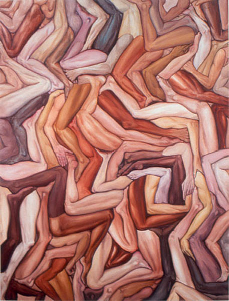
Photo A: Dream Object by Jim Shaw
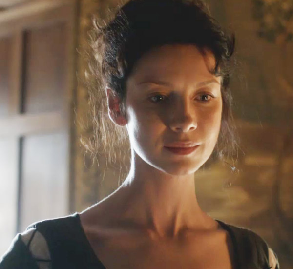
We all know that Colum’s legs captured Claire’s attention from the get-go. Here, her glass face shows what she thinks “plain and clear” (Starz episode 102, Castle Leoch). She’s surprised when the laird catches her riffling through his personal letters and books and then she is startled by his unusual physique.
Herself records Claire’s thoughts (Outlander book):
“At the moment, though, my discomfort arose from the fact that the beautifully modeled head and long torso ended in shockingly bowed and stumpy legs. The man who should have topped six feet came barely to my shoulder. … ‘I welcome ye, mistress,’ he said, with a slight bow. ‘My name is Colum ban Campbell MacKenzie, laird of this castle.’ ”
Colum’s bowed and twisted legs burden him with two “gifts that keep on giving:” pain and discomfort. Scientific explanations for his disability are coming up soon but first we must learn normal anatomy of the leg. So let’s get a leg up and start trotting!
In Anatomy Lesson #7, “Jamie’s Thighs or Ode to Joy,” we learned that anatomists define the leg differently than is common. In anatomy, the lower limb is divided into thigh, the region between hip and knee joints; leg, the region between knee and ankle joints; and foot, the remainder of the limb (Photo B). Colum’s entire lower limbs are affected by his disability but as that entails too much anatomy for a single lesson, we will focus on the anatomical leg.
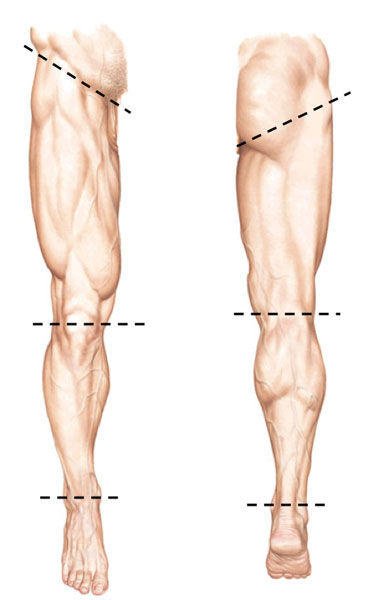
Photo B
As always, we begin with the bony foundation. Each lower limb contains 29 bones, one fewer than each upper limb: the thigh contains one bone, the leg contains two and the foot has 26.
The femur or thigh bone is the longest and by most measures the strongest bone of the human body (Photo C – right femur). Its bony details (and Jamie’s thighs) were covered in Anatomy Lesson #7 so we will skip them today.
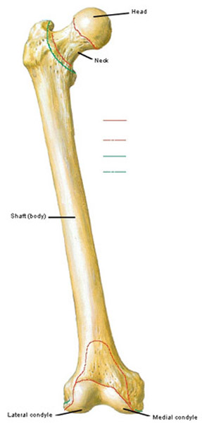
Photo C
Each leg contains two bones, the tibia situated medially (closer to body midline) and the fibula positioned laterally (away from body midline). Although not shown in Photo D, strong ligaments bind tibia and fibula to each other and also to femur and a few foot bones.
Parts of the sturdy tibia are subcutaneous thus its sharp anterior border and medial surface are easily palpated. Its proximal (near) end forms a bony plateau for the knee joint and its distal (far) end forms the medial malleolus or inner ankle bone (Photo D).
The needle-shaped fibula (Latin meaning clasp – as the pin of a brooch) is difficult to palpate except at its ends: the head forms a bony knob at the outside of each knee and the lateral malleolus is the outer ankle bone (Photo D). Fibulae are useful because surgeons can harvest most of their lengths for bone grafts (e.g. mandibular reconstruction) with little deficit as long as both fibular ends are left intact.
Try this: Place fingers on the front of your leg. Palpate the sharp anterior border of tibia or shin bone, the part that gets barked on projecting surfaces. Ouch! Move fingers toward the inside of your leg and feel the hard, flat medial surface of tibia. Move fingers to the inner ankle and tap the bony medial malleolus of tibia.
And try this: Next, move fingers to the muscle mass at the side of your leg; it covers most of fibula except at each end. The bony knob you feel near the knee is the head of fibula. Find the bony outer ankle or lateral malleolus of fibula. Together, medial and lateral malleoli (pl.) form a strong box-like frame for a foot bone which will be covered in a later lesson (Photo D- green arrow).
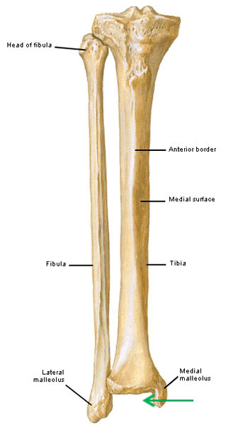
Photo D
The leg contains 12 muscles; we’ll cover four. The first muscle is tibialis anterior (Anatomy Lesson #9), the muscle was savaged by a boar’s tusk at the tynchal (Starz episode 104, The Gathering). Tibialis anterior is a strong fleshy muscle that hugs the outer surface of the tibia. It arises from the tibia and inserts into a foot bone (Photo E – right tibialis anterior). Contraction dorsiflexes the foot meaning it lifts the toe-end of the foot toward the sky, an action critical for walking and running. It also helps invert the foot, meaning to turn the sole of foot toward the midline.
Try this: Plant a shoeless foot on the floor. Leave the heel planted but lift the toe-end of your foot skyward. This is dorsiflexion. Replant the foot and turn sole inward to face the midline; this is inversion (some practitioners call this supination). Return the foot to dorsiflexion and palpate the tense fleshy muscle just lateral (to the side) to your tibia; this is tibialis anterior.
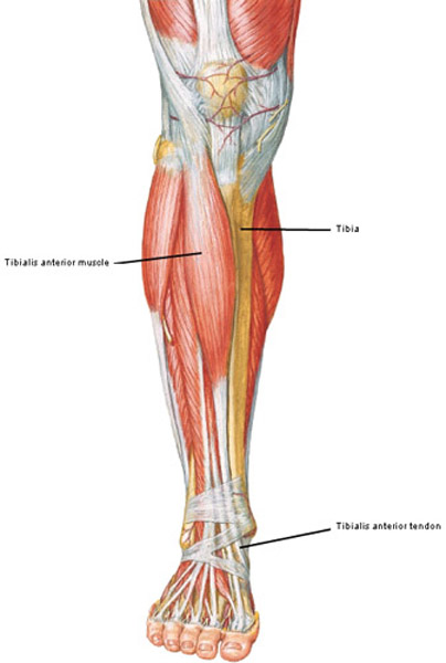
Photo E
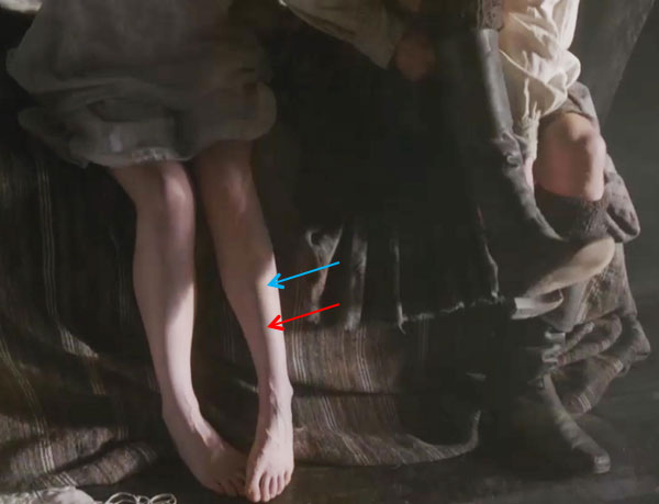
Want to see a really cute tibia (red arrow) and tibialis anterior muscle (blue arrow)? Of course ye do! Here is Claire the morning after (Starz episode 107, The Wedding). Yep…her curvaceous legs are beautiful and her crossed toes make her look soooo innocent but we all ken what those toes did last night! Jamie, weel, he’s hungry. How about a bite, Claire?
At the side of the leg is fibularis longus (aka peroneus longus). A long spindle-shaped muscle that arises from head of fibula, its tendon passes under the foot and inserts on a bone near the instep (Photo F). Fibularis longus contracts to evert the foot meaning it turns the sole away from the body midline. Think of a novice ice skater: ankles knock together and soles turn outward – this is eversion of the feet. Fibularis longus also helps plantar flex (point) the foot.
Try this: Plant a shoeless foot on the floor. If you can, turn your foot so the sole faces to the side and your inner ankle moves closer to the ground. This is eversion (some practitioners call this pronation) of the foot. With the foot in this position, feel the tense muscle running down the outside of your fibula; this is fibularis longus. Now point the foot, this is plantar flexion also aided by fibularis longus.
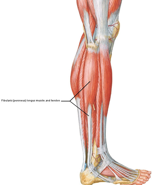
Photo F
The back of the leg contains seven muscles but we will cover only two. First is gastrocnemius (Latin and Greek meaning stomach of the leg); this odd name reflects the bulging shape of the calf created by gastrocnemius. This muscle spans two joints: knee and ankle. It also has two heads, one arising from each side of the femur (Photo G). The heads unite high in the leg but about midway down the calf the muscle gives way to the calcaneal or Achilles tendon. The longest and strongest tendon of the body, it inserts into the calcaneus or heel bone (Photo G, green arrow).
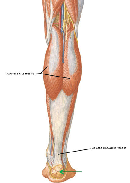
Photo G
Contraction of the gastrocnemii (pl.) flexes the legs at the knee joints and plantar flexes (points) the feet. These are powerful muscles and each is active during fast movements such as running, jumping and dancing.
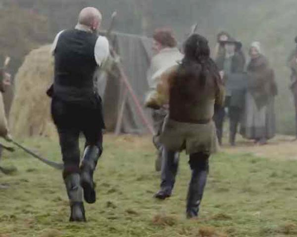
Gastrocnemii are verra active during a shinty game (Starz episode 104, The Gathering). Run ruaidh Jamie! Big bad Uncle Dougal is hot on your handsome Highland heels! Run, run as fast as you can; you can’t catch the ginger-haired man!
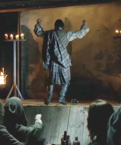
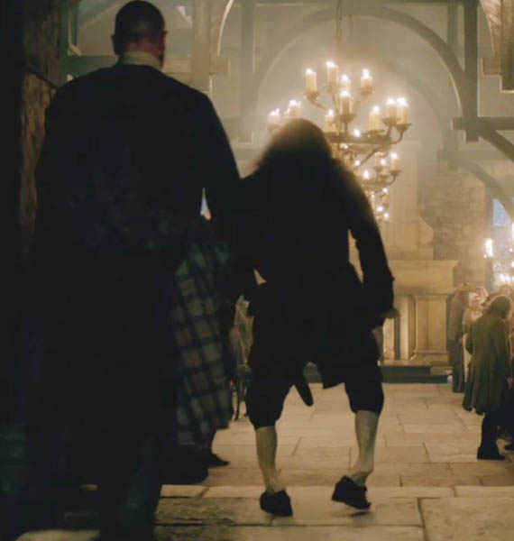
Covered by boots, we canna see the gastrocnemii but they are also busy as Murtagh, um, “executes” his version of the Highland sword dance (Starz episode 114, The Search).
The soleus (Latin meaning sandal) is a large, flat muscle deep to each gastrocnemius. Soleus arises from the back of tibia and fibula and joins gastrocnemius to form the Achilles tendon (Photo H). Because gastrocnemius and soleus share the Achilles tendon, some anatomists consider them a single muscle, the triceps surae (Latin meaning three-headed calf muscle). Soleus crosses only the ankle joint so it helps plantar flex the foot. But equally important, soleus is a postural muscle that helps us stand aright; if not for its constant backward pull, we would do a face-plant!
Now, here’s an interesting factoid: besides plantar flexion and posture maintenance, soleus helps prevent stasis of venous blood. A large leg vein (posterior tibial) passes deep to soleus so when the muscle contracts, venous blood is moved towards the heart against gravity. For this contribution, the soleus is also called the sural pump. This is so important that some health providers recommend soleus exercises for inactive patients to help prevent deep venous thrombosis (DVT or blood clots). And, smart passengers on long air flights should flex and point their feet every couple of hours (activates the soleus pump), walk around, wear loose-fitting clothes, etc., to help prevent blood clots.

Photo H
A comment or two about the calcaneal/Achilles tendon, the most commonly injured tendon of the human body! Explosive actions such as jumping or a sudden pushing off can tear the tendon or avulse it from its insertion on the heel bone. Achilles tendon injury is much more common in males than females and accompanies increased athletic activity (especially after years of sedentary lifestyle), the effects of aging or the use of some antibiotics.
And, just so we understand its origins, the Achilles tendon is named for the mythical Greek hero Achilles who was slain during the Trojan War when Paris’ poisoned arrow hit the heel, his only vulnerable body part (Photo I). As a wee lad, his mom held him by the heel and dipped him in the River Styx to grant him indestructibility but, sadly, that heel didna get baptized!
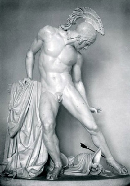
Photo I
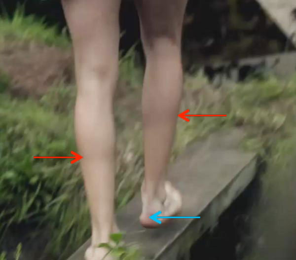
Want to see a manly example of an Achilles tendon (Starz episode 112, Lallybroch)? Weel…Jamie has one! First off, the lad has long tibiae and fibulae (pl.) which lend his calves a lean, lengthy leg-look. Red arrows mark the junctions between Jamie’s gastrocnemius muscles and his Achilles’ tendons which continue down the legs to end at the heel bones (blue arrow). Nice runner’s legs, laddie! Getting into that mill pond? Take good care; Claire willna like it if ye freeze off her fav body part!
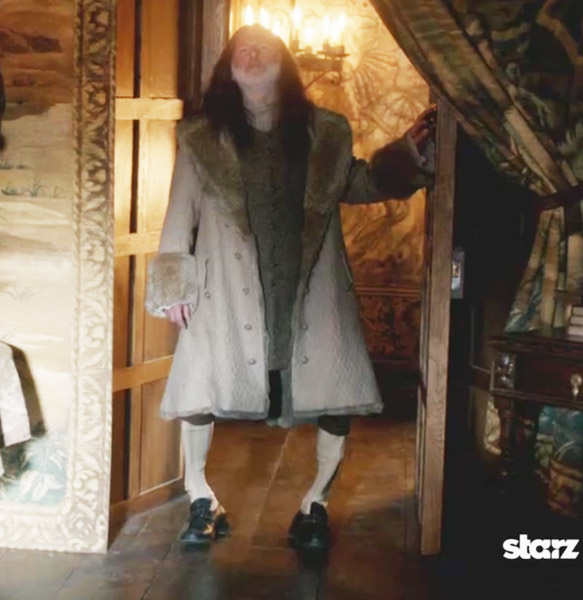
Now, we are done with normal anatomy, so let’s apply it to Outlander! Returning to Colum MacKenzie (Starz episode 104, The Gathering), Claire’s considers Colum and his brother, Dougal (Outlander book):
“Now there was a strange man. A cultured man, courteous to a fault, and thoughtful as well, with a reserve that all but hid the steely core within. The steel was much more evident in his brother Dougal. A warrior born, that one. And yet, to see them together, it was clear which was the stronger. Colum was a chieftain, twisted legs and all. Toulouse-Lautrec syndrome. I had never seen a case before, but I had heard it described. Named for its most famous sufferer (who did not yet exist, I reminded myself), it was a degenerative disease of bone and connective tissue.”
Question #1: Does Claire correctly diagnose Colum as suffering from Toulouse-Lautrec syndrome? Let’s take a closer look.
Henri de Toulouse-Lautrec (aka Henri Marie Raymond de Toulouse-Lautrec-Monfa, 1864-1901) was a 19th century artist who garnered fame as a painter, printmaker, illustrator and draughtsman. A contemporary of Gauguin and van Gogh, his work embraced the colorful, daring and glaring life of Bohemian Paris and his subjects were often members of that social stratum. His works are highly valued. Ten years ago, his painting of a young laundress (La blanchisseuse, 1889) sold for a record US $22+ million (Photo J)!
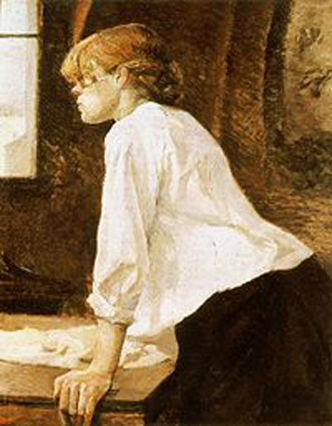
Photo J
Toulouse-Lautrec was the first born child of an aristocratic French family whose parents were first cousins. By eight years of age, his talent for drawing and painting had surfaced, but problems with growth and development soon followed. At 13, Henri fractured his right femur and a year later, his left, but the breaks never healed properly. His legs ceased to grow such that as an adult, he stood 4’ 8” (1.42 m). His trunk was adult-sized but his legs remained those of a child (Photo K).
Fast forward to 1962; a pair of French physicians (Maroteaux and Lamy) described a rare clinical entity (1-1.7/1 million births) dubbed pycnodysostosis (Greek meaning dense, defective condition of bone) or Toulouse-Lautrec syndrome. Pycnodysostosis is inherited if a child receives a recessive gene for the disease from each parent, an unlikely outcome unless parents are closely related. However, because genetic testing was unknown during Henri’s lifetime, it is surmised that he suffered from this disease (there are other possibilities).
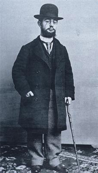
Photo K
Now, lace up your shoon, gobble down your haggis and bear with me for a bit of microscopic anatomy. We humans think of our skeletons as static structures but this is so not so! Each bone is a living organ that is restructured and remodeled throughout life by action of two bone cells: osteoblasts (bone-forming cells) that make bony matrix and osteoclasts (Greek meaning bone + broken) that breakdown bone. Osteoclasts (Photo L – blue dashed line) are multinucleated giant cells many times larger than our red blood cells (Photo L – red dashed line). A third bone cell, the osteocyte, also has bone activity but we will not cover this cell today.
Our bones are remodeled as we grow, age, gain or lose weight, experience pregnancy, change exercise regimes, or mend broken bones; all possible because of osteoblasts and osteoclasts.
It turns out that pycnodysostosis/Toulouse-Lautrec sufferers cannot make an enzyme (Cathepsin K) essential for protein breakdown. Because osteoclasts lack this enzyme these cells cannot break down bone; thus, bones neither remodel nor mend properly. Make sense? 🤔
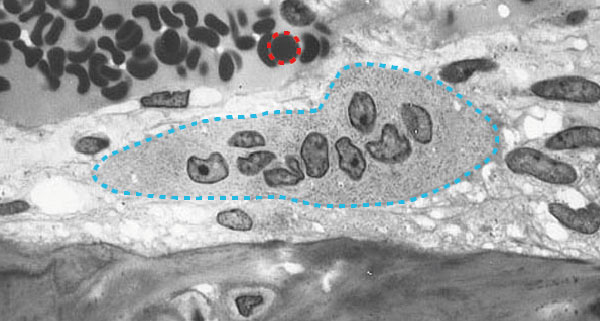
Photo L
Lacking just ONE enzyme, Toulouse-Lautrec sufferers experience these painful and debilitating symptoms:
- Stature: adult males stand less than 150 cm (4’ 11’); adult females are shorter.
- Bones: bones are dense because osteoclasts cannot resorb them. Bones are brittle and fracture easily especially weight-bearing bones of the lower limbs, mandible (Anatomy Lesson #26) and clavicles (Anatomy Lesson #3).
- Skull: bones of forehead (frontal bone), back of head (occipital bone), nose and mandible as well as the teeth do not form properly. The anterior fontanel (‘soft spot” on top of the head) remains widely open, possibly why Toulouse-Lautrec consistently wore a hat.
- Hands: skin over the back of fingers is deeply wrinkled; nails are flat and grooved. Distal phalanges (finger and toe bones) are short (Anatomy Lesson #22 & Anatomy Lesson #23).
- Spine: defective vertebrae cause the spine (Anatomy Lesson #15) to curve laterally (scoliosis).
- Impotency and/or sterility: Toulouse-Lautrec purportedly contracted syphilis from a prostitute who served as his model – his history suggests he was not impotent but he is not known to have sired children. Alcoholism (absinthe for pain), tertiary syphilis and his weakened constitution took his life at 37. Colum was apparently sterile because Dougal ensured his bloodline by begetting Hamish with Letitia (Starz episode 109, The Reckoning). Dougal also assured Colum “that she was tender and sweet as a ripe peach and all that a man could want in a woman” (Outlander book) implying that Colum did not know his wife sexually (impotent).
Answer to Question #1: Yes, Claire very likely correctly diagnosed Colum’s disability as Toulouse-Lautrec syndrome (pycnodystosis).
Question #2: Does Diana’s description of Colum’s symptoms match with those of Toulouse-Lautrec syndrome? You be the judge. Claire reflects in Outlander book:
“Victims often appeared normal, if sickly, until their early teens, when the long bones of the legs, under the stress of bearing a body upright, began to crumble and collapse upon themselves. The pasty skin, with its premature wrinkling, was another outward effect … that characterized the disease. …. As the legs twisted and bowed, the spine was put under stress, and often twisted as well, causing immense discomfort to the victim. … Because of the poor circulation and the degeneration of connective tissue, victims were invariably sterile, and often impotent as well.”
And another quote from Outlander book: Here, Ned Gowan explains to Claire:
“Colum was a whole man to the age of eighteen or so,” … “but soon after the marriage he had a bad fall, during a raid. Broke the long bone of his thigh, and it mended poorly.” …“And then,” Mr. Gowan went on with a sigh, “he rose from his bed too soon, and took a tumble down the stairs that broke the other leg. He lay in his bed close on a year, but it soon became clear that the damage was permanent.
Okay, let’s all be the judge!
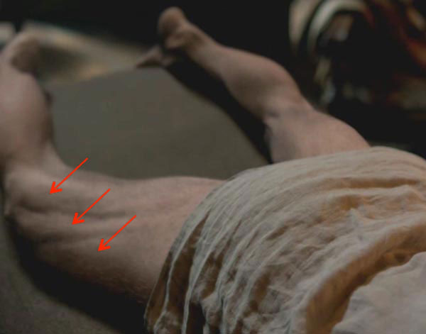
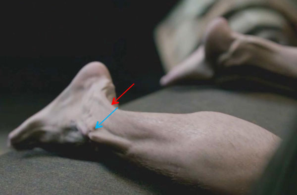
Answer to Question #2. My opinion: Claire’s description and the visuals of Colum’s legs almost exactly match the symptoms of Toulouse-Lautrec syndrome or pycnodysostosis. Colum’s thigh muscles are abnormally twisted and grooved (above photo – red arrows) due to his poorly mended femora (pl.). His right fibula also appears to have been broken and mended badly (below photo – blue arrow) and his twisted Achille’s tendon (below photo – red arrow) is displaced laterally. Remember Claire’s offer to massage the base of Colum’s spine rather than his legs? Nerves innervating the legs emerge near the base of the spine explaining why spinal massage of that region might provide temporary relief. Colum suffered greatly with his laird’s legs but make no mistake: “this clan remains under the charge of this man!”
The visual presentation of this terrible syndrome was very convincing and well done. Way to go Colum, Claire, Diana and CGI team!
The only inconsistency with the Colum’s disabilities is that most individuals with Toulouse-Lautrec Syndrome exhibit normal sexual development and fertility.
Here is an interesting and apropos tidbit about a cute cat (Photo M). The Scottish fold cat (aka Highland Fold, Longhair (Laoghaire?), Longhair Fold) is a domestic cat with ears that fold forward toward the nose. They also suffer from distorted limb bones and arthritis. The breed is the result of a genetic mutation in a single barn cat named Susie (Perthshire, Scotland, 1961). But most pertinent here, the Scottish fold syndrome belongs to the same group of cartilage and bone disorders as Toulouse Lautrec syndrome. Yep, it does!

Photo M
Now, let’s end this lesson with some fun frivolous facts about legs (here meaning the entire lower limb)!
- Michael Flatley (Riverdance) insured his legs in 1999 for US $40 million.
- In 1836, Mexican General Santa Anna held an elaborate state funeral for his amputated leg.
- The average person takes 6,000 -9,000 steps per day: 28,000 – 42,000 miles in a 70 year lifespan. This exceeds the circumference of the globe!
- The “legs up the wall” yoga pose relieves anxiety, stress and tired legs (lie on back near a wall. Lift lower limbs up the wall and scoot butt against the wall). It feels marvelous as blood drains from the lower limbs!
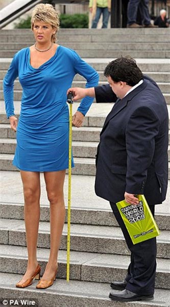
- Svetlana Pankratova of Russia purportedly has the longest legs of any woman in the world – at 1.32 m or 4.33 ft. (Does the Guinness rep realize that she is wearing heels? Not exactly a scientific measurement 😵💫). Her hip joints and my oxters are at the same height!
Be grateful! Kick your own legs up in the air if ye can and offer a hand to those whose legs dinna work quite so well!
A Deeply Grateful,
Outlander Anatomist
Photo creds: Netter’s Atlas of Human Anatomy, 4th ed., Starz, www.greekmyths-greekmythology.com (Achilles), www.montessoricats.com (Buddha cat), www.news.asiatown.net (Svetlana Pankratova), www.praz-delavallade.com (Jim Shaw painting), www.reddit.com (Scottish Fold cat), www.wikipedia.org (La blanchisseuse and Toulouse-Lautrec photos), Robert M. Hunt (B&W osteoclast)
Thanks for sharing your knowledge. I have learnt a lot. I look forward to your blog on my news feed. Makes my day. Very interesting!
Thank you, Donna! I am so glad you find the lessons interesting. It is great fun to put together the topics!
Thank you, for taking the time.
And giving me something worth while too read. It was a easy read and very well illustrated. It made me think of Tuesday morning conference …
Hi Glenda:
You are most welcome and I am so happy that the lesson was worth your time to read.
Tuesday AM conference? Grand rounds, maybe? You must be in the medical field!
Love it! As usual. Great explanation and tie in with the series. TY!
Hi Moz! Great to hear from you. Glad you like the lesson; very interesting stuff.
Thank you for writing!