Anatomy Lesson 8: “Jamie Takes a Beating and Claire’s Healing Touch”

Anatomy Lesson #8:  Solar plexus, Respiratory Diaphragm, Kidneys and Cheekbone

Now, that’s a whole lot of anatomy, but dinna get into a swivet; this lesson is dictated by Rupert’s beating of Jamie. Well, now, Jamie is a warrior who has a knack for getting himself into a mess o’ messes. Claire is a warrior too, only her battle field is healing!

Getting started, let’s review the healing Claire has given thus far for young Jamie – showing that she is a real Healer-Dealer and how that rascal Rupert undoes some of her warrior work!

In Starz episode #1, Sassenach, Murtagh captures Claire on Saturday – likely the same day Jamie dislocates his shoulder. Several hours pass before Claire reduces Jamie’s shoulder (see Anatomy Lesson #2). “Tá Dia (I think this is what he says) – it doesna hurt anymore!” exclaims Jamie. Ha, the Highlanders hadna a clue that a feral Sassenach cat could perform such miracles!

image

Claire and her captors ride through Saturday night and into Sunday when Jamie is shot at Cocknammon Rock (See Anatomy Lesson #3). Later that night, after fainting from lack of blood, Claire securely binds his wound so he doesna have to stay and determine his own fate wit’ a loaded pistol (Starz episode #1, Sassenach).

image

On Monday they arrive at Castle Leoch (Starz episode #2, Castle Leoch). Claire insists on properly cleaning and dressing Jamie’s gunshot wound (See Anatomy Lesson #3), all the while conducting a thorough counseling session fer the sad-lad! She also binds his right arm to his chest (thorax in anatomy) to immobilize the freshly reduced shoulder joint. What a caregiver!

image

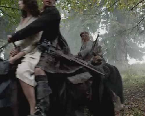
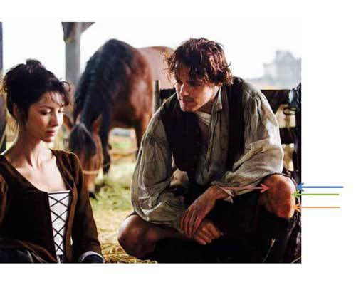
On Tuesday (Starz episode #2, Castle Leoch), Claire brings Jamie comfort in the form of lunch and fresh bandages but accidentally upsets his work at the stables. Jamie falls to his knees. Oooh, this clearly hurts his pride and mayhap messes a wee bit wit’ his injured shoulder? Oops, Claire better watch that hip padding – it gets in the way! After lunch, true confessions and a good deal of “facetime,” Claire begs Jamie not to get stabbed or flogged today.

image

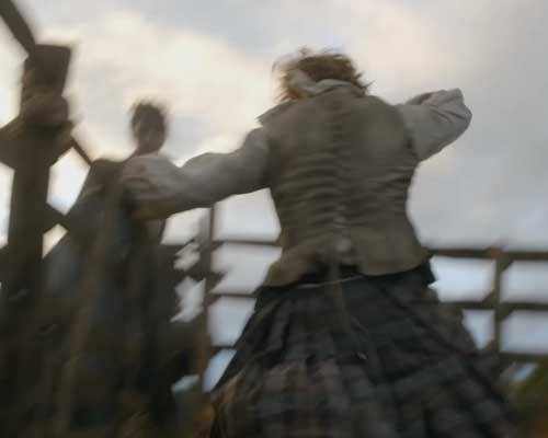
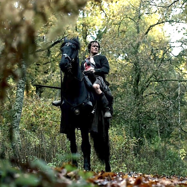
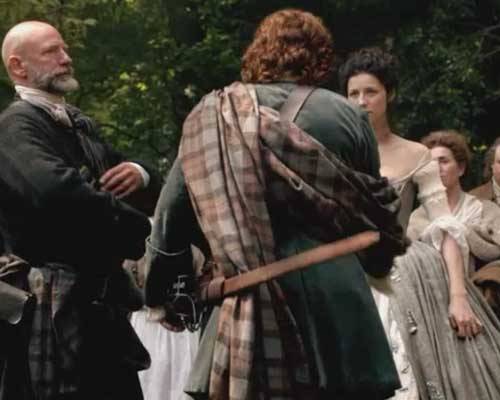
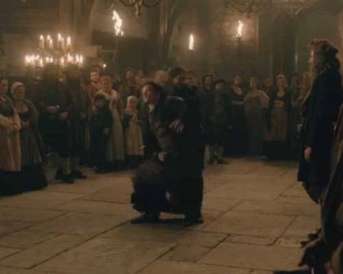
By Friday, after having been a good lad fer far too long Jamie gallantly steps forth at the Hall to take punishment for “damn-her-eyes” LiarHair who clearly has been a verra bad lassie (Starz episode #2, Castle Leoch). Ye’ll ken this is about six days after his shoulder dislocation and only five days since his gunshot wound! This is important because at this point new blood vessels are growing (angiogenesis), cells are dividing (mitosis) and collagen is forming (fibrogenesis) to help heal pulled ligaments and strained muscles; everything in Jamie’s shoulder is tender and sore. But, braw showman that he is, he chooses fists no the strap!

image

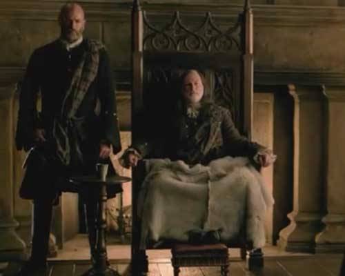
Laird Colum allows it.  Rupert will administer the fisticuffs delivering two consecutive blows to Jamie’s belly!

Now for some anatomy! Both blows strike Jamie in the pit of the stomach (Photo A). This type of blow affects three major regions of the body: the belly wall, the solar plexus and the diaphragm.
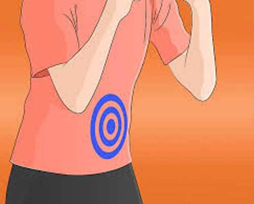
image

Photo A

As I will cover the belly wall in a future post, I won’t describe its full anatomy now. Suffice it to say that it is defined by the blue area seen in Photo B. In a warrior like Jamie, whose belly is very strong, two punches will not do much real damage. But consider the gut punch BJR gives Claire (Starz episode 6, The Garrison Commander) along with the belly kick by his weenie gopher. I suspect that Claire’s corset stays, several undergarments and that fabulous pleated tartan gown kept any real damage to a minimum; ye ken that each pleat includes three layers of wool? (Aye, Terry and her team create such splendid garments!)
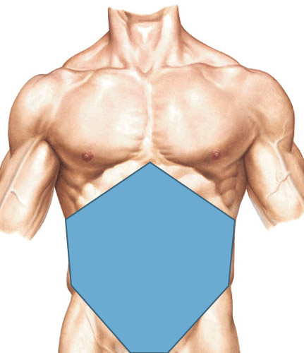
image

Photo B

Now, just behind the stomach lies the solar plexus (Photo C – surface anatomy), so named because it resembles a sunburst; anatomists call it the celiac (coeliac) plexus.  
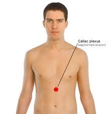
image

Photo C

To understand the celiac plexus, let’s first visit the aorta the largest artery of the human body (Photo D – posterior abdominal wall – from the front with organs removed). It begins in the chest, pierces the diaphragm and enters the abdominal cavity where it lies in front of the spine.
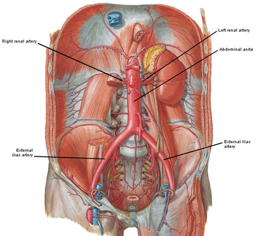
image

Photo D

The celiac plexus (Photo E – black arrows) wraps the front and sides of the abdominal aorta. The white blobs in Photo E are ganglia, swellings that house nerve cell bodies. The white strings are nerves; arising from the cell bodies they connect with other ganglia and supply organs (viscera). The nerves cause viscera to contract or secrete and also carry pain sensation back to the central nervous system. A blow to the belly can bruise the stomach, shock the celiac plexus, cause pain and potentially interfere with visceral function.
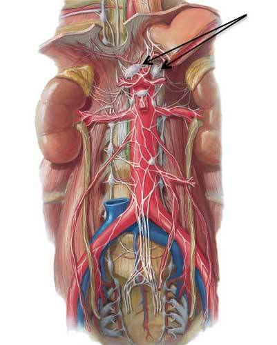
image

Photo E

Finally, the last body part vulnerable to a belly blow is the thoracic diaphragm, a dome-shaped sheet of muscle and tendon between the chest and the abdominal cavities that sits surprisingly high under the rib cage  (Photo F – from the front showing cut edge of diaphragm). It is attached to the inner surfaces of breast bone, ribs and vertebrae and is a powerful muscle of respiration: as we breathe in (inspire) it descends, as we breathe out (expire) it rises. Blows to the belly wall cause temporary paralysis of the thoracic diaphragm making breathing difficult – the sensation of “getting the wind knocked out of you” and perhaps the most uncomfortable part of a gut punch! The spasm is accompanied by pain, anxiety and difficulty sitting or standing. But, as with Jamie, the spasm passes, breathing resumes and equanimity is restored.
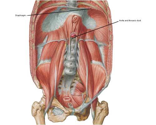
image

Photo F

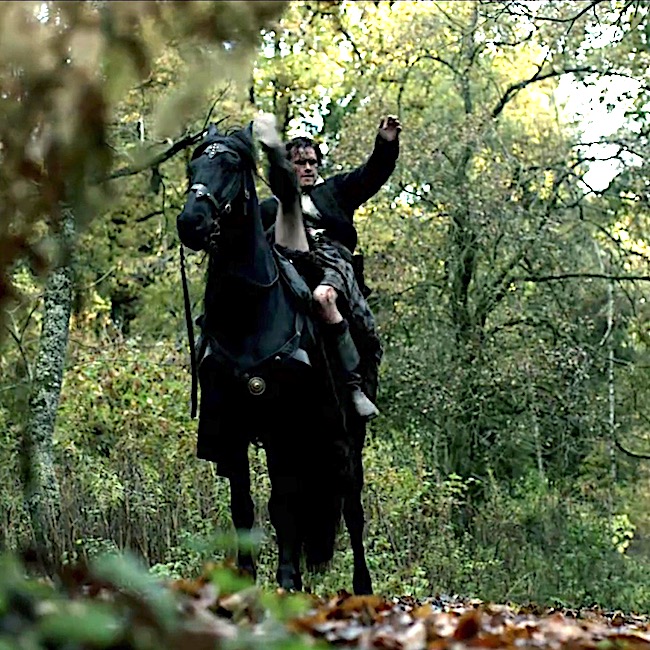
Moving on…Rupert circles around to Jamie’s back and delivers a third blow causing him to stagger! As ye well ken, this is a kidney punch and it is verra painful!

image

The kidneys are paired bean-shaped organs that produce urine, extract wastes, balance body fluids and are vital for normal life! Each day they filter about 189 liters (200 qt) of blood but produce only about 1.89 liters (2 qt) of urine because normally most all the fluid is resorbed. The urine flows into paired ureters then to the urinary bladder and is subsequently voided through the unpaired urethra (Photo G).

image

Photo G

A blow to the kidney is painful but not always serious (unless, of course, Claire’s stabbing ye in the back wit’ her sgian dubh!). Why? Weel, because the 11th & 12th ribs protect much of the left kidney and 12th rib protects part of the right (Photo H – from the back; kidney outlines shown as dashed lines). Also, strong back muscles absorb energy from a kidney punch. Kidney injury from blunt trauma ranges from bruising to serious tearing of tissues. Just so ye ken, blood in the urine is a good indicator of kidney trauma and the kidney punch is one of the meanest places to whack an opponent – although our fiery hero doesn’t seem that worse for wear.
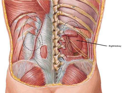
image

Photo H

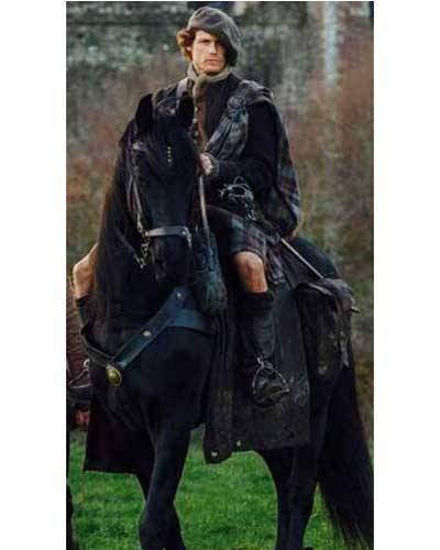
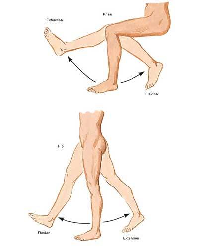
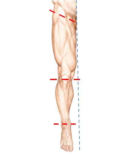
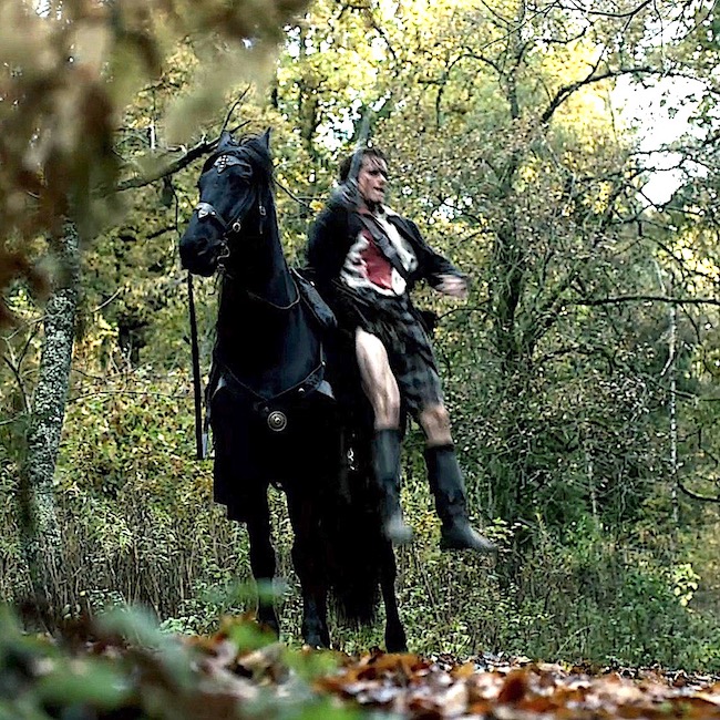
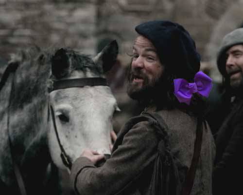
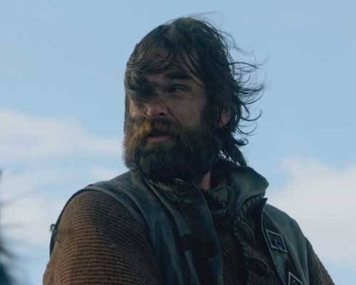
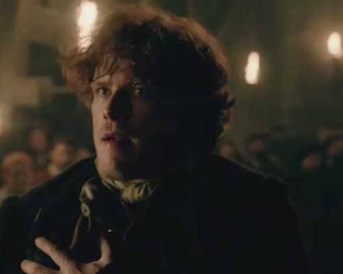
By now Rupert is sweating. So he checks wit’ his heid, “Dougal the Devil Man” ta see if it’s time to stop. Now, we all ken that Dougal is Colum’s war chief. So, take a peek at his menacing stance – splayed legs all covered wit’ his shiny high top boots. But, Dougal has no problem wi’ his nephew getting hurt and hurt badly. After all, the lad is still standing at this point and standin’ in the way of his best-laid plans. So Uncle Dougal directs Rupert to land a 4th blow – this time to Jamie’s face and blood splatters the slates. As there is another punch coming to Jamie’s face, I’m saving facial anatomy until blow #6! Read on!

image

Hey, now, I thought the beating was supposed to stop once blood was drawn. Geillis says so and we all know she never lies. Next, Rupert hauls back and lands a mighty one ta Jamie’s right shoulder!

So, what was Claire thinking through all this physical plundering? Herself writes in Outlander:

“I was in agony of apprehension, lest one of the blows re-damage the wounded shoulder…”

Too late Claire…Och, Rupert, that was brutal, man!

image

Time fer more anatomy, heh. The shoulder blow is bad news because there’s nay been enough time for the wounds to have healed properly. The next photo shows the proper alignment of a normal glenohumeral joint (Photo I:  x-ray right shoulder). Part of the humeral head (red arrow) sits in the glenoid cavity of the scapula (blue arrow).
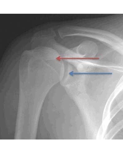
image

Photo I

Nearly a week earlier Jamie suffered an anterior dislocation of the glenohumeral joint (Photo J: x-ray right shoulder). Here, the humeral head (red arrow) is typically displaced forward (anterior) and down (inferior) from the glenoid cavity (blue arrow) as shown in the following x-ray. Claire restored the normal anatomy of the joint, but as we learned earlier, the whole area remains inflamed and tender. Ouch! It hurts just ta look at it.
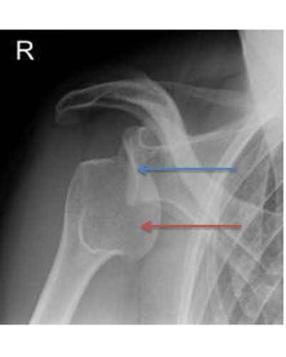
image

Photo J

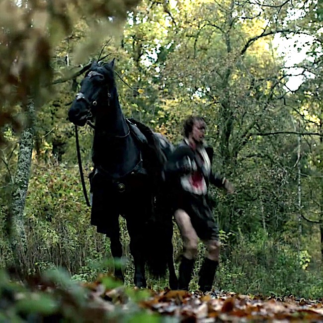
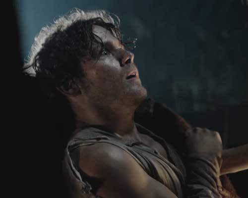
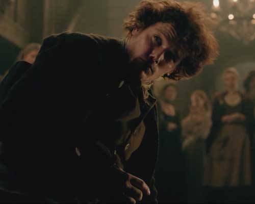
Finally, Rupert delivers the last punch to Jamie’s face and knocks him to the ground!

image

Now, ye best understand the damage these facial blows can cause! In anatomy, the cheek bone is the zygomatic arch (Photo K). With color-coded skull bones, ye can clearly see that the zygomatic arch is made of two bones: the pink part belongs to the temporal bone and the orange part to the zygomatic bone. The two bones meet at a jagged suture (non-moveable joint). Although strong, the suture is at risk for fracture.
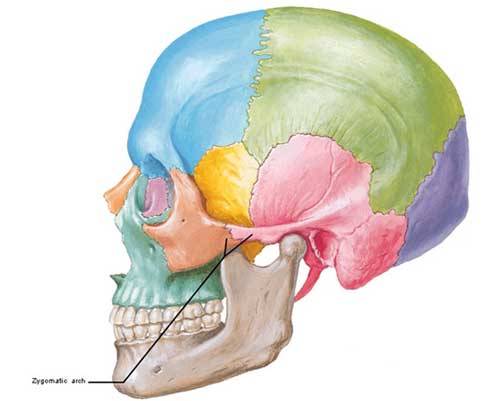
image

Photo K

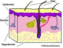
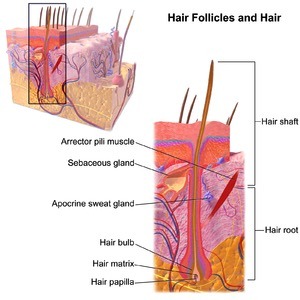
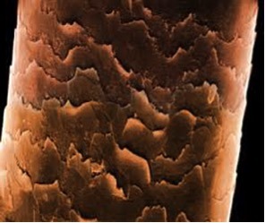
Also, the zygomatic bone forms the outer rim of the bony orbit where the eye is housed (Photo L). Both zygomatic arch and bony orbit are subcutaneous structures (see Anatomy Lesson #5 – The Skin) and vulnerable to injury. The fact that Rupert’s blow didn’t fracture either of these bony areas is probably due to the fact that Jamie’s head is harder than an iron pot – or so says his sister Jenny!
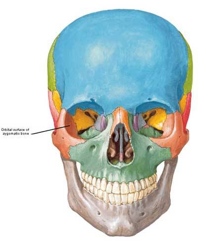
image

Photo L

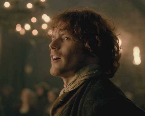
After the beating, Murtagh helps our hero out of the hall but not afore Jamie casts a look at Claire: “Sassenach., I need you!” Well, that does it. Claire has ta’ git outta that Hall – NOW! Make way, Geillis! Ah, have patience Claire, a few more weeks and ye’re going ta get all the healing touches of Jamie ye want. Lucky Lady!

image

Soon enough, Claire tenderly soothes Jamie’s bruises: he’ll be sore for a couple of days, but he’s no really damaged. She is dying ta know why he took Miss LongHare’s punishment! In Outlander book Jamie answers Claire:

“Why not me?”…Why not? I wanted to say. Because you didn’t know her, she was nothing to you. Because you were already hurt. Because it takes something rather special in the way of guts to stand up in front of a crowd and let someone hit you in the face, no matter what your motive.”

Friends, if you have yet to read the marvelous Outlander books, I urge ye to do so as well as follow the Starz episodes! There are lots more to these vignettes than can fit into a 60 minute episode!

image

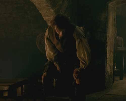
Then Claire delivers the sad news that tomorrow (Saturday) she is headed back to Inverness with Pitre the tinker! Jamie groans as he stands to say farewell. He is devastated. Are ye sobbin’ yet? He is!

Ah…breathe a sigh of relief – fate has a surprise in store fer Mistress Beauchamp. Yep, Colum and his lil’ Big Bad Bro willna let her go wit’ the tinker. Claire has to stay and take her own Beaton – er, the surgery I mean – that little “chamber of horrors” needs her healing touch too!

image

Now, looking fer remnants of Jamie’s beating – were the continuity detailers attending to details? Yep, they were. Many films show heroes beaten within an inch of their lives but they emerge moments later wit’ nary a scratch – keeps me up at night!

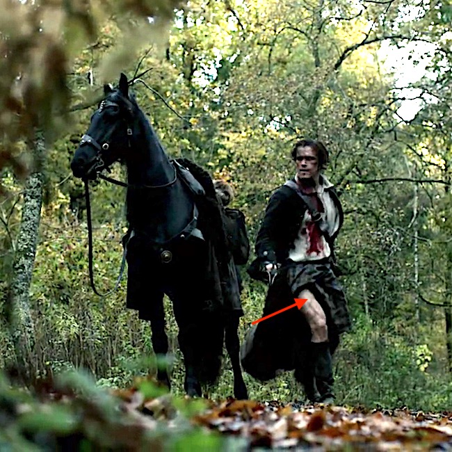
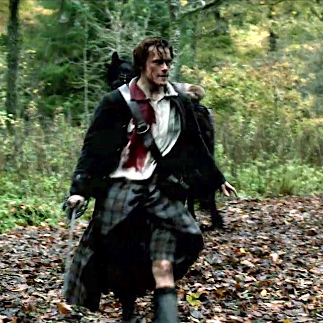
Here’s a great example. Did ye see Jamie hobble along wit his right arm drooping as the Teenage Trollop hauls him into an alcove ta have her way wit’ him (Starz episode 3, The Way Out)? Yep, he’s still hurting after that thrashing!

image

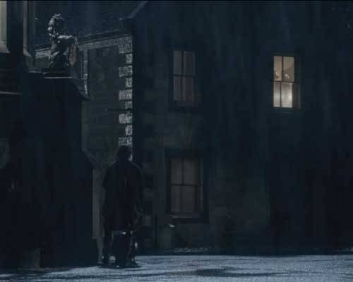
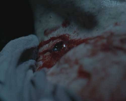
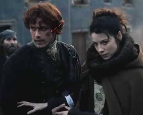
At the pillory (Starz episode 3, The Way Out), we glimpse a dark shadow over his zygomatic arch (red arrow) and mayhap a bit o’ black eye as he helps Claire from a feigned swoon – she who canna stand the sight of blood. Hah. Looks like a big old nasty bruise ta me! Verra nice.

image

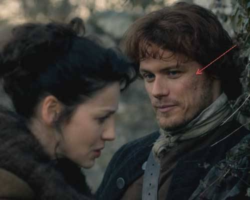
Finally, at the Black Kirk, Claire gives Jamie a botany 101 class explaining the difference between wood garlic and its deadly look-a-like, lily of the valley. This time we see up close the mottled bruising o’er Jamie’s zygomatic arch. Ahhh…he does have a lovely arch does he not?

image

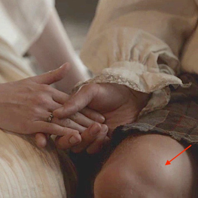
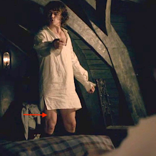
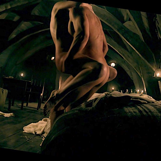
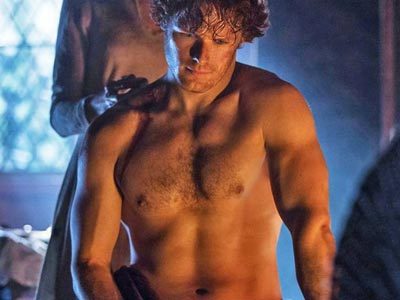
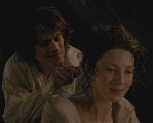
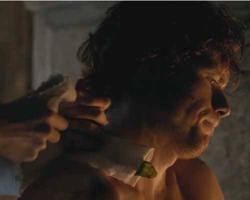
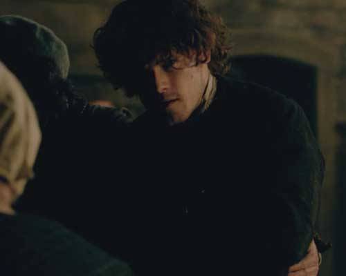
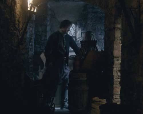
I’ll leave ye now with three wonderful images from Starz episode 3, The Way Out. At Gwyllyn the Bard’s performance, Claire is deep into Colum’s Rhenish. Ever watchful Jamie escorts her safely to her Beaton dungeon under the guise of needing help wit’ his bandage. Jamie, ye are the man! Claire decides to check his gunshot wound anyway and starts ta take off his clothes -weel, she just unties his stock and unbuttons his shirt but…Gah!

image

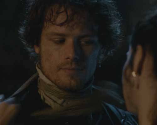
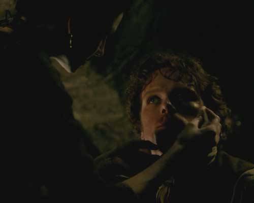
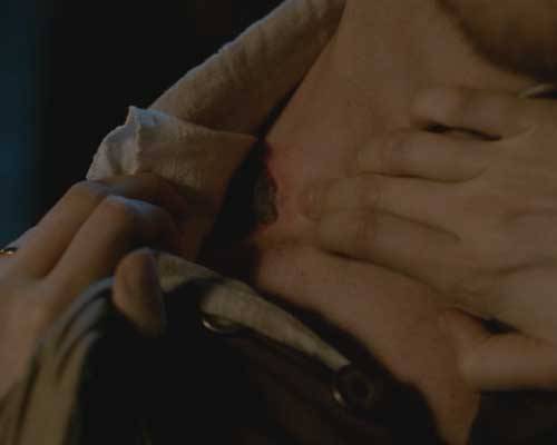
She pulls back the bandage: “Scabbed over nicely; no drainage.” She isna going ta let Jamie leave her surgery without feeling his fair, fresh skin. After all she is the healer and she is in charge!

image

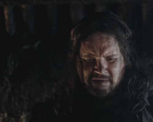
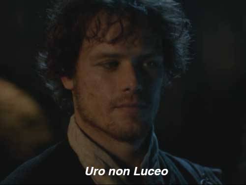
And, what is young Jamie’s thinking wit’ Claire half undressing him and putting her bonny fingers on him? Weel, look at his face. Ha’ mercy, lass! Ye are making it verra hard for the puir man. Snort! Jamie, he is silently taking’ the MacKenzie oath – only backwards. Jamie, man, ye BURN not shine!

image

OK, that does it fer this anatomy lesson. Thanks for joining me as we explored how Jamie’s wounds ne’er fail to bring on Claire’s healing touch! Hope ye all are hunkering down as the winter solstice approaches (in the Northern Hemisphere, anyway).

And fer those who are new to my blog, a note of explanation: I follow the convention of using the character’s names rather than the Starz actor’s names. This gives the cast at least one degree of separation and a wee bit o’ respect as I dissect their bodies with words! I hope ye all understand!

You can now follow me on Facebook and Twitter!

The deeply grateful,

Outlander Anatomist

Images courtesy of: Starz, www.wikihow.com, www.wikipedia.org, Kidney Health Australia, radiopaedia.org, Netter’s Atlas of Human Anatomy 4th ed., Hollingshead’s Textbook of Anatomy, 5th ed.

Anatomy Lesson 7: “Jamie’s Thighs – Ode to Joy!”

Anatomy Lesson #7, Jamie’s Thighs, is a true ode to joy! I hope Ludwig won’t be offended by my reference to his 9th, but the title perfectly suits this lesson! Perfect timing with the upcoming US Thanksgiving Holiday.  I’m down on my knees giving thanks because Jamie’s knees also appear in this lesson! 🙏🏻

Are you ready? Let’s go!

Just to prove my goodwill, take a gander at Jamie’s knees in the image below. May this tide you over as our lesson proceeds!

image

Definitions: Let’s begin with a few basic tidbits, otherwise much of this lesson might remain obscure!

  • Joint – Site where bones meet and move relative to each other
  • Muscle – Muscles attach to bones (some exceptions); these are voluntary meaning we can contract them at will – a.k.a. skeletal muscle
  • Contraction – As muscles contract, a joint moves in various ways (Image A)
  • Origin – Attachment site of muscle to bone – site moves little or not at all during contraction
  • Insertion – Attachment site of muscle to bone – site moves during contraction
  • Flexion – Muscle contraction closes (approximates) bones of a joint
  • Extension – Muscle contraction opens (straightens) bones of a joint

image

Image A

Lower limb: Our lower limbs are the body parts from hip joints to toes. Anatomists don’t use the terms upper and lower leg. Rather, the lower limb is divided into (Image B):

  • Thigh – segment between hip and knee joints
  • Leg – segment between knee and ankle joint
  • Foot – segment beyond ankle joint

BTW, the dashed blue line in Image B represents a vertical midline through the body. A medial structure lies closer to this midline – lateral structure lies further from this midline. That’s the gist of it!

Try this: Locate your thigh, leg, foot and midline.

Test Q: Are your tiny toes medial or lateral to your middle toes?

image

Image B

Test A: They are lateral. Great job!

Outlander Time! The first time we rencounter Jamie’s thighs is in Outlander book where Claire straddles-his-saddle on the way to Castle Leoch. Herself writes:

My companion seemed to be having little trouble, in spite of being unable to use his right hand. I could feel his thighs behind mine, shifting and pressing occasionally to guide the horse. I clutched the edge of the short saddle in order to stay seated; I had been on horses before, but was by no means the horseman this Jamie was.

And yet another quote from Outlander book – this one from randy-dandy, Jamie:

But then that ride through the dark together….with that lovely broad arse wedged between my thighs…

Well! Starz Claire doesn’t sport a broad arse, but it surely is curvaceous and pert and we all ken where it was wedged during this ride!  Mmphm!

image

Back to anatomy and more about bones!

Femur: The thigh contains one bone, the femur. I love superlatives, so here’s the first one for this lesson: the femur is the longest bone of the human body (Image C – front of right femur). The top of each femur ends in an angled neck and head that fits securely into the acetabulum, a deep socket of the hip bone Together, they form the acetabulo-femoral (hip) joint, a ball and socket joint.

Fun Facts: You may recall from Anatomy Lesson #2, When Claire Meets Jamie or How to Fall in Love While Reducing a Dislocated Shoulder Joint!:  although the glenohumeral (shoulder) joint is very moveable it is also less stable due to a shallow ball and socket joint! Conversely, each hip joint is very stable but less moveable due to a deep ball and socket joint. The hip joint needs to be stable to support our weight against gravity. Very interesting!

At the knee, the femur ends in two sturdy knobs, medial and lateral condyles (Greek for knuckle); both knobs help form the knee joint (Image C).

image

Image C

Tibia and Fibula: The leg contains two bones, tibia and fibula (Image D – front of right leg bones). The larger tibia is medial to the smaller, more lateral, fibula. The top of tibia ends in two flat surfaces, the medial and lateral condyles; lower down, it bears a midline knob, the tibial tuberosity. Only tibia helps form the knee joint; fibula plays no role.

image

Image D

Patella: The small patella or knee cap is critical to knee anatomy. Patella is the largest sesamoid bone of the body (there are others) meaning it is enveloped in tendon. Its deep surface slides in a groove between medial and lateral femoral condyles as the knee joint extends and flexes (Image E – front of right knee joint).

image

Image E

Thigh Muscles: Yes! Thigh muscles are massive because they support much of our weight and help maintain our bipedal stance against gravity.

There are 13 thigh muscles divided into three compartments. To keep this lesson shorter than a bloody master’s thesis, we will cover only quadraceps femoris (Latin meaning four-headed muscle of the femur) with a wee bit about iliopsoas muscle.

Quadraceps: The quads (as trainers call them) are four muscles at the front of each thigh:

    • rectus femoris
    • vastus lateralis
    • vastus intermedius
    • vastus medialis

Image F (front of right thigh) shows the three of the quad muscles. A fourth, vastus intermedius, is visible only after removal of rectus femoris  (Image G).

image

Image F

image

Image G

Origins: Each quad muscle has a different origin (Image H –  red marks). Rectus femoris originates from hip bone and crosses both hip and knee joints . The three vasti (pl.) muscles take origin from different sites on the femur and cross only the knee joint.

Insertion:  All four muscles  fuse into a common quadraceps tendon that engulfs the patella and then continues as the patellar ligament to insert (Image H – blue mark) on the tibial tuberosity.

Actions: Acting together, all four heads of each quad extend the knee joint but only rectus femoris also flexes the hip joint.

Because quads are the only muscles that extend the knee joints, they are crucial for walking, running, jumping and squatting. Quads are also called anti-gravity muscles because they contract as we rise from a seated position or lower our bodies in reverse, holding our weight against gravity (think of thighs during snow skiing or Jamie lowering Claire onto the marriage bed. Yum!).

image

Image H

Iliopsoas: One last muscle… Although recti femoris (pl.) are decent hip flexors, iliopsoas muscles are the strongest. Like the quads, iliopsoas is a compound muscle formed by fusion of iliacus and psoas (pronounced soas) muscles.

Iliacus arises from the hip bone and psoas arises from the lumbar vertebrae (Image I – right front). They fuse into a single tendon that inserts into the femur. These muscles draw the femur closer to the torso, assisted by rectus femoris and a few other minor hip flexors. Or, with thighs held stationary, they draw torso closer to thighs (think sit-ups).

Image I

Try this: To test your quads, place back against a wall.  Drop the tush while walking your legs away from the wall until you assume a squat position (Image J). Thighs and legs should be at 90° to each other (don’t drop lower – this is bad for knee joints!). Now hold your torso in place for 30 seconds and then gradually straighten (extend) the knee joints. If you feel wobbly, then you may need quad work as these muscles quickly lose mass and strength due to inactivity, sedentary jobs or aging. Quads can be strengthened by wall squats or by any exercise that adds resistence while extending the knee joint!

image

Image J

Knee Joint: Now on to the knee jointlargest joint of the human body (Image K – right side and front)! Here medial and lateral femoral condyles (knuckles) ride atop a flat plateau formed by the medial and lateral tibial condyles – no stable joint here!

All bony surfaces of knee joint are covered with articular cartilage, a bloodless, firm connective tissue that allows for smooth movement. This odd joint also has medial and a lateral meniscus (different type of cartilage) that create two shallow sockets atop the tibial plateau – one for each femoral condyle; these also act as shock absorbers.

As mentioned above, the patella glides between the femoral condyles during flexion and extension.

image

Image K

Knee Ligaments: The knee is stabilized by very strong ligaments designed to secure the tibia and femur (Image L – right knee in full flexion – patella absent):

  • medial (tibial) collateral ligament
  • lateral (fibular) collateral ligament
  • anterior cruciate ligament
  • posterior cruciate ligament (and, there are others!)

Overall, the knee joint is at risk due to our sports-crazy cultures and because supporting the body weight while in motion is a challenge for these relatively flat surfaces. Hence, the über-strong ligaments.

image

Image L

Now, for the fun stuff!  I promised in Anatomy Lesson #3 that I would be returning to the scene where Jamie dismounts to grab Claire (Starz episode 101, Sassenach).  Yep, that’s the one!  Let’s relive that scene wherein we first spy Jamie’s thigh – aye, readers, there’s a first time for everything.  Hah!

Wait fer it…………….”Lost yer way?”

Wait fer it……….. Jamie’s shifts his weight. Um…..he looks a wee bit fashed!

Wait fer it.…. With a high kick Jamie’s right thigh clears the steed’s neck. Yay, iliopsoas!

Wait is over! Jamie’s drops.  Gad, this lad has loooooong legs!!!!  Thank gawd that kilt didna do its job here! (That impressive thigh muscle is vastus lateralis!)

A firm landing and further evidence of those thighs and knees!

This just keeps getting better and better! Geez, Jamie is a perfect anatomical model!

Three of four quad heads are visible as he strides towards Claire. The red arrow points to the head of rectus femoris! (Claire! why aren’t ye laid out in a swoon?)

OK, Claire, now you are in fer it! Ye’re gonna get what ye deserve!  Oops, sorry, wrong  Starz episode.  (Thinking 109, The Reckoning). Snort!

Okey dokey then, are you ready for a pop quiz? Let’s go for it!

Can you identify one of Jamie’s quads in the next image (Starz episode 7, The Wedding)?

Q: Name the major bulge (no, not that one! <G>) on the inside of his thigh (red arrow).

A: Right vastus medialis muscle. Good job!

Q: The light is dim, but name the quad at the red arrow.

A: Left vastus lateralis muscl– just peeking out from under his wedding sark. He is sooo modest!

Back to anatomy!

Knees: Now for those gorgeous knees!  I know gazillions of you have been awaiting another glimpse of Jamie’s knees!  Sorry to mess them up with a quiver of arrows, but just so you ken:

  • Blue arrow marks his lateral femoral condyle
  • Red arrow is his medial femoral condyle
  • Green arrow marks his patella
  • Orange arrow is his tibial tuberosity.

So now you can name all the knuckles and knobs o’ Jamie’s knees!

Claire, lift your eyes lass! He squatted down to show off his gorgeous gams! He kens he is one damn fine-looking Scot – grubby sark or no!image

Almost done, so hang with me!

IT Band: Lastly, thigh muscles are wrapped in a strong sleeve of connective tissue, the fascia lata (Image M – right lower limb). Fascia lata thickens at the side to form the iliotibial (IT) tract or IT band (Anatomy Lesson #1, Jamie’s Tush).

image

Image M

Now, for more applied anatomy! The red arrow marks a thick ridge of tissue at the back of Jamie’s thigh (Starz episode 101, Sassenach). Yep, that’s his right IT band! You can see it plain as Rupert swigging that raw whisky!

Fascia lata Function: You all should understand the importance of the fascia lata and IT band. The heart faces a long haul to pump venous blood against gravity from the foot back to the chest. Thigh muscle contractions help milk venous blood back toward the heart. Fascia lata aids this process by compressing contracting muscles against the deep thigh veins.

Danger!  If thigh muscles get seriously injured, they swell and the inelastic fascia lata does not stretch to accommodate the swelling. When this happens, it is a medical emergency because swollen muscles restrict the return of venous blood to the heart and diminish the flow of oxygen-rich arterial blood to the lower limb – and, as you ken, tissue dies if deprived of oxygen!

This condition is known as compartment syndrome and the next photo (Image N) shows the result of medical intervention. This is the right thigh of a former students (he gave permission to post). After injuring his thigh muscles, the swelling threaten necrosis (death) of his thigh muscles so surgeons slit his fascia lata (near the IT band) to relieve pressure and re-establish blood flow. Now, when he contracts his right quads, they bulge through the slit in the fascia lata! An excellent visual of how the fascia lata compresses the thigh muscles!

image

Image N

Okey dokey, with this example, we come to the end of our thigh and knee lesson! But, I’ll not leave you bereft. Here is one last image designed to increase your admiration and respect for Jamie’s anti-gravity quads and iliopsoas muscles!

In this, ahem, modest scene from Starz episode 7, The Wedding, Jamie bears Claire’s entire weight (9 stone or 126 lb – Herself records in Outlander). Then, he slowly lowers their combined weight (about 23 stone/322 lb?) to the bed afore he flips her over! That takes a whole lot of quad strength!  Are you impressed?  <G>

Ok, mukkers, that’s it for now. Please stay tuned for our next lesson.  Still gobs of anatomy to cover as we work our way through the first eight episodes of Outlander!

The lessons are deliberately slow-paced to keep us occupied until Starz episode 109 makes its way into our hearts and minds!

Oh, almost forgot. You can now follow me on Facebook, Tumblr, IG and Twitter.

The deeply grateful,

Outlander Anatomist

Image credits: Starz, Grant’s Atlas of Anatomy, 10th ed., Netter’s Atlas of Human Anatomy, 4th ed., Clinically Oriented Anatomy, 5th ed, www.AuthenticFX.com, www.gouletballet.com, Wikipedia, OA archival photos, AAOC Website, Tumblr.com

Anatomy Lesson #6: “Claire’s Hair – Jamie’s Mane” or “Jesus H. Roosevelt Christ!”

Hallo again, friends of Outlander Anatomy! Today’s Anatomy Lesson #6:  The Skin – Part 2, Hair, will continue with skin but, today, will focus on hair, hair follicles, arrector pili muscles and sebaceous glands, all of which you learned from Skin Part 1 are made by skin and are therefore appendages of this organ.

Now, before we get on with today’s lesson, I must confess that I did a quiet switcheroo on you in the last anatomy lesson. My first four lessons were confined to that part of human anatomy known as gross anatomy, the field revealed by human dissection.

image

Nay…not that kind of gross, Rupert!  It is termed “gross” not because it is yucky, but because it deals with structures visible to the naked eye. In Anatomy Lesson #5, I switched (without telling) to another field of human anatomy, that of microscopic anatomy.

Microscopes are used to magnify structures too wee for us to see with eyes unaided by magnifying lenses. Many of today’s images are drawings made from images observed with a compound microscope such as this one (photo A):

image

photo A

Once again there are 3-D images taken with powerful SEM/scanning electron microscope (Photo B). I have used both types of microscopes many times in teaching and various research projects!

image

photo B

Now, getting in the mood for today’s Anatomy Lesson: Skin 2 – the Hair! As with skin, Herself often writes about hair in the Outlander books, offering her audience a more intimate glimpse into characters and situations through vivid use of this physical trait. So, once again, I begin our lesson with images from the Starz Outlander series and with words from the Outlander books.

Let’s begin with our heroine. Early in Starz episode 1, Sassenach, Claire emerges from the roadster standing in the picturesque village of Inverness.  We can clearly appreciate her dark brown hair – very full and very curly.

image

Later, during a lighting storm, Herself writes

The wind was rising and the very air of the bedroom was prickly with electricity. I drew the brush through my hair, making the curls snap with static and spring into knots and furious tangles!

The humid air makes Claire’s hair wildly curly and disobedient (Starz, episode 101, Sassenach) to which she exclaims: Jesus H. Roosevelt Christ!!!

image

All the while, someone is awatching her futile struggles through the window of her room.

image

Nay, it isn’t a peeping tom, it is a keeking Jamie! Ha!

This next image of Claire always makes me laugh! In Starz episode 102, Castle Leoch, Mrs. Fitz unceremoniously rouses Claire from her sleep, seats her in a chair and hands her a cup ‘o brakfast fer her empty belly. Mrs. Fitz then whisks it away afore Claire even finishes! Look at Claire’s hair! It is absolutely fabulous! She certainly looks like the “wee milkweed” Jamie affectionately calls her later in the Outlander book.

“Fretful porpentine, was it?” he asked. He tilted his head, examining me inquisitively. “Mmm,” he said, running a hand over his head to smooth down his own hair. “Fretful, at least. You’re a fuzzy wee thing when ye wake, to be sure.” He rolled over toward me, reaching out a hand. “Come here, my wee milkweed.

image

With these great images to set the mood, it is time for our anatomy lesson on hair and with it a lot of  juicy tidbits to share!

First, the length of body hair varies a lot – from less than 1 mm (.04 in) on the forehead to well over 1 m (3.3 ft) with long scalp hair (Photo C)! But, the wee hairs of the eyelids (not the eyelashes) are so short they barely reach the skin surface! And, you should know that most hair grows very rapidly, about 0.3 mm/day or 1 cm/per month.

image

photo C

You should also know that hair does not grow straight out of the skin; it emerges at a slant (Photo D).

Try this: Check the angle of growth of your own hair: place your forearm on a flat surface with the palm down. Examine your forearm hairs and see that they are angled toward the little finger side of the forearm. That’s the slant I’m a talking about.

image

photo D

Hair is also denser in some skin areas than in others: the face has about 600 hairs/cm2 (.16 in2) compared to about 60 hairs/cm2 on the rest of the body. 

Hair diameter also varies greatly but even the coarsest hair is only about .5 mm (.02 in) in diameter (Photo E). Even so, a scalp hair is strong enough to support the weight of 100 gm (3.5 oz)!

image

photo E

Another interesting tidbit: Human hair grows autonomously; each hair cycles at its own pace through periods of growth and periods of quiescence. If all our hair were on the same cycle, we would molt!

image

And sometimes our hair does unspeakable things and we just have to pull it outta the way like Angus here who does prefer a wee bit o’ purple ribbon fer his scalp hairs!

Now back to microscopic anatomy! Using the same image from Skin – Part 1, I’ll be reminding ye that skin is divided into a thin outer epidermis that overlies a thicker dermis. And, although not part of skin the hypodermis lies deeper still. The dermis and hypodermis also anchor structures that we’ll cover in this anatomy lesson: hair, hair follicles, arrector pili muscles, and sebaceous glands (Photo F).

image

photo F

Hairs emerge from hair follicles which are down growths of the epidermis (Photo G). The internal anatomy of each follicle is verra complex so I’m simplifying it: the hair and its follicle are divided into a hair root and a hair shaft. At the root is a bulb where cells divide and push older cells toward the surface to form the hair shaft!

image

photo G

Along the way, hair cells harden and get plastered together so by the time the hair clears the skin surface, the cells are dead, flat and stiff with their free edges pointing toward the hair tip. They also overlap each other like shingles on a roof (Photo H). This is a SEM image of a single hair!

image

Photo H

Ye should also ken that hair follicles are verra sensitive to the influence of hormones! These chemicals produce secondary sex characteristics such as hair distribution. In fact, the distribution of hair between the two sexes play an important role in socio-sexual communications!

In women, estrogens (oestrogens) cause most body hair to develop as short, thin vellus hairs that are anchored in the dermis. Both genders exhibit the coarse terminal hairs of scalp, eyelashes, eyebrows, axilla and pubis that are embedded deep in the hypodermis.

In men, androgens (testosterone being the most important) also convert facial and chest hairs into terminal hairs. Now then, isna this the right place to offer praises to Dougal MacKenzie who won Saturday’s Starz contest with his comely beard? Congrats! It looks mighty fine on ye, man! Tulach Ard!

image

And, no anatomy lecture is ever complete without at least one image of a half-dressed Jamie! So here is his chest hair just in case ye be forgettin’!  No verra damn likely! Gawd!

image

Something else: When viewed by SEM, straight hair has a round shaft as seen in this photo of scalp hair (Photo I –computer generated color); the surrounding dead skin cells look like scatter leaves on a forest floor.

image

Photo I

Murtagh’s scalp offers a perfect example of straight hair – here he is explaining to Claire why Jamie is nowhere to be seen (Starz episode 5, Rent)! Plus, he has mighty fine eyes and braw eyebrows just in case ye been so focused on Jamie that ye havena been noticing!

image

Scalp hair that is curly like Claire’s…

image

…has a shaft that is flattened in cross-section as shown in this SEM image (Photo J). The flatter the shaft, the curlier the hair!

image

Photo J

Now, onto a couple of other structures associated with the hair follicle. First, stretched between the follicle and the dermis is a thin band of tissue, the arrector pili muscle. Second, between the hair follicle and the arrector pili muscle lays one or more sebaceous glands with ducts opening into the hair follicle (Photo K). Sebaceous glands produce sebum, a complex mixture of fats, waxes and other materials.

image

photo K

The arrector pili muscles are made of smooth muscle cells that are not under conscious control. They contract in response to cold or the fright, flight, fight reflex! Contractions of this muscle elevate the hair, forming goose bumps or goose flesh and help squeeze sebum from the sebaceous glands into the hair follicle and onto the hair shaft (Photo L).

image

Photo L

Contraction of the arrector pili muscles in animals traps air between the erect hairs to retain body heat or to help the creature appear more fierce (Photo M)! This adaptation isn’t of much use to us short haired humans but the release of sebum does help lubricate and protect the hair itself.

image

Photo M

Finally, on to hair color! Like the epidermis, hair color requires the presence of melanin; melanocytes in the hair bulb synthesize melanin and package it into granules that move up the hair shaft as it forms. Now, it turns out that there are a couple of different types of melanin!

Like Claire, most hair color is due to the presence of varying amounts of brown or black eumelanin. But, now, ye are in fer a BIG surprise! I bet ye dinna ken this! Flaming red hair in one such as our Great Scott, Jamie, contains a chemically different type of melanin known as pheomelanin and this molecule is red (or red-brown)! Thus, Jamie’s gorgeous mane of red hair is due to the presence of pheomelanin as seen from the back in this image (Starz episode 7, The Wedding)!

image

And just so ye won’t ferget it, here is Jamie’s hair from the front! We can literally see the words Herself wrote in Outlander about his hair:

…a mass of auburn, copper, cinnamon and gold all gleaming together in the morning sun…

image

And one last point fer yer eddycation: Check out both upper corners of Jamie’s forehead. See how the hair line is squared off? This is known as the temporal notch; it is a secondary sexual characteristic in men brought about by the influence of testosterone. Women typically have an oval hairline in the corresponding areas of the forehead!

And now, folks, our journey through the skin and its appendages has come to an end! I do hope you have enjoyed learning about the skin ye are in and that of the Outlander cast while we are at it! At some point in the future, I will post Skin 3 – The Breast.

In the meantime, I’ll be leaving ye with these lovely words from Herself in the Outlander book and an image from Starz episode 7 (The Wedding):

You’ve the loveliest hair,” said Jamie, watching me.  ….”But it’s so .…curly,” I said, blushing a little….“Aye, of course.” ….He sat up and tugged gently on one curl, stretching it down so that, uncurled, it reached nearly to my breast…

And:

 “Mo duinne?”…“It means ’my brown one.’ ”He raised a lock of hair to his lips and smiled, with a look in his eyes that started all the drops of my own blood chasing each other through my veins. Rather a dull color, brown, I’ve always thought,”….”No, I’d not say that, Sassenach. Not dull at all.”  He lifted the mass of my hair with both hands and fanned it out. “It’s like the water in a bern, where it ruffles over the stones. Dark in the wavy spots, with bits of silver (auburn on Starz) on the surface where the sun catches it.”

image

Gah, this man has a way with words! Does he ever say anything wrong?  Just look at the look on Claire’s face! She’s both enchanting and enchanted!

Psst…next time, I will be writing about someone’s thighs and knees (guess whose?)! Stay tuned!

The deeply grateful,

Outlander Anatomist

Follow me on:

Photo credit: Starz, Cat photo from goosecam Edmonton Journal, Goosebumps from genius.com, Basic Histology by Junqueira and Carneiro, 11th ed., University of Leeds, Rochester education Foundation, Wikipedia, WebMD, Loyola University Dermatology website, Histology Guide, University of Leeds, Wikimedia.org. CSIR – Council for Scientific and Industrial Research, South Africa. OA archival photos, Aersol Research – Washington University St. Louis