Hallo again, friends of Outlander Anatomy! Today’s Anatomy Lesson #6: The Skin – Part 2, Hair, will continue with skin but, today, will focus on hair, hair follicles, arrector pili muscles and sebaceous glands, all of which you learned from Skin Part 1 are made by skin and are therefore appendages of this organ.
Now, before we get on with today’s lesson, I must confess that I did a quiet switcheroo on you in the last anatomy lesson. My first four lessons were confined to that part of human anatomy known as gross anatomy, the field revealed by human dissection.
Nay…not that kind of gross, Rupert! It is termed “gross” not because it is yucky, but because it deals with structures visible to the naked eye. In Anatomy Lesson #5, I switched (without telling) to another field of human anatomy, that of microscopic anatomy.
Microscopes are used to magnify structures too wee for us to see with eyes unaided by magnifying lenses. Many of today’s images are drawings made from images observed with a compound microscope such as this one (photo A):
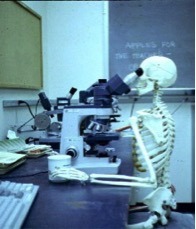
photo A
Once again there are 3-D images taken with powerful SEM/scanning electron microscope (Photo B). I have used both types of microscopes many times in teaching and various research projects!
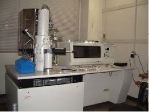
photo B
Now, getting in the mood for today’s Anatomy Lesson: Skin 2 – the Hair! As with skin, Herself often writes about hair in the Outlander books, offering her audience a more intimate glimpse into characters and situations through vivid use of this physical trait. So, once again, I begin our lesson with images from the Starz Outlander series and with words from the Outlander books.
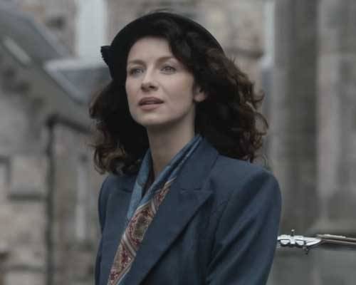
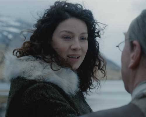
Let’s begin with our heroine. Early in Starz episode 1, Sassenach, Claire emerges from the roadster standing in the picturesque village of Inverness. We can clearly appreciate her dark brown hair – very full and very curly.
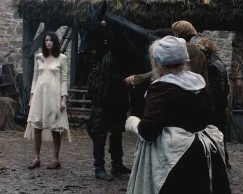
Later, during a lighting storm, Herself writes
The wind was rising and the very air of the bedroom was prickly with electricity. I drew the brush through my hair, making the curls snap with static and spring into knots and furious tangles!
The humid air makes Claire’s hair wildly curly and disobedient (Starz, episode 101, Sassenach) to which she exclaims: Jesus H. Roosevelt Christ!!!

All the while, someone is awatching her futile struggles through the window of her room.
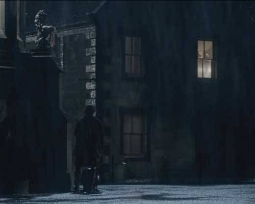
Nay, it isn’t a peeping tom, it is a keeking Jamie! Ha!
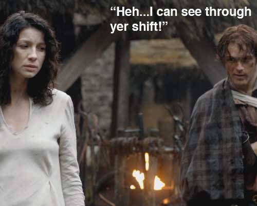
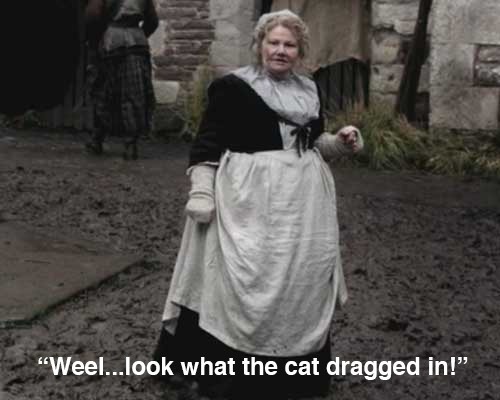
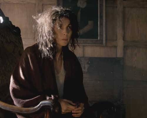
This next image of Claire always makes me laugh! In Starz episode 102, Castle Leoch, Mrs. Fitz unceremoniously rouses Claire from her sleep, seats her in a chair and hands her a cup ‘o brakfast fer her empty belly. Mrs. Fitz then whisks it away afore Claire even finishes! Look at Claire’s hair! It is absolutely fabulous! She certainly looks like the “wee milkweed” Jamie affectionately calls her later in the Outlander book.
“Fretful porpentine, was it?” he asked. He tilted his head, examining me inquisitively. “Mmm,” he said, running a hand over his head to smooth down his own hair. “Fretful, at least. You’re a fuzzy wee thing when ye wake, to be sure.” He rolled over toward me, reaching out a hand. “Come here, my wee milkweed.”
With these great images to set the mood, it is time for our anatomy lesson on hair and with it a lot of juicy tidbits to share!
First, the length of body hair varies a lot – from less than 1 mm (.04 in) on the forehead to well over 1 m (3.3 ft) with long scalp hair (Photo C)! But, the wee hairs of the eyelids (not the eyelashes) are so short they barely reach the skin surface! And, you should know that most hair grows very rapidly, about 0.3 mm/day or 1 cm/per month.
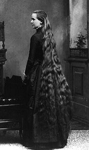
photo C
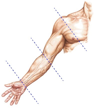
You should also know that hair does not grow straight out of the skin; it emerges at a slant (Photo D).
Try this: Check the angle of growth of your own hair: place your forearm on a flat surface with the palm down. Examine your forearm hairs and see that they are angled toward the little finger side of the forearm. That’s the slant I’m a talking about.
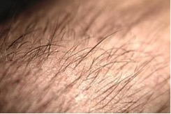
photo D
Hair is also denser in some skin areas than in others: the face has about 600 hairs/cm2 (.16 in2) compared to about 60 hairs/cm2 on the rest of the body.
Hair diameter also varies greatly but even the coarsest hair is only about .5 mm (.02 in) in diameter (Photo E). Even so, a scalp hair is strong enough to support the weight of 100 gm (3.5 oz)!
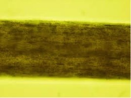
photo E
Another interesting tidbit: Human hair grows autonomously; each hair cycles at its own pace through periods of growth and periods of quiescence. If all our hair were on the same cycle, we would molt!
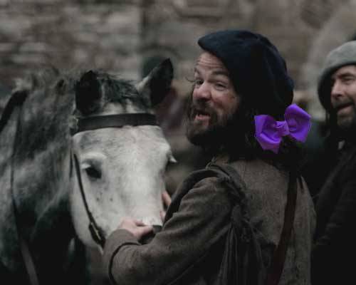
And sometimes our hair does unspeakable things and we just have to pull it outta the way like Angus here who does prefer a wee bit o’ purple ribbon fer his scalp hairs!
Now back to microscopic anatomy! Using the same image from Skin – Part 1, I’ll be reminding ye that skin is divided into a thin outer epidermis that overlies a thicker dermis. And, although not part of skin the hypodermis lies deeper still. The dermis and hypodermis also anchor structures that we’ll cover in this anatomy lesson: hair, hair follicles, arrector pili muscles, and sebaceous glands (Photo F).
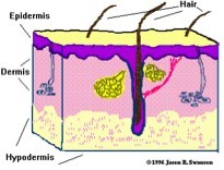
photo F
Hairs emerge from hair follicles which are down growths of the epidermis (Photo G). The internal anatomy of each follicle is verra complex so I’m simplifying it: the hair and its follicle are divided into a hair root and a hair shaft. At the root is a bulb where cells divide and push older cells toward the surface to form the hair shaft!
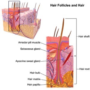
photo G
Along the way, hair cells harden and get plastered together so by the time the hair clears the skin surface, the cells are dead, flat and stiff with their free edges pointing toward the hair tip. They also overlap each other like shingles on a roof (Photo H). This is a SEM image of a single hair!
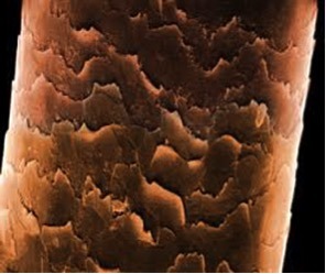
Photo H
Ye should also ken that hair follicles are verra sensitive to the influence of hormones! These chemicals produce secondary sex characteristics such as hair distribution. In fact, the distribution of hair between the two sexes play an important role in socio-sexual communications!
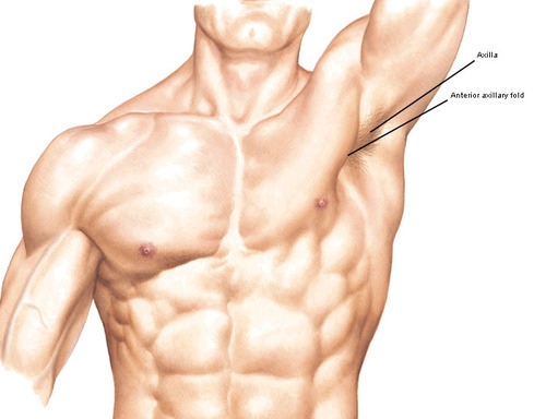
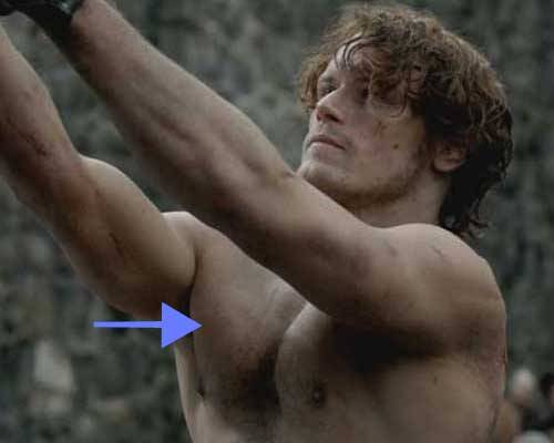
In women, estrogens (oestrogens) cause most body hair to develop as short, thin vellus hairs that are anchored in the dermis. Both genders exhibit the coarse terminal hairs of scalp, eyelashes, eyebrows, axilla and pubis that are embedded deep in the hypodermis.
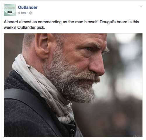
In men, androgens (testosterone being the most important) also convert facial and chest hairs into terminal hairs. Now then, isna this the right place to offer praises to Dougal MacKenzie who won Saturday’s Starz contest with his comely beard? Congrats! It looks mighty fine on ye, man! Tulach Ard!
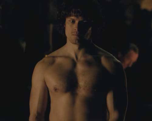
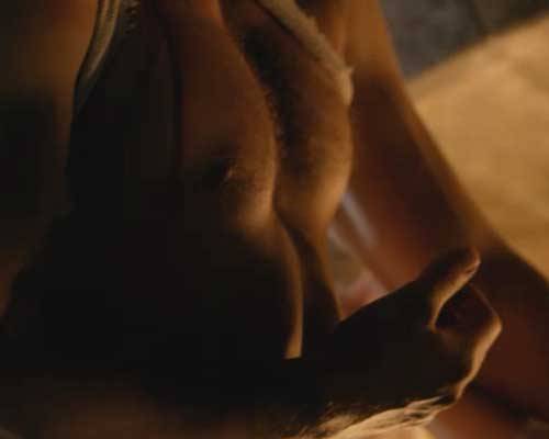
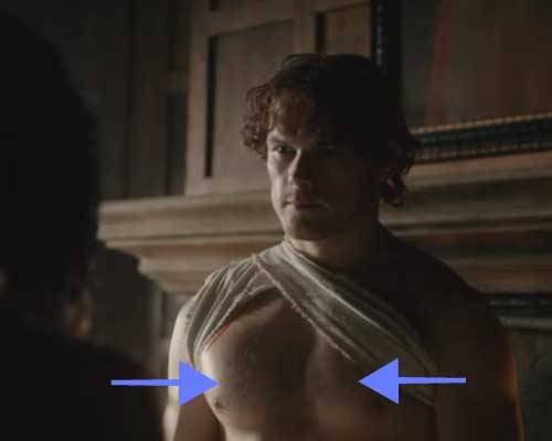
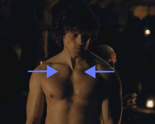
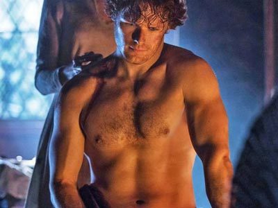
And, no anatomy lecture is ever complete without at least one image of a half-dressed Jamie! So here is his chest hair just in case ye be forgettin’! No verra damn likely! Gawd!
Something else: When viewed by SEM, straight hair has a round shaft as seen in this photo of scalp hair (Photo I –computer generated color); the surrounding dead skin cells look like scatter leaves on a forest floor.
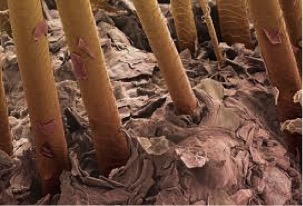
Photo I
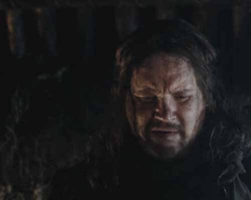
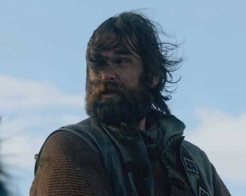
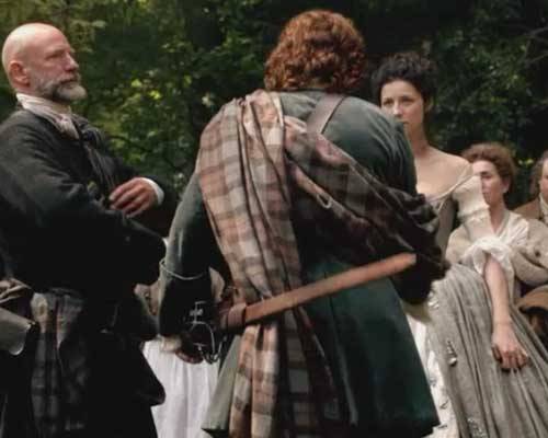
Murtagh’s scalp offers a perfect example of straight hair – here he is explaining to Claire why Jamie is nowhere to be seen (Starz episode 5, Rent)! Plus, he has mighty fine eyes and braw eyebrows just in case ye been so focused on Jamie that ye havena been noticing!
Scalp hair that is curly like Claire’s…
…has a shaft that is flattened in cross-section as shown in this SEM image (Photo J). The flatter the shaft, the curlier the hair!
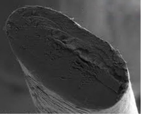
Photo J
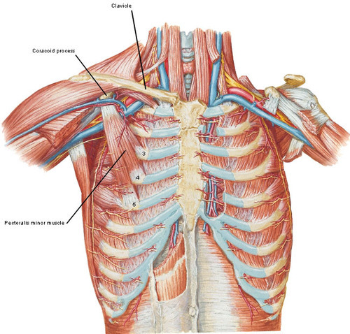
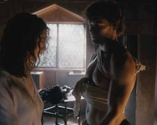
Now, onto a couple of other structures associated with the hair follicle. First, stretched between the follicle and the dermis is a thin band of tissue, the arrector pili muscle. Second, between the hair follicle and the arrector pili muscle lays one or more sebaceous glands with ducts opening into the hair follicle (Photo K). Sebaceous glands produce sebum, a complex mixture of fats, waxes and other materials.
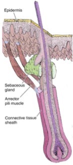
photo K
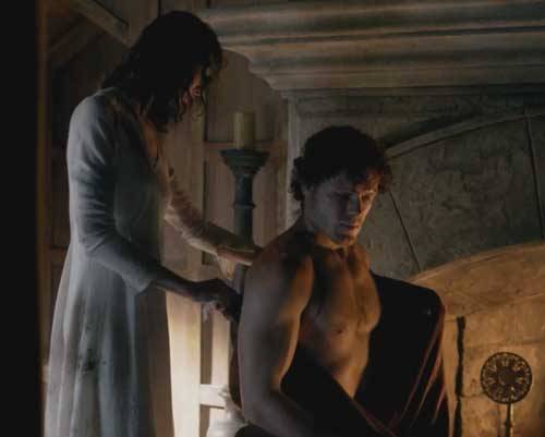
The arrector pili muscles are made of smooth muscle cells that are not under conscious control. They contract in response to cold or the fright, flight, fight reflex! Contractions of this muscle elevate the hair, forming goose bumps or goose flesh and help squeeze sebum from the sebaceous glands into the hair follicle and onto the hair shaft (Photo L).
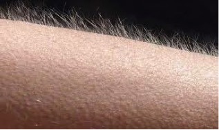
Photo L
Contraction of the arrector pili muscles in animals traps air between the erect hairs to retain body heat or to help the creature appear more fierce (Photo M)! This adaptation isn’t of much use to us short haired humans but the release of sebum does help lubricate and protect the hair itself.
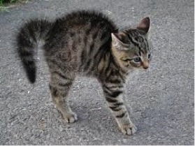
Photo M
Finally, on to hair color! Like the epidermis, hair color requires the presence of melanin; melanocytes in the hair bulb synthesize melanin and package it into granules that move up the hair shaft as it forms. Now, it turns out that there are a couple of different types of melanin!
Like Claire, most hair color is due to the presence of varying amounts of brown or black eumelanin. But, now, ye are in fer a BIG surprise! I bet ye dinna ken this! Flaming red hair in one such as our Great Scott, Jamie, contains a chemically different type of melanin known as pheomelanin and this molecule is red (or red-brown)! Thus, Jamie’s gorgeous mane of red hair is due to the presence of pheomelanin as seen from the back in this image (Starz episode 7, The Wedding)!
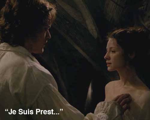
And just so ye won’t ferget it, here is Jamie’s hair from the front! We can literally see the words Herself wrote in Outlander about his hair:
…a mass of auburn, copper, cinnamon and gold all gleaming together in the morning sun…
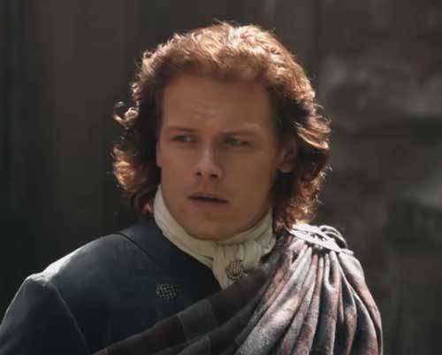
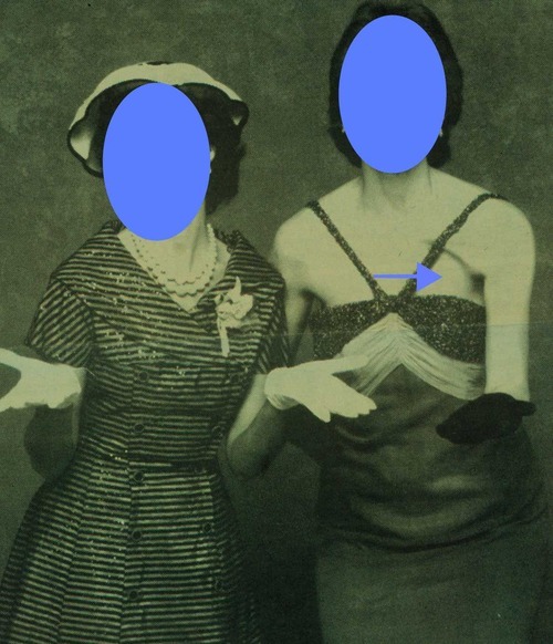
And one last point fer yer eddycation: Check out both upper corners of Jamie’s forehead. See how the hair line is squared off? This is known as the temporal notch; it is a secondary sexual characteristic in men brought about by the influence of testosterone. Women typically have an oval hairline in the corresponding areas of the forehead!
And now, folks, our journey through the skin and its appendages has come to an end! I do hope you have enjoyed learning about the skin ye are in and that of the Outlander cast while we are at it! At some point in the future, I will post Skin 3 – The Breast.
In the meantime, I’ll be leaving ye with these lovely words from Herself in the Outlander book and an image from Starz episode 7 (The Wedding):
You’ve the loveliest hair,” said Jamie, watching me. ….”But it’s so .…curly,” I said, blushing a little….“Aye, of course.” ….He sat up and tugged gently on one curl, stretching it down so that, uncurled, it reached nearly to my breast…
And:
“Mo duinne?”…“It means ’my brown one.’ ”He raised a lock of hair to his lips and smiled, with a look in his eyes that started all the drops of my own blood chasing each other through my veins. Rather a dull color, brown, I’ve always thought,”….”No, I’d not say that, Sassenach. Not dull at all.” He lifted the mass of my hair with both hands and fanned it out. “It’s like the water in a bern, where it ruffles over the stones. Dark in the wavy spots, with bits of silver (auburn on Starz) on the surface where the sun catches it.”
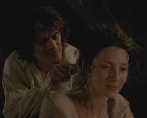
Gah, this man has a way with words! Does he ever say anything wrong? Just look at the look on Claire’s face! She’s both enchanting and enchanted!
Psst…next time, I will be writing about someone’s thighs and knees (guess whose?)! Stay tuned!
The deeply grateful,
Outlander Anatomist
Follow me on:
-
- Twitter: @OutLandAnatomy
- Facebook: OutlandishAnatomyLessons
- Instagram: @outlanderanatomy
- Tumblr: @outlanderanatomy
- Youtube: Outlander Anatomy
Photo credit: Starz, Cat photo from goosecam Edmonton Journal, Goosebumps from genius.com, Basic Histology by Junqueira and Carneiro, 11th ed., University of Leeds, Rochester education Foundation, Wikipedia, WebMD, Loyola University Dermatology website, Histology Guide, University of Leeds, Wikimedia.org. CSIR – Council for Scientific and Industrial Research, South Africa. OA archival photos, Aersol Research – Washington University St. Louis
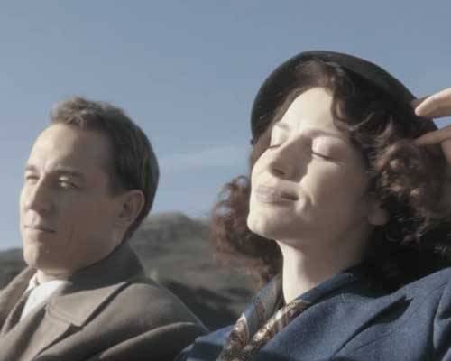
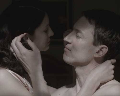
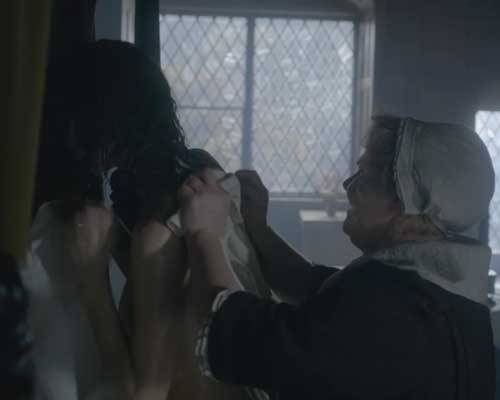
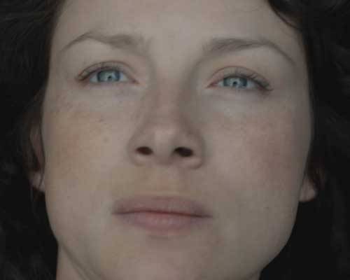
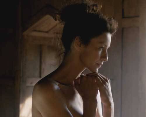
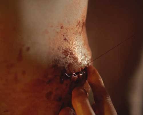
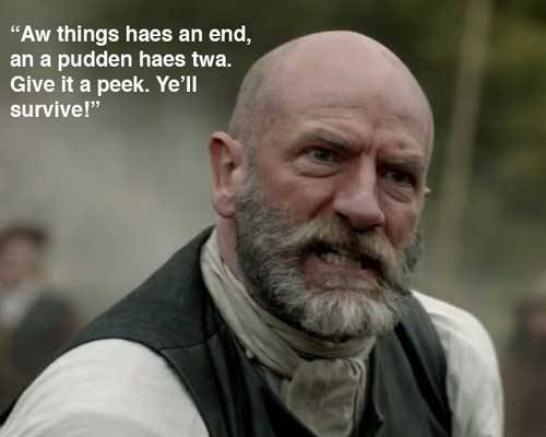
 Photo A
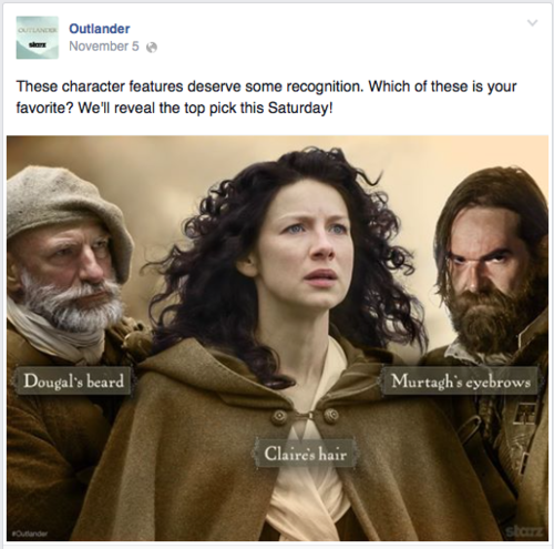
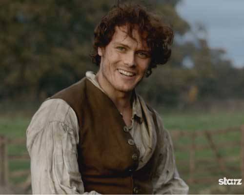
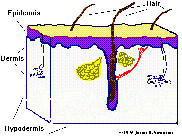 Photo B
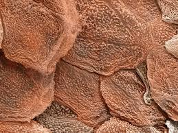 Photo C
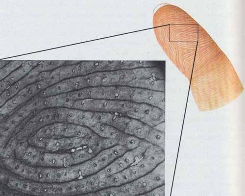
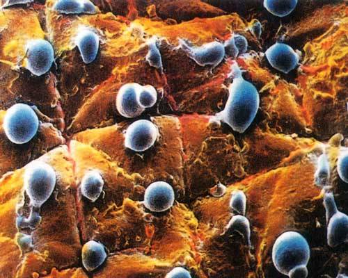

 Photo G
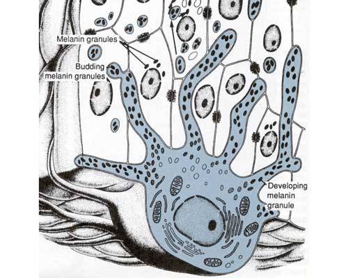 Photo H
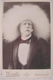 Photo I