Anatomy Def: Cricothyroidotomy is an incision through the cricothyroid ligament. Wait!!! Whaaaat???
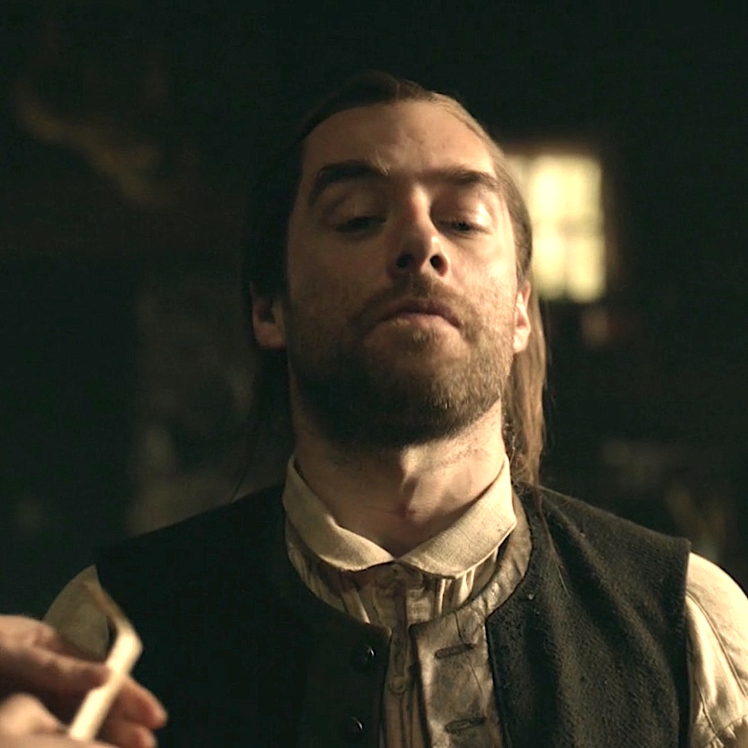
Outlander Def: Puir Roger gets a pipe stem stuck in his neck, not his mouth!
Learn more about the cricothyroid ligament in Anatomy Lesson #42, The Voice!
Here we go! What is the cricothyroid ligament?
Bear with me, here…. In order to understand, we must examine the anatomy of the larynx. This requires a longer Fun Fact than usual – more like a mini-anatomy lesson. 🤓
Larynx: The larynx is what people often call the voice box. Regardless of the name, it is an anatomical wonder formed of a whopping nine different cartilages squished into the upper neck! Together, these cartilages create a hollow conduit for air to pass from nose/mouth/pharynx to the trachea and thence into the lung airways.
We will investigate two of these cartilages and the ligament that unites them.
-
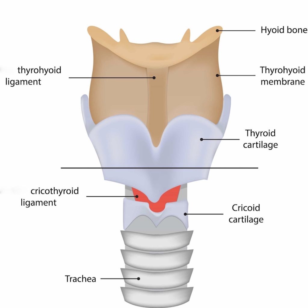
- Thyroid cartilage – shaped like a shield
- Cricoid cartilage – shaped like a signet ring turned backward
- Cricothyroid ligament – tissue connecting thyroid and cricoid cartilages
The below figure shows these structures from a frontal view 👇🏻. Notice that the trachea begins directly below the cricoid cartilage.
If the upper airway is obstructed, a temporary hole may be cut in the conduit allowing air into and out of the lungs.
Bingo! A cricothyroidotomy is an incision in the cricothyroid ligament to reestablish airflow after an obstruction. Yay!
This procedure is also known as a cricothyrotomy. In Roger’s case, the obstruction is caused by swelling of tissues following a short-drop hanging and subsequent strangling 😱.
But, wait! Is there a correct spot to cut the neck? After all, it contains a host of crucial structures:
-
- Carotid arteries (supply blood to most of head)
- Recurrent laryngeal nerves (cut these and the vocal cords are paralyzed)
- Thyroid isthmus (bridge) between right and left lobes of thyroid gland (cutting causes major bleeding)
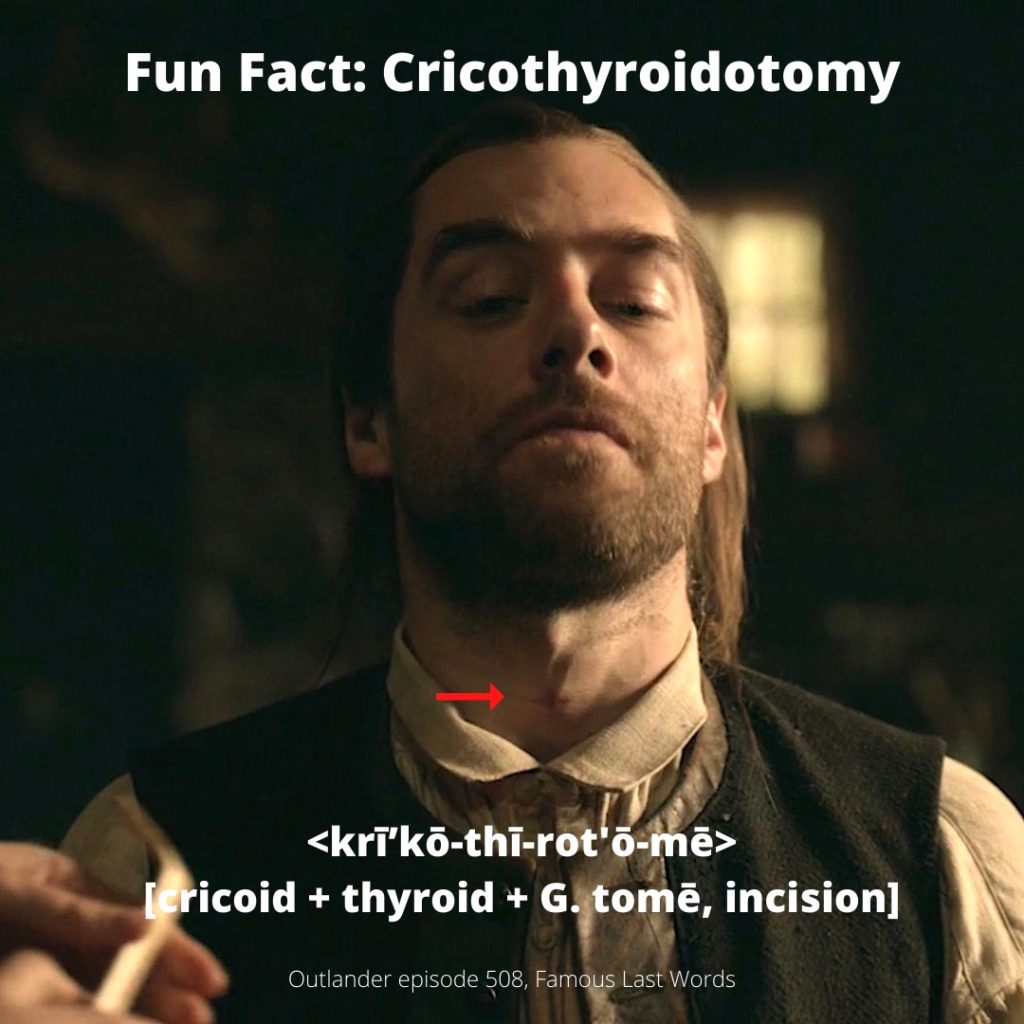
But, of course there is a correct spot! A skilled physician makes the cut using important anatomical landmarks! Let’s use Jamie’s neck to demonstrate👇🏻!
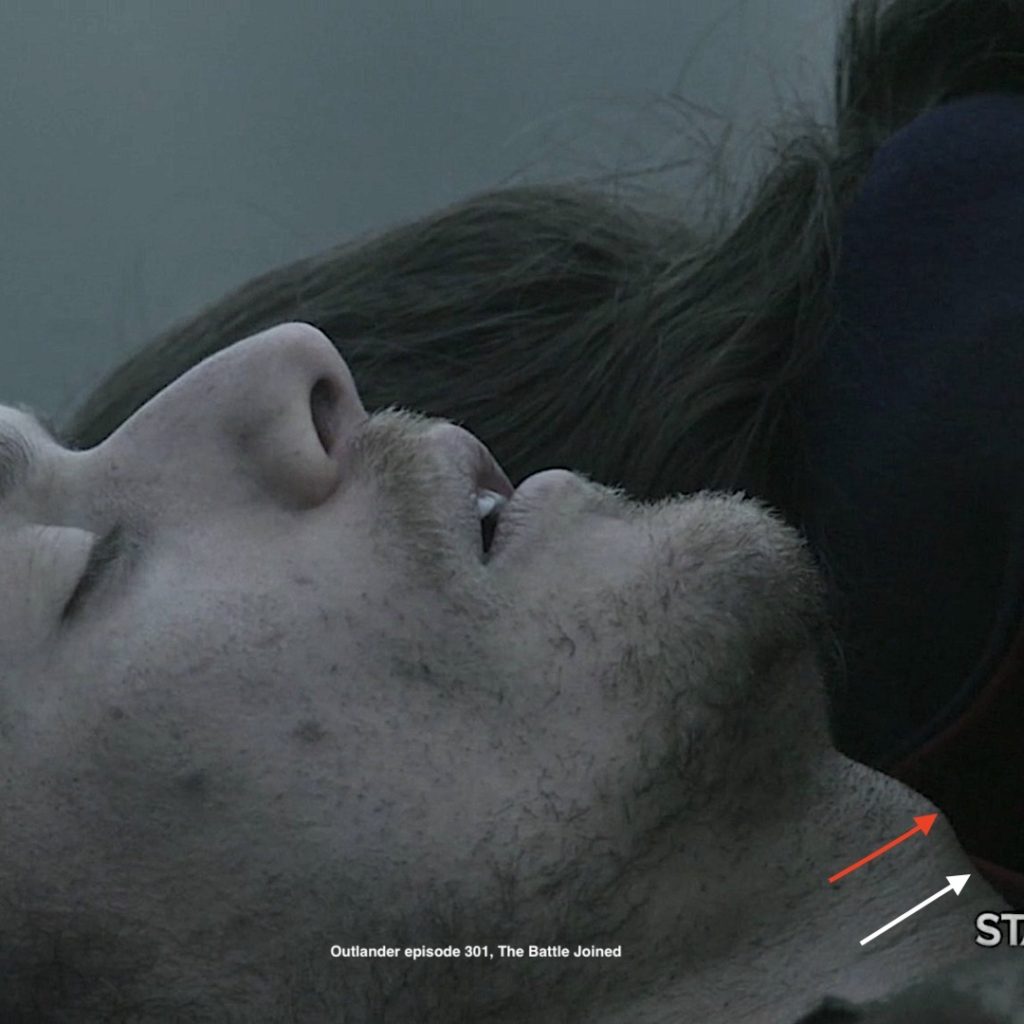
The red arrow points to Jamie’s Adam’s apple, anatomically known as the laryngeal prominence, a mound on the thyroid cartilage. A finger is placed on the laryngeal prominence (red arrow) and then moved downward until it enters a dip. The dip marks the location of cricothyroid ligament (white arrow); the cricoid cartilage lies below. A knife tip can be safely inserted in this dip but only in the midline to avoid injury to the above structures.
Try This: Lay face up. Relax your throat. Locate your laryngeal prominence. Slowly move finger downwards until you feel a dip, the cricothyroid ligament. It won’t be far, roughly an inch or so. You will also feel a very firm structure below the dip, the cricoid cartilage. Oh! Well done, all!!!🥇
The episode did not show Claire locating the cricothyroid ligament, but obviously, she found it because Roger is still with us!
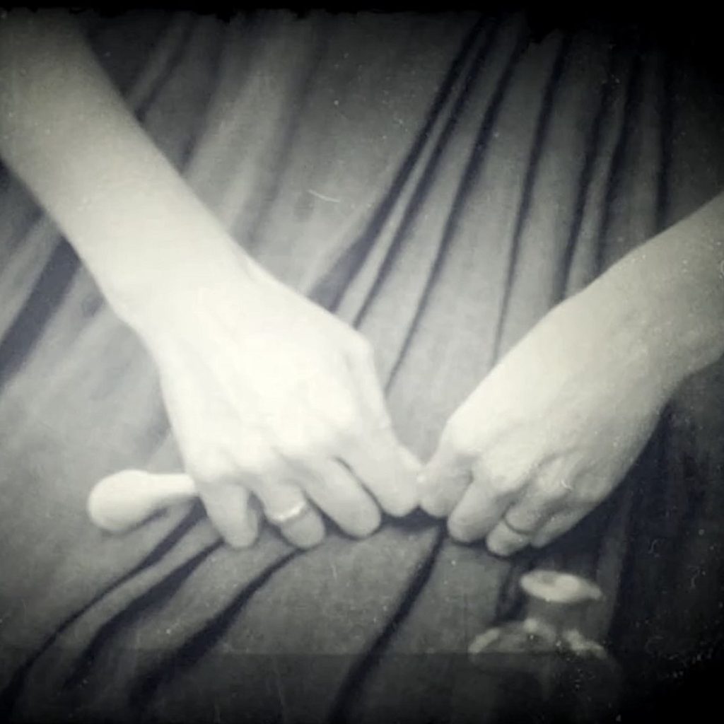
Claire makes the incision!
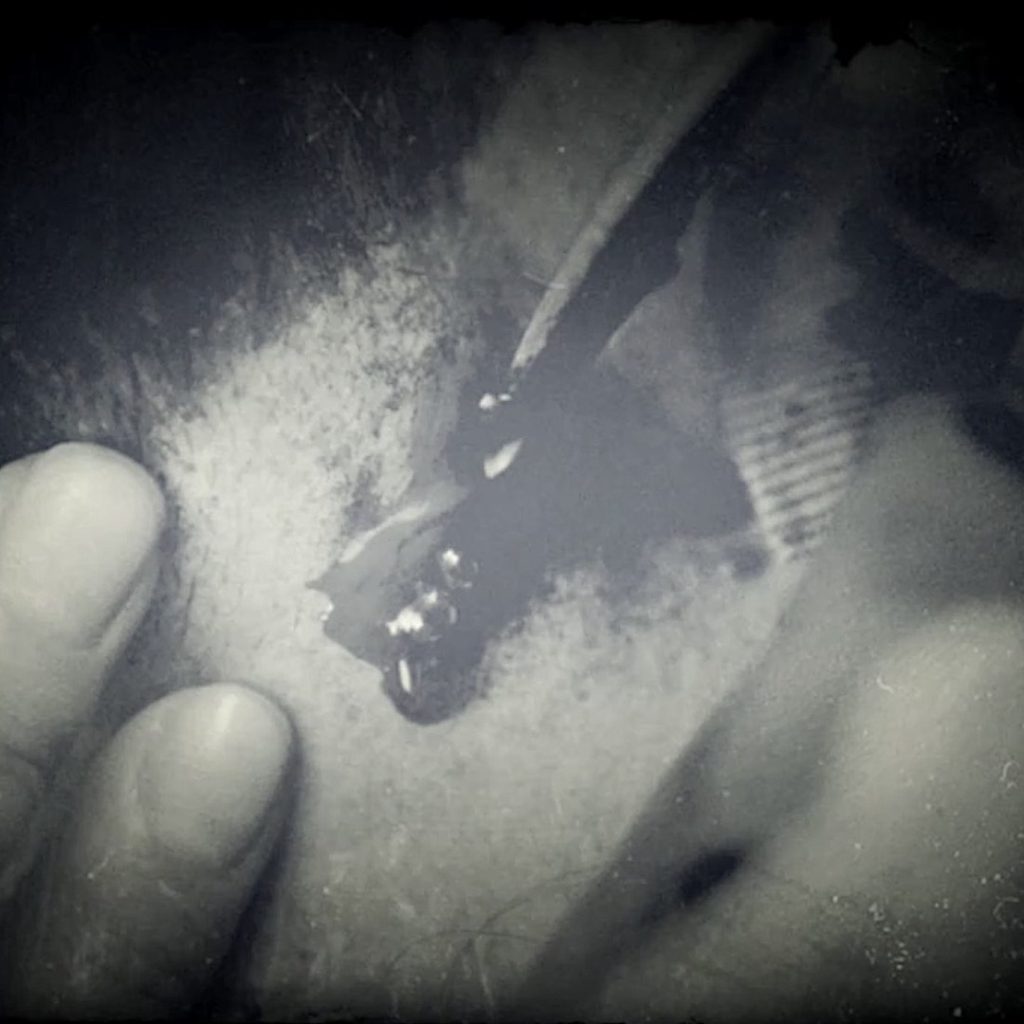
The incision will collapse unless it is held open. So, Claire temporarily inserts what appears to be a wooden peg into the cricothyroidotomy.
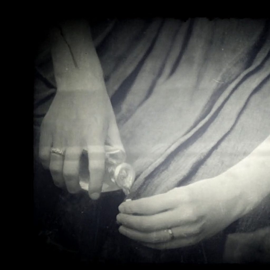
But, a wooden peg doesn’t permit air flow, so a hollow tube is needed, STAT! Brilliant surgeon that she is, Claire commanders Mr. Caswell’s imported English pipe, ruthlessly breaking off the hollow stem!
Rinsing it with alcohol to disinfect……
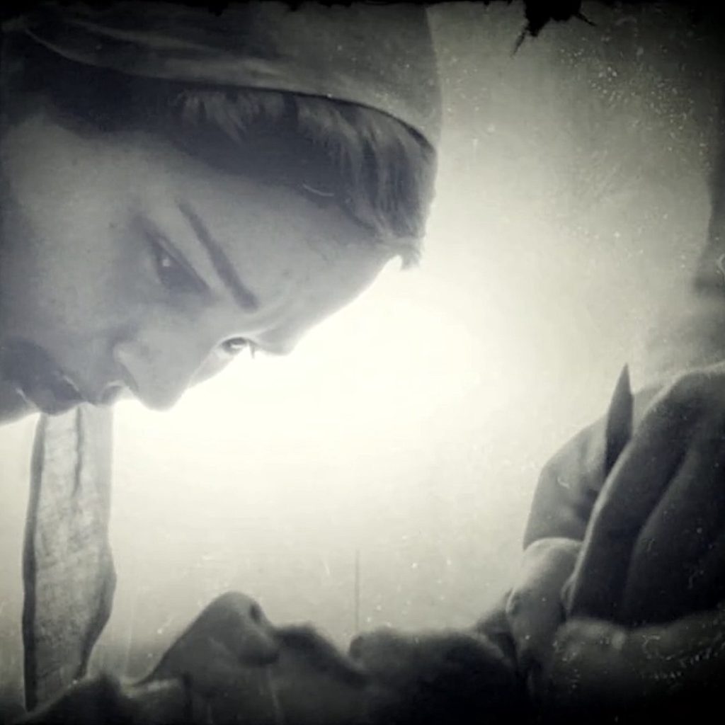
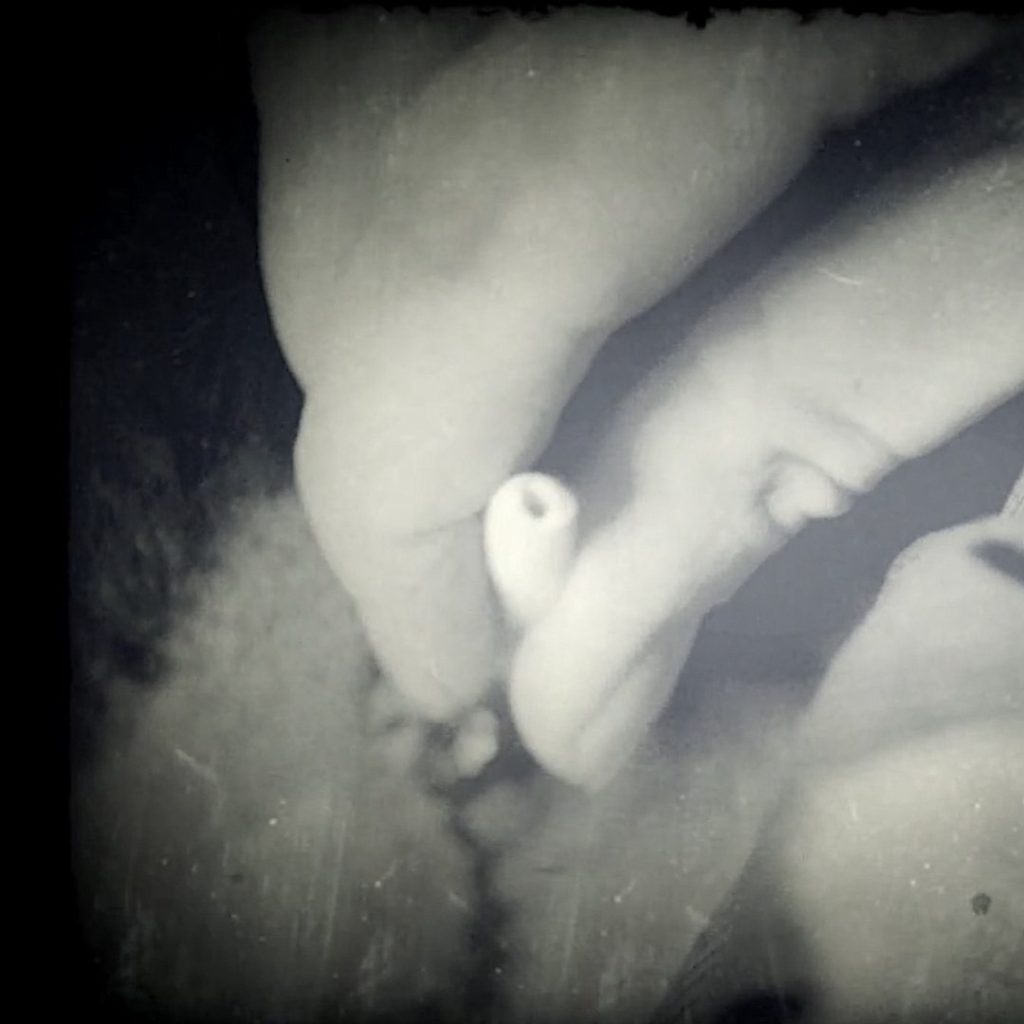
And successfully inserting the cleaned stem into the cricothyroidotomy.
Voilà! Roger breathes through the tube thus bypassing swollen and mangled tissues of his upper airway.
Read about Roger’s throat surgery in Diana’s fabulous tome, The Fiery Cross. In the book, Claire gives Roger a tracheotomy, not a circothyroidotomy.
A tracheotomy is a bit more perilous because it is done very near the thyroid isthmus, the landmarks are less sure, and the neck must be hyperextended – not as good for a “hanged man!” 😳
I took a moment, hands on his neck, eyes closed, feeling for the faint throb of the artery, the slightly softer mass of the thyroid. I pressed upward; yes, it moved. I massaged the isthmus of the thyroid, pushing it out of the way, hard toward his head, and with my other hand, pressed the knife blade down into the fourth tracheal cartilage.
…The cartilage here was U-shaped, the esophagus behind it soft and vulnerable; I must not stab too deeply. I felt the fibrous parting of skin and fascia, resistance, then the soft pop as the blade went in. There was a sudden loud gurgle, and a wet kind of whistling noise; the sound of air being sucked through blood. Roger’s chest moved. I felt it, and it was only then that I realized my eyes were still shut.
…He was breathing, though; she could hear the faint whistle of air through the tube in his throat. Claire had commandeered Mr. Caswell’s imported English pipe, ruthlessly breaking off the amber stem. Rinsed hastily with alcohol, it was still stained with tobacco tar, but seemed to be functioning well enough.
See Roger’s cricothyroidotomy scar in Outlander episode 508, Famous Last Words.
Roger-the-dodger, dodged an awful fate. Kudos to MIL! 👏🏻👏🏻👏🏻
Wow! Dr. Claire continues to impress! 😲
The deeply grateful,
Outlander Anatomist
Follow me on:
-
- Twitter @OutLandAnatomy
- Join my Facebook Group: OutlandishAnatomyLessons
- Instagram: @outlanderanatomy
- Tumblr: @outlanderanatomy
- Youtube: Outlander Anatomy
Photo credits: Sony/Starz; www.thenakedvocalist.com