Anatomy Lesson #43 – “Hamstring – You Make My Heart Sing!”

Hi, anatomy students! So great to meet with you again. Today’s Anatomy Lesson #43 uncovers (ha) the posterior (back) thigh. We already met the anterior thigh waaaay back in Anatomy Lesson #7, “Jamie’s Thighs or Ode to Joy!” where we studied the quadraceps. Thus, we have much more to learn about the thigh.

Our object is to study muscles of the posterior thigh with two other thigh muscles thrown in for good measure. Some folks don’t find muscles all that compelling (unless they are Jamie’s, of course!); mayhap this lesson will change their minds. Anatomical tidbits are added to hold your attention.

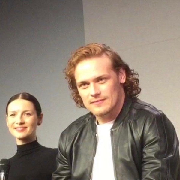
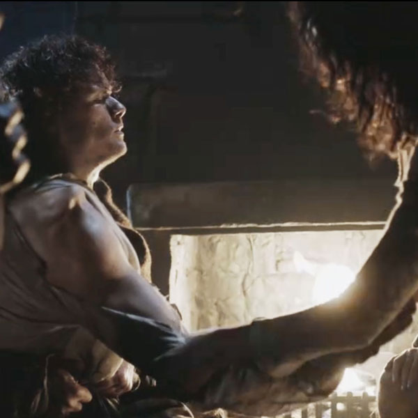
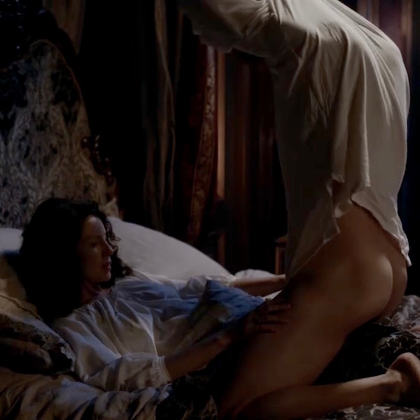
As always, Outlander books and Starz episodes are blatantly scattered throughout. Let’s start with a great view of Jamie’s posterior thighs (Starz episode 112, Lallybroch). Och! Lad, best tuck in those flapping shirt tails! Oops, nothing to tuck into. Where’s a kilt when you need one?

ep-112-thigh-03

Ah, there’s that plaid (Starz episode 109, The Reckoning). Gird up your loins, Jamie! Hee, hee.

ep-109-kilt

Lower Extremity: This lesson starts with essential definitions. Anatomists define the lower limb or lower extremity as the appendage from hip joint through toes. Folks (including me, at times) refer to the lower extremity as the upper leg and lower leg but, in anatomy, this is not so. The thigh is that part of the lower limb between hip and knee joints (Image A – between dashed lines). The leg lies between knee and ankle joints and the foot is distal to (beyond) the ankle joint.
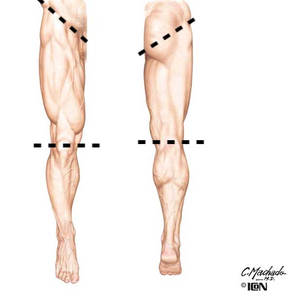
figure0467-thigh-01

Image A

Thigh Overview: Today’s lesson focuses on posterior thigh so let’s define this area. The human thigh is divided into compartments based on connective tissue septa (partitions) and actions; these are best visualized in cross-section (Image B). By convention, cross-sections are interpreted is as if you stand at a person’s feet and look toward the head.

A plane crossing the femur, reveals three compartments, each bearing different muscle groups: anterior (Image B – red), medial (Image B – pink; OK, OK – it’s dusty rose!) and posterior (Image B – lavender). Anterior means front; medial means toward the midline; posterior means back. We will study three muscles of the posterior compartment, one of the anterior and one of the medial.
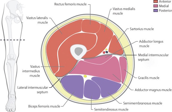
thigh-x-section

Image B

Posterior Thigh Muscles: Three muscles make up the posterior thigh compartment; all are typically long and strong.

  • Semitendinosus
  • Semimembranosus
  • Biceps femoris

Try This: Open the space between thumb and forefinger and grip your posterior thigh about mid-femur. Your entire hand should grasp a sturdy muscle mass containing the three posterior compartment muscles. Appreciate the same anatomy with your other thigh or someone else’s. <g>

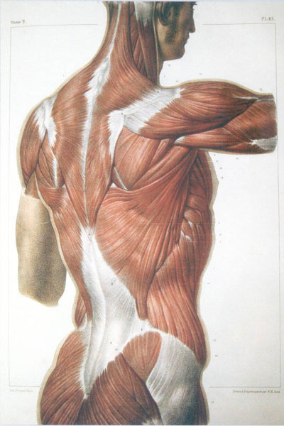
Topographical Anatomy: Looking at topography or surface anatomy of an intact posterior thigh, is it possible to see grooves and ridges created by its muscles? Yes, if the subcutaneous fat layer is not too thick and if the thigh is well-muscled. Posterior thigh (Image C – right lower extremity) bears three muscles in succession: Blue arrow marks semimembranosus, red arrow marks semitendinosus and green arrow shows biceps femoris.
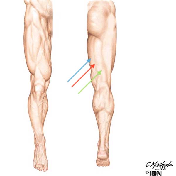
figure0467-thigh-01-c

Image C

Not easy to see? OK, let’s look again using a male body builder (Image D). He is not mooning us, rather, he demonstrates posterior muscles of the lower extremity. Same color coding as Image C and not a challenge to recognize the long columns of posterior thigh muscles. Impressive!

Understand that semitendinosus (red arrow) and semimembranosus (blue arrow) fill the medial (inner) side of posterior thigh and biceps femoris (green arrow) occupies the lateral (outer) side.
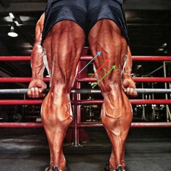
hamstrings-01

Image D

Book Quote: Speaking of long columns of thigh muscles, Diana obligingly (yay!) provides a description of Jamie’s own fine muscular columns, here from Dragonfly in Amber. In case you don’t recognize the scene, these are Claire’s poignant thoughts the eve before she passes through the stones (sob!), back to Frank (gah!). Dinna ken if  “seasoned oakwood in the columns of his thighs” refers to anterior or posterior thigh muscles but, knowing Claire, it was very likely both. Snort!

I touched each soft hollow, the hidden places of his body. Felt the grace and the strength of each curving bone, the marvel of his firm-knit muscles, drawn lean and flexible across the span of his shoulders, smooth and solid down the length of his back, hard as seasoned oakwood in the columns of his thighs.

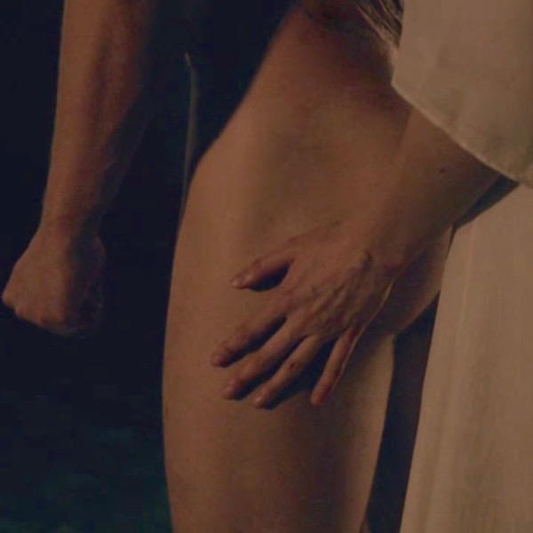
Book quote and photo (Starz episode 204, La Dame Blanche) aren’t a match, but will do in a pinch…the pinch being a love bite on Jamie’s thigh. Man, you are in BIG trubble! Yep, seasoned oakwood it is. Ha, ha!

ep-204-thigh-01

Remove the skin and, voila, three posterior thigh muscles pop into view (Image E – posterior right thigh).

Semitendinosus: Superficial semitendinosus courses down the medial side of posterior thigh. The muscular part ends roughly midway down the femur, transforming into a long, round tendon and curving behind the knee joint to end in the tibia. Did you know, part of semitendinosus tendon can be harvested during knee reconstructions to replace the ACL or anterior cruciate ligament (Anatomy Lesson #7, “Jamie’s Thighs” or “Ode to Joy!”)? Yep, a very useful muscle.

Semimembranosus: Semimembranosus (Image E) is a broad, flat muscle so named because it has an unusually membranous tendon of origin. Although it lies deep to semitendinosus, it is the most medial of the posterior thigh muscles. Semimembranous also drops across the knee joint to end in tibia. Semimembranosus is variable; it may be reduced, enlarged, duplicated or absent.

Biceps Femoris: Longtime students will ken that we studied biceps brachii back in Anatomy Lesson #20, “Arms! Arms! Arms! – Redux.” Well, like the arm, posterior thigh also has a biceps muscle, biceps femoris. Biceps derives from Latin, meaning two heads and femoris reflects its close association with femur. Indeed, biceps femoris (Image E) does have two heads of origin: the long head courses the length of the thigh and the short head is roughly half as long. Both muscle parts unite as a single tendon ending in the fibula. The short head of b. femoris may be absent but otherwise, the muscle demonstrates few variations.

Clinical Correlation: Recently, a professional soccer player suffered major posterior thigh pain. Imaging revealed a large, anomalous (duplicated?) right semimembranosus which was crossed over and compressed by the tendon of semitendinosus. This unusual anatomy created what is known as entrapment syndrome of the posterior thigh, which compresses the popliteal artery (see below) restricting blood flow. This is especially acute during exercise and hence, pain.  Muscles object when deprived of oxygen….. “blood of my blood.”  We know the drill.
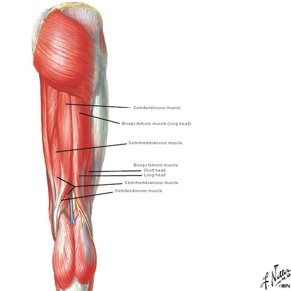
figure0477a-thigh-03

Image E

Remove gluteus maximus and posterior thigh muscles spring into view (Image F). Remove semitendinosus and long lead of b. femoris and, ta da,  semimembranosus and short head of b. femoris say hello! Image F reveals the long, flat membranous tendon of origin for semimembranosus. Short head of b. femoris arises from lower half of femur.

figure0477b-thigh-04

Image F

Moment of Silence, please!: In Image E, the tops of posterior thigh muscles are not visible because gluteus maximus covers them. G. maximus is a large, flat quadrangular-shaped muscle honored in Anatomy Lesson #1, “Jamie’s Tush” and was the original inspiration for Outlander Anatomy lessons! Thus, it is only fitting that we interrupt this lesson to pay homage to lovely body curves created by these, the body’s most massive muscles (Starz, episode 107, The Wedding). Gasp, thud!

ep-107-thigh-06

Muscle Attachments: Next, let’s suss out bony attachments of the posterior thigh muscles – know these and muscle actions make sense. Image G of the posterior hip bone and femur is busy but nicely shows muscle origins (red) and insertions (blue). Origin means the body site (almost always a bone) giving rise to a muscle. Insertion means the site where the muscle ends by attaching to a different bone. Muscles usually cross one or more joints (there are exceptions) – a site where bones meet to allow for movement. Make sense? Dandy! Succinctly put:

  • Origin:
    • Proximal (nearer the body center)
    • Fixed point (least moveable site)
  • Insertion:
    • Distal (further from the body center)
    • Movable (moves with muscle contraction)

Origins and Insertions: Long head of biceps femoris, semitendinosus, and semimembranosus, all take origin from the ischial tuberosity (Image G) of the respective hip bone, cross the hip joint and pass down the posterior thigh. As noted, the short head of b. femoris takes origin from the lower half of femur (Image G). BTW, the sturdy ischial tuberosities are known at gyms as “sits bones” and, indeed, we do sit on them!

After coursing down the posterior thigh, each muscle claims a different insertion site.

Semimembranosus crosses knee joint to end in medial tibia (Image G – inner leg bone).
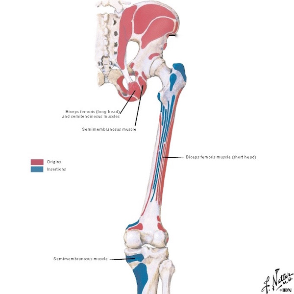
figure0473-thigh-02

Image G

The tendon of semitendinosus swings behind the knee joint and inserts into anteromedial (front/inner) surface of tibia (Image H).

Long and shorts heads of biceps femoris unite as a single tendon which crosses the knee joint to end in the head of fibula (outer leg bone) Image H.
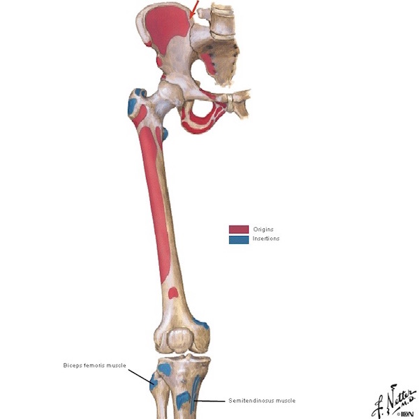
figure0472-use

Image H

Actions: Now for the nitty-gritty: What actions do these muscles perform? Well, they are critical for successful lower limb ambulation: walking, running, climbing, kicking, squatting, jumping, etc. Semitendinosus, semimembranosus and long head (not short head) of b. femoris cross the hip joint so as they contract, the hip joint extends or straightens (Image I – bottom right). All three muscles (including short head) cross the knee joint so as they contract, the knee flexes or bends (Image I – top right). All three muscles counteract forward bending at the hips to keep us from doing a face plant. Whew, busy muscles!
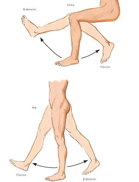
figure0525b

Image I

Hamstrings: Now, here’s an interesting tidbit: Anatomists call semitendinosus, semimembranosus and long head of b. femoris true hamstrings; “ham,” from Old English hom, meaning the hollow or bend of the knee and “string” referring to tendons located there. To receive this “anatomical blessing,” a posterior thigh muscle must meet the following criteria:

  • Take origin from the ischial tuberosity (ergo, short head of b. femoris doesn’t quality)
  • Cross the hip joint causing extension (short head of b. femoris doesn’t quality)
  • Cross the knee joint causing flexion
  • Same innervation (short head of b. femoris has a different innervation)

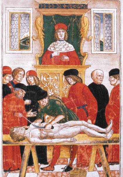
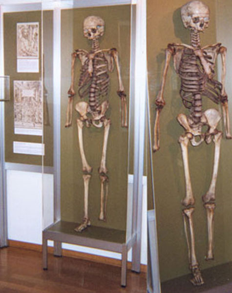
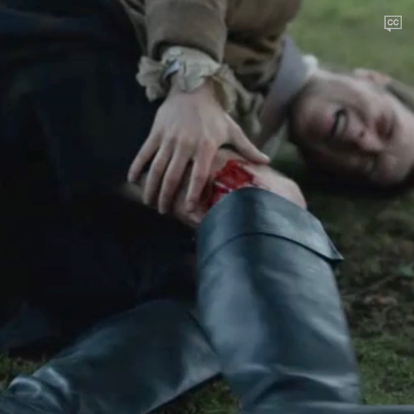
Hamstringing is an dreadful event wherein a victim is incapacitated by severing hamstring tendons at the back of the lower thigh (Image J – in cartoon form, thank goodness!). This laceration was crippling, painful and often caused death by exsanguination from severed vessels at back of the knee (see below). Hamstringing was a time-honored (eek!) method of permanently crippling animals and humans so they could not reengage in future warfare.

Hamstringing is a very old practice. Identified as houghing in the King James Version of the Bible, both Joshua and David ordered hamstringing of chariot horses. Carthaginians hamstrung their Roman enemies as did Germanic tribes. Romans hamstrung elephants and the practice has been revived most recently in Zimbabwe!
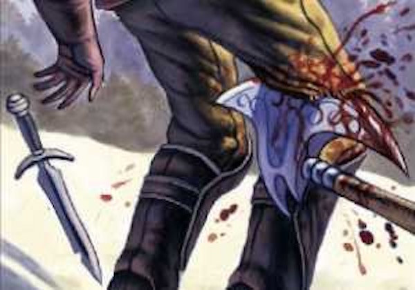
hamstringtcg

Image J

After he is stabbed in the torso by the mouthy MacDonald, Jamie doesn’t fail us! Pulling a dirk with his left hand, he prepares to strike his attacker (Starz episode 110, By the Pricking of My Thumbs). Who’s the cowering coward behind the tree? The Duke, of course. Off with his head!

ep-110-thigh-04

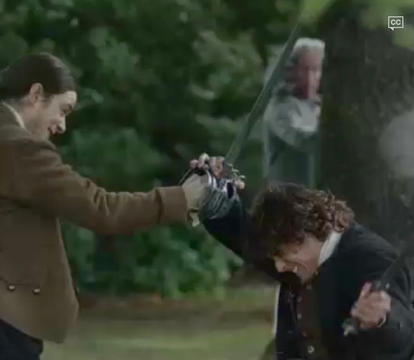
And, strike he does! Down goes MacDonald as Jamie’s blade slices into the unguarded right biceps femoris tendon, hamstringing his opponent. Given the state of 18th century medicine, this MacDonald willna be farming his croft. Ei, Ei, Ohhhhh….. Well, Jamie is a warrior – even if he is cute!

Try This: Want to feel your own hamstrings? Sit in a chair or on a sofa with knees bent and right foot turned outward. Place right fingers beneath outer right thigh (near knee). Pull right heel back against chair rung or couch base. Feel the tendon? This is biceps femoris. Relax and place fingers of left hand behind inner lower thigh, this time with turn foot inward. Pull heel back again. Feel two tendons? The superficial one belongs to semitendinsus and the deep is semimembranosus! A+ for you!

ep-110-thigh-05

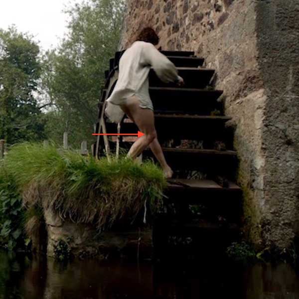
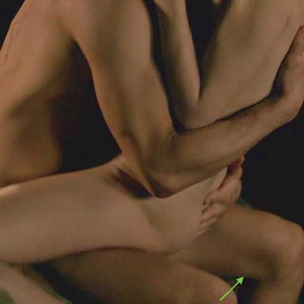
“Down by the old mill stream,” the tendon of Jamie’s right biceps femoris muscle is clearly visible as he shifts weight to the mill wheel. See the taut tendon behind his right knee? Oops, lost some of you! Please focus on the hamstring. Don’t see it? Bummer! <G>

ep-112-thigh-04

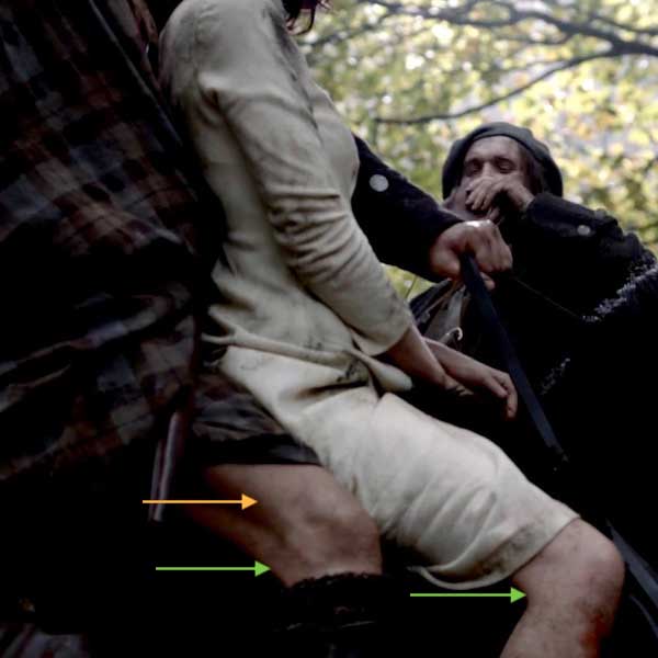
Let’s try again. How about both Jamie and Claire this time (Starz episode 101, Sassenach)? “Here’s to you lass, for tipping us to the villains in the rocks, and gieing us a wee bit of fun,” boasts and toasts Rupert. Green arrows mark tendons of biceps femoris muscles. Can you spy a second band of taut tissue, just forward of Jamie’s biceps tendon? This is the iliotibial tract or IT band (orange arrow – Anatomy Lesson #7, “Jamie’s Thighs or Ode to Joy!”). Yep, keep your eyes peeled for all sorts of Outlander anatomy!

BOOK QUOTE: Both Claire and Jamie have something to say about rubbing thighs during that intimate ride together, although Claire’s is slightly more high-brow. From Outlander book:

My companion seemed to be having little trouble, in spite of being unable to use this right hand. I could feel his thighs behind mine, shifting and pressing occasionally to guide the horse. I clutched the edge of the short saddle in order to stay seated; I had been on horses before, but was by no means the horseman this Jamie was….

but then that ride through the dark together – with that lovely broad arse wedged between my thighs…with a bum like that… What does it matter if she’s a f-face like a ah-ah- sheep?”

Baa, baa, bleats Claire! 🙂

ep-101-thigh-01

It took more than a bit of sleuthing to find an Outlander image showing inner hamstring tendons. Probably the best comes from The Wedding (Starz episode 107). Squee! Shift eyes, ahem, to Jamie’s left inner thigh; green arrow marks a ridge created by tendons of semitendinosus and semimembranosus. Flexed knee brought to you by power of the mighty hamstrings!

Although a bit later in their relationship, this quote from Dragonfly in Amber book says it all!

I scooped out a good bit of the salve and spread it down the long muscle of the thigh, pushing Jamie’s kilt above his hip to keep out of the way. The flesh of his leg was warm; not the heat of infection, only the normal heat of a young male body, flushed with exercise and the glowing pulse of health. I massaged the cream gently into the skin, feeling the swell of the hard muscle, probing the divisions of quadriceps and hamstring. Jamie made a small grunting sound as I rubbed harder. “Hurt?” I asked. “Aye, a bit, but don’t stop,” he answered.

ep-107-thigh-05

Hamstring Injury: Moving on… anyone who has suffered a hamstring pull understands just how much we rely on these muscles. You can scarcely engage in a single lower-limb activity without feeling the burn. Yeow! This is a short list of things which contribute to hamstring injury:

  • Sitting for long periods of time
  • Sitting on a hard surfaces, putting pressure on the hamstrings
  • Kicking any ball (football, soccer)
  • Hurdles
  • Splits
  • Poorly conditioned folks suddenly trying things they once did as teenagers!

Hamstring Stretch: Because we are so dependent on hamstrings, it is important to keep them supple by stretching. This can be done sitting, standing or reclining (Image K). Anatomically, the reclining stretch is safest because shoulders, head, spine and hips are supported by the floor; sitting and standing stretches can result in hunched backs, arched necks and hips out of alignment. Here are some pointers:

  • Keep hips and shoulders flat and aligned
  • Contract abdominal muscles to support the lower back
  • Flex both ankles
  • Grip hands behind the thigh (NOT behind the knee)
  • Gently lift one limb until resistance is met – then stop! Don’t force the lifted limb
  • Don’t arch the neck or back
  • Alternatively, flex the down knee to release tension on the raised hamstrings
  • Alternatively, pass a towel behind the up thigh and gently pull on towel to lift
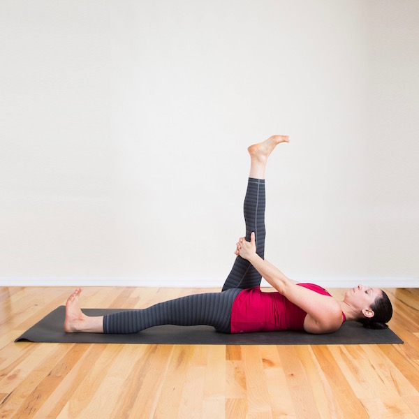
hamstring-stretch

Image K

Videos can be helpful, so the next one demonstrates a good hamstring stretch. As with the above photo, hands go behind the thigh with the interesting addition of flexing alternating with not flexing the ankle joint. Some yoga practitioners grip the great (peace) toe, which works wonderfully if one is a seasoned hamstring stretcher, otherwise avoid!

Sciatic Nerve: A note about the sciatic nerve, the largest of the body (Image L). This behemoth (in adults, it rivals the size of my thumb!) courses down the back of the thigh hidden by gluteus maximus and hamstrings. It gives off twigs to innervate all the hamstring muscles, crosses the knee joint and supplies most of the leg and foot. It is a very busy nerve, hence its size. Sciatic nerve pain is a common affliction but must wait for another lesson. So sorry!

Popliteal Vessels: Behind the knee lies the popliteal fossa, a potential space filled with fat and blood vessels. The large popliteal artery and vein descend through this space to supply knee, leg and foot. These are at risk during hamstringing. If severed, the victim will bleed out, very quickly!
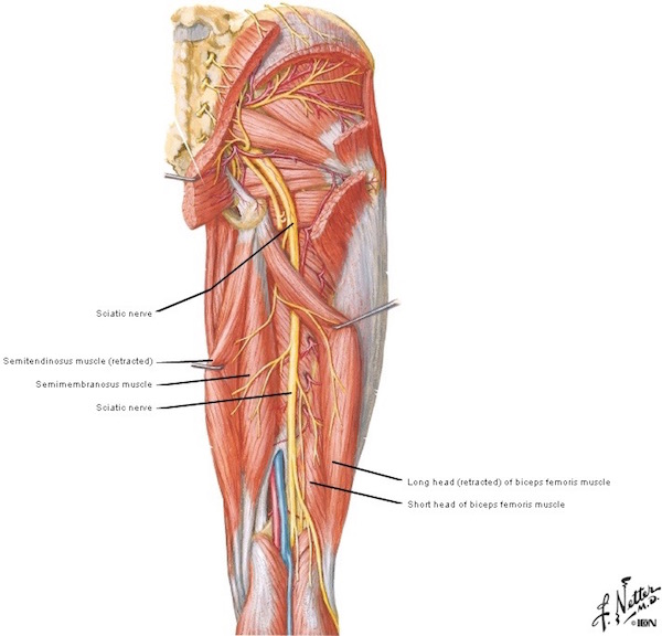
figure0484-sciatic-nerve

Image L

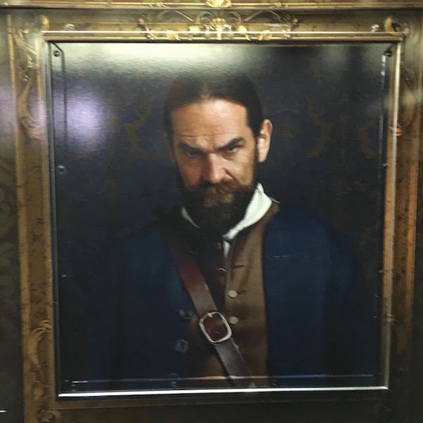
Switching gears, do you recall this terrified wee fellow? He served as Colum’s tailor-of-the-day (Starz episode 103, The Way Out). His wife was a MacKenzie, but kinship won’t protect him from Colum’s dirk! The MacKenzie chieftain was mightily p.o.ed at the tailor because his newly minted frock coat was too long. Did the tailor mean to hide Colum’s legs (Anatomy Lesson #27, “Colum’s Legs and Other Things too!”)? Well, erm, aye, he did, but he hadn’t planed to lose his larynx over a Heiland fashion statement (Anatomy Lesson #42, “The Voice – No, not that One!”).

What in the world does a tailor have to do with the posterior thigh? Read on!

ep-103-tailor-01

Perhaps you recall at lesson start, I mentioned we would cover two other muscles, one from anterior and one from medial thigh compartments (review Image B). Here we go!

Sartorius: The sartorius muscle spirals down the anterior thigh. Its name derives from the Latin sartor meaning, tailor. Why? Because, back when garments were hand sewn, many tailors assumed a cross-legged posture so the knees could support a garment under construction (Image M). Sartorii (pl.) are engaged in adopting the cross-legged position, hence the name.
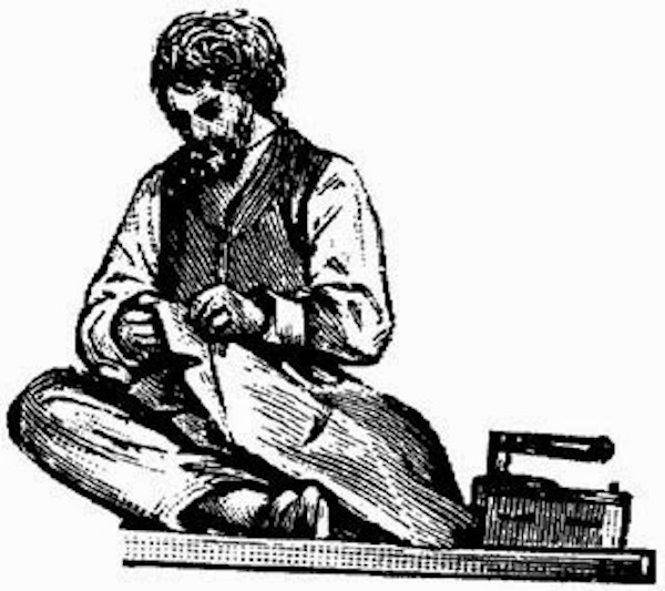
tailor

Image M

The striking sartorius muscles are very apparent in this female body builder (Image N). Thin, flat and superficial, we rarely see sartorii except in folks who are heavily muscled and/or express little subcutaneous fat (Anatomy Lesson #5, “Claire’s Skin – Opal, Ivory and White Velvet”).
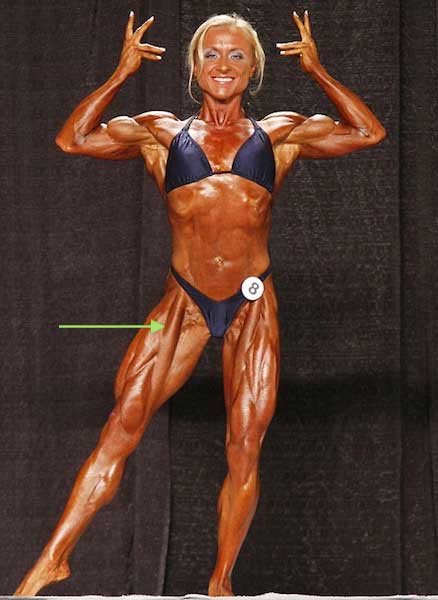
sartorius-01

Image N

Spiraling down the thigh but superficial to the quadriceps (Image O), sartorius is the body’s longest muscle (love superlatives)! Sartorii can be pulled during activities which require a forcefully push off, as with sprinting, jumping and running. Thus, sports, such as hockey, rugby, football or basketball, place one at higher risk of injuring this muscle, although improperly-performed squats can also do the trick. Pain can occur anywhere along the length of sartorius with groin and inner knee as common sites of complaint.
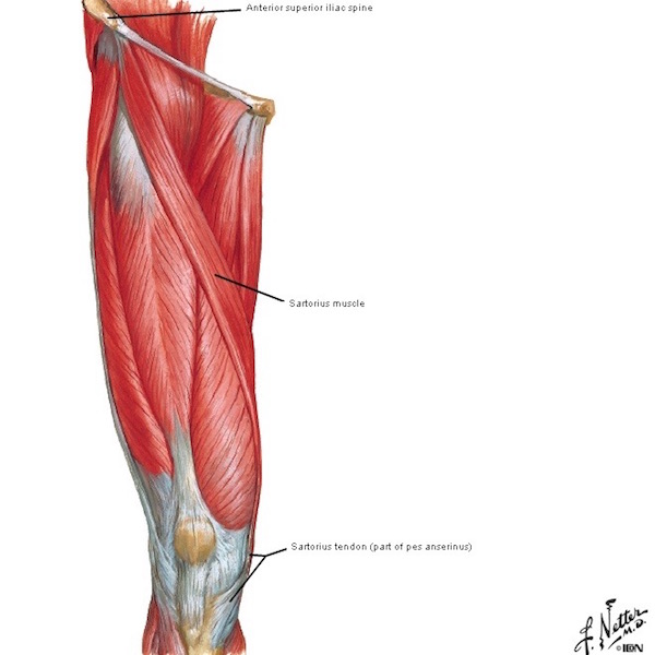
figure0474a-sartorius

Image O

Sartorius belongs to the anterior thigh compartment. It takes origin from a knob of hip bone, the anterior superior iliac spine or ASIS (Image P). Crossing both hip and knee joints, the muscle arcs across the front of the thigh to end in the anteromedial tibia.
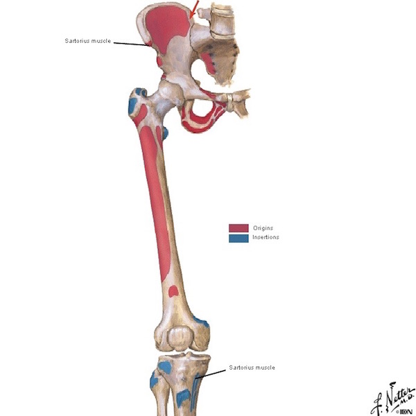


figure0472

Image P

As each sartorius contracts, hip and knee joints flex and knee rotate laterally (to the side) as in yoga’s lotus pose (Image Q). Sartorii don’t appear in Image Q because the feet cover them.
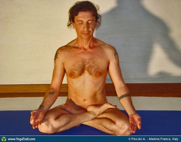
lotus-pose

Image Q

However, this image of a yoga practitioner entering half-bound lotus pose nicely shows the left sartorius (Image R – double arrow); its tendon creates a skin groove just above the arrow.
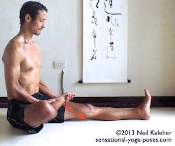
half-bound-lotus

Image R

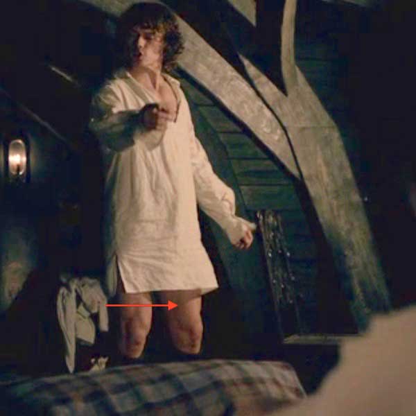
And, Jamie reveals a near perfect sartorius groove (of course) as he scolds new wifey: Lassie, I tell you the truth about Ned Gowan and that strumpet! Ned paid a high price for your splendid wedding dress; a dirty job against which he fought, valiantly. Ha!

See the thigh groove created by Jamie’s left sartorius (Starz episode 107, The Wedding)? Look closely, a red arrow marks the groove directly behind his vast vastus medialis, (Anatomy Lesson #7, “Jamie’s Thighs” or “Ode to Joy!”). Got it? Yay!

ep-107-thigh-03

Gracilis: Graceful gracilis belongs to the medial thigh compartment  (review Image B). Derived from the Latin gracilis meaning slender, this is the most superficial muscle of the medial thigh (Image S). It is thin and flat, broad above, narrow and tapering below. Like true hamstrings and sartorius, gracilis also crosses both hip and knee joints.
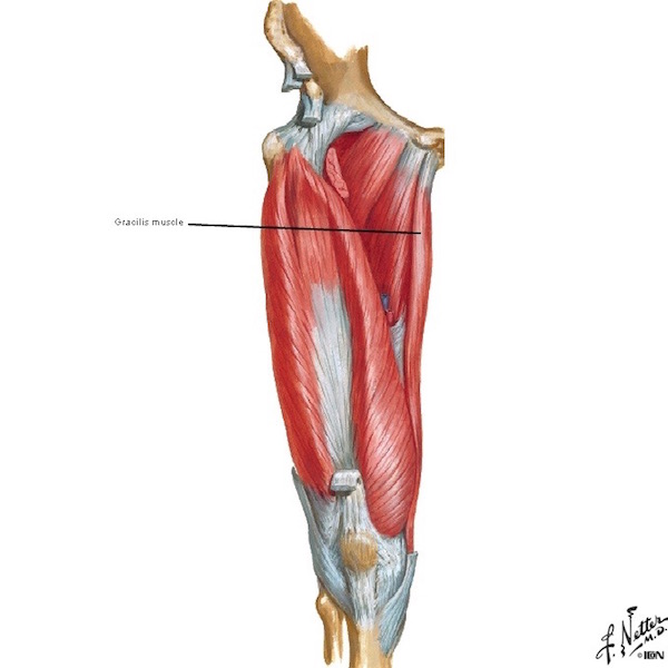
figure0474b-gracilis

Image S

Gracilis takes origin from the pubic bone and inserts on anteromedial tibia (Image T). It helps adduct the thigh (draw toward body midline) and is a weak flexor of both hip and knee joints. Interestingly, gracilis is widely used in reconstructive surgery as a replacement for facial muscles, hand muscles or the external anal sphincter. Loss of the gracilis is not disabling as larger, stronger muscles of the medial compartment are able to compensate.

Clinical Correlation: A few years back, surgeons teaching in my anatomy course presented a case wherein they replaced the external anal sphincter with gracilis muscle, performed on a young woman who had suffered a horrific injury in an auto accident. So shocking and graphic, one of our first year medical students fainted, undoubtedly from vasovagal syncopy wherein heart rate and blood pressure suddenly drop due to extreme emotional distress. Yes, that brought this memorable lecture to a halt! But, the student soon regained consciousness and composure and this gave the surgeon a teaching moment to explain what had happened. I am pleased to report that not only student recovery but the reconstructive surgery were both successful. Surgeons I worked with were very resourceful docs and imaginative thinkers. Their motto: A chance to cut is a chance to cure! Very confident folks, the surgeons.

figure0472-gracilis

Image T

Pes Anserinus: Now, perhaps you spy an insertion theme: tendons of sartorius, gracillis and semitendinosus all converge and insert as a unit into anteromedial tibia. At insertion, they form a webbed configuration known as the pes anserinus, Latin for goose foot. The name, “goose foot,” arises from a three-pronged appearance assumed by the three tendons, each from a different thigh compartment (Image U).  The order of insertion from front to back can be recalled using the acronym for sergeant: SGT for sartorius, gracilis, semitendinosus. Now, this seems especially appropriate because sergeant comes from the Latin servier meaning “one who serves”; an apt moniker for these three hardworking “guy ropes” as they are sometimes called!

All three pes anserinus muscles are primarily knee flexers but studies show that the peculiar three-pronged insertion provides rotatory stability to the knee, meaning together they stabilize external (outward/lateral) rotation of the knee. Hallelujah! Why? Remember, the knee joint is inherently unstable because its constituent bones do not enjoy a ball and socket type of interaction (Anatomy Lesson #7, “Jamie’s Thighs or Ode to Joy!”). Ergo, the joint is provided with numerous ligaments and tendons, including pes anserinus, to ramp-up stability. Go anatomy!
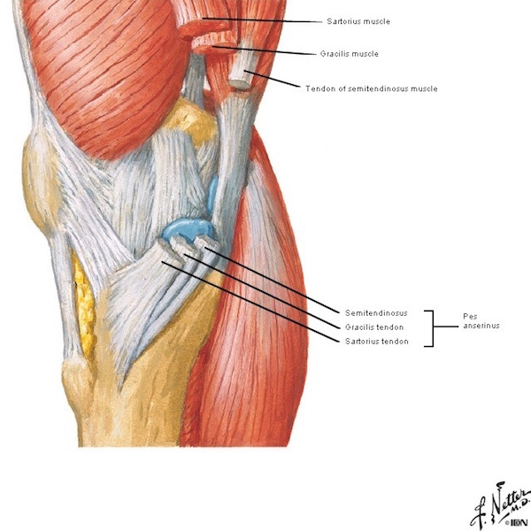
figure0488b

Image U

Goose Foot: Did you see a goose’s foot in the pes anserinus? No? OK, then, let’s look at a goose’s foot! How about now (Image V) – see the three pronged toes joined by webbing? Still not convinced? Understand that early anatomists didn’t have magazines, internet, social media, TV or movies for creative word imagery so they drew on the natural world to devise a new language describing body parts and passions (Lesson #34, “The Amazing Saga of Human Anatomy”). Many of their words may seem quaint but, some 500+ years ago, they were cutting edge!

pes-anserinus-01

Image V

That’s it for the hamstrings and its two closely allied pals. Remember, these muscles are important in virtually all types of ambulation. The next time you stride through a mall seeking new fashions, kick a soccer ball or chase down an errant child, please give a brief nod of thanks for these wonderful muscles!

Do you love art? I love art. Created by Steel Neal and installed at Cooper Square, NYC, “The Agony of Man” is a 1200 pound, 3 times life-size rendition of the human form (Image W). It is constructed entirely of salvaged scrap metal: I-beams, railroad track, rebar, boilers, New York City garbage cans. Teeth (Anatomy Lesson #26, “Jamie’s Chin – Manly Mentus”) were repurposed from road resurfacing equipment. Bony orbits (Anatomy Lesson #30, “Aye, Eye – The Eyes – Part 2!”) were salvaged from original park benches at Madison Square Park. Ribs (Anatomy Lesson #15,“Crouching Grants – Hidden Dagger”) are from the original concrete island of Worth Square.

Sartorii? Yes, there they are, spiraling across the front of thighs – these are made from an extinct type of rebar salvaged from the original foundation of the Union Square Subway Station. Indeed, an evocative rendering of human agony!
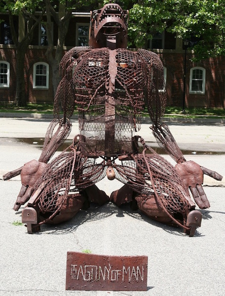
agony-of-man

Image W

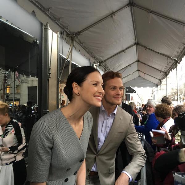
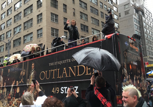
How does human agony fit into a lesson about “hamstring – you make my heart sing?” Well, because we, the wretched, are suffering from drrrrrouuuuughtlander (Photo X)! Gasp! Ron.com, must have a glass of OUTLANDER soon! Dying here!

droughtlander

Image X

All together now:

Hamstring!

You make our hearts sing!

You make everything,

groovy!!! (shamelessly lifted from the Troggs)

A deeply grateful,

Outlander Anatomist

Photo Creds: Starz, Netter’s Atlas of Human Anatomy, 4th edition (Images A, C, E, F, G, H, I, L, O, R, S, T), Outlander Anatomy photo collection, www.christineshipjoint.blogspot.com (Image M), www.getbig.com (Image N), www.keyword-suggestions.com (Image U), www.mnn.com (Image W), www.popsugar.com (Image K), www.sartoriusmusclepainigi.wordpress.com (Image V), www.seannal.com (Image D), www.sensation-yoga-poses.com (Image Q), www.thelancetnorway.com (Image B), www.wowwiki.wikia.com (Image J), www.yogatrail.com (Image P)

My Tartan Affair 2016!

My apologies this post took so many days to go live. Today, April 18th, is tax day in the states; otherwise, the taxman cometh, so I had to set aside my blog for the IRS!

This year, my Tartan Affair week began under the sunny blue skies of LA where I attended the Writer’s Guild Theater’s, “A Tribute to Outlander” on March 31. Many fans stood in line for hours in order to secure good seats. Gah, concrete is rough on the metatarsals!

LA event 01

The WGT panel included (from L to R – next two images): Tobias, Sam, Caitriona, Matt Roberts, Toni Graphia, Anne Kenny, Maril Davis, Ron D. Moore, and the panel moderator, Anne Thompson. Sadly, we wished Ira Steven Behr could have been there.

What followed was a riveting hour of fascinating exchanges, mostly between the writers/producers. We learned many things about them including that writers often advocate for their fav character (e.g. Matt for Geillis).

The writer’s war room boasts a variety of whiskies, presumably as calming anodynes. The group starts with one of Diana’s books broken into segments that are posted on a story board. Together they re-build a story segment that is compatible with the visual medium. Surprise! They often disagree!

LA panel 01

We learned that Dragonfly in Amber for S.2 was more challenging to adapt than Outlander for S.1. Cait expressed delighted that the series gives the actors a wonderful range of acting opportunities to enjoy. Sam commented that it is almost as if he learns a new skill for each episode, and Ron noted that because the story goes in so many different directions, there is no cliché term to describe Outlander!

We heard a very charming Tobias tale about when he was bit by the acting bug…as a young lad, he attended a theatrical production of Wind in the Willows. Intermission found him in the bathroom where Badger was standing at a urinal (loved the way Tobias pronounced this word, ur-y-nal). This was a lightbulb-moment as he realized that Badger was a human actor and this set him on his path to stardom!

We learned that a production featuring magic by Merlin was the impetus for Sam to pursue an acting career and Cait was influenced by a school play in which she donned her Father’s coat and a pillow as a costume.

This event did not include a meet and greet for the admiring fans, but we did enjoy hearing from the writers about how the whole production thing works. Last but not least, we learned that Anne thinks Jamie is a bit of a caveman! what? No way, Anne! <G>

LA panel 02

Headed back to the Pacific Northwest for one day and then off to NY Tartan Week! Let’s dissect Tartan Week, one day at a time!

Day 1 (Sunday, April 3)

Arrived in NYC. Met my roommate, and then dinner with pals at St. Andrew’s pub on W. 46th Street where I tentatively had my first haggis hookup! I felt a little Laoghaire…ooops, I mean leery, but it was surprisingly tasty! I kid you not!

Haggis 01

Photo by JoKc

Day 2 (Monday, April 4)

Evening and we headed to the LeFrak IMAX Theater at the American Museum of Natural History for the Outlander S.2 Premier! We were pummeled by a cold rain but were quickly ushered inside. Fans without tickets waited patiently in the elements but were eventually admitted. So happy for them!

Inside the theater, Starz interviewed many fans. Turns out the interviewer reads Outlander Anatomy blog, bless her heart and its ventricles…but I did not catch her name. ?

Premier interview 03

Photo by JoKc

The evening portended well because I found myself seated next to the Outlander Herbalist! Then, we were quickly greeted with the “guess whose coming to dinner” image on the big screen!

Premier 01

Entertainment followed with a beautiful rendition of The Skye Boat Song, Season 2 version, performed by Bear McCreary on accordion, wife Raya Yarbrough on vocals, a bodhran and violin (sorry, don’t know the names of the last two artists).

The Premiere began with Claire on Craigh na dun (no photography allowed). By now, you all have seen ep 201 and like me, probably found yourself weeping in tune with Claire. She sure does emote a weepy, wobbly chin worthy of her grief! The first half of the episode contains some darned intense material and I couldn’t help drawing parallels between Frank and BJR. Utter silence from the audience such that you could have heard a pin drop until Jamie appeared grasping Claire’s fingers and then the entire theater erupted in shouts of joy and wild applause! Everyone I spoke with loved the adaptation of the episode…including me!

Premier 02

Day 3 (Tuesday, April 5)

As soon as stores opened, I promptly went for a shop in the garment district – beads and ribbons required. When what to my wondering eyes should appear but Outlander bus speeding down 7th Avenue! I ran but no one batted an eye as a grey-haired gal in snappy, red boots gave chase – it is NYC after all!

Outlander bus 01

Then, three blocks later, Eureka! There it was, Outlander bus, taking a brunch break at curbside! Much more effective than a sandwich board!

Outlander bus 02

That evening we braved the cold for a few more hours outside NYU Skirball Center for the Performing Arts awaiting the Outlander “From Scotland to Paris” event presented by the Television Academy (folks that bring us the Emmys!). After all invitees were seated, the overflow fans were welcomed in and we found wonderful balcony seats.

Skirball Outlander-panel

We were blessed with an excellent panel discussion on teamwork, collaborations, production, costume design, set creation, and music development. Beautiful slides flashed on the screen as Tobias, Cait, Sam, Ron, Terry, Maril, Jon Gary Steele, (Production Design) and Bear added commentary. The lively discussion flowed so easily and naturally between the panelists that it felt as if we were watching a large, diverse and friendly family at work: Outlander family, that is. Thrilling! Here is a link to the panel discussion and well worth watching!

Photo by Television Academy

Outside the Skirball Center, Tobias, Caitriona and Sam patiently gave autographs and selfies to avid fans.  I am not very tall, so I had to stretch to get my eyeballs in this photo!

Guess who?

BJR and Me

Day 4 (Wednesday, April 6)

My first ever trip to see Lady Liberty; she had me at “hello”! Tears flowed over this one. I canna help but feel sad for her puir right arm… her deltoid muscle must be exhausted after years of holding up that weighty torch!

Lady Liberty

Afternoon found us headed to the elegant Apple Store, SoHo for this event:

Apple event 02

After waiting for several hours to be admitted to the theater (yes, it has one), we gave a rousing fan welcome to Maril, Cait, Sam and Tobias. A few narrator questions and then the audience got its chance.

I was chosen and promptly forgot my question. Hee, hee, I wonder why!

Apple event 03

Photo by Joanne

IMG_3067

Photo by Rachel

My question to the stars was: had they ever been surprised by a scene that ended up on the editing room floor (apparently, they don’t see scenes until edited), and if yes, which one surprised them the most? Two great answers. Cait wished a S.1 scene had been retained wherein Frank talked about how WW II had shaped him. Maril added that a scene with Fergus has been omitted from S.2, but didn’t elaborate. Thinking caps on: which scene do you think she had in mind? The next video captures some of that Q&A.

Video by Joanne

Turning girlie for a just a moment, everyone loved Cait’s classy black pumps with the wavy straps (and those awesome pins)! Oh là là là là!

After a few more audience questions, the quartet closed with a group-selfie! Quick autographs and more photo ops outside and then they were whisked away in a sleek SUV!

      Apple event 04

Day 5 (Thursday, April 7)

Rain again! Headed for Saks Fifth Avenue in the early afternoon for Outlander Season 2 window unveiling and costume display! Four windows featuring Terry Dresbach’s costuming genius were up for all of Tartan Week. Every costume was splendid with inventive details, superb workmanship, and stunning design!

A fine example is Claire’s Dior-inspired Bar Suit! Pssst: the following videos have black-outs near the bottom of each costume as the image is briefly lost…..this is where window signs video-bomb the images. You can also hear street noise. So sorry about this, but I’m a novice. Hand me a scalpel and I’m in my element. <G>

Details of the beautiful, curved Bar Suit sleeve! The oyster-shell color is both sophisticated and spectacular! Terry just posted a wonderful article about her inspiration and process; I highly recommend reading: Dior!

Saks Dior sleeve

Next, let’s follow a video of Claire’s yellow-gloved garden number!

The next image shows a close-up of the embroidered skirt. A few images down is a video of Terry talking about this amazing fabric!

Saks embroidery

Photo courtesy of JoKc

Next is a video of Jamie’s court duds from trapezius to toes! Verra manly indeed!

This is a closeup of Jamie’s embroidered waistcoat and buttons. Love the spectacular but subtle color scheme!

vest embroidery

But, today, the fifth and final window was unveiled to reveal THE RED DRESS!

The red-dress bodice features a flattering under-the-bosom criss-cross, a feature that is cleverly echoed in sleeve details! Add the drop-waist piping and itsy-bitsy, teenie-weenie pleats and, voila, scarlet perfection! A thin band of wispy stuff follows the neckline….very tasteful, very feminine, very alluring!

Saks Red Bodice

Mayhap Claire’s gold-colored fan will provide a modicum of modesty to that plunging neckline… a perfect 10!

Saks Red Fan

Claire’s beautiful pumps complete the incomparable ensemble. Featuring five stone encrusted buckles and four straps (skirt covers the top strap), the shoe design again echoes the below-bosom banding of this absolutely breath-taking gown. Canna wait to see Jamie’s face as he spies Claire laced up in this daring dress. OK, I saw his face in ep 202 and was so happy because it appropriately registered shock and awe!

red dress shoes

I asked the charming Mark Briggs, Executive Vice President of Saks Fifth Avenue, if partnering with future Outlander costume events lay on the horizon. He answered that it is a distinct possibility as Saks is always seeking the “finest in fashion.” I think Saks got it in spades with Terry’s designs and this flawless window display! What say you?

Saks Mark Briggs

Next, I asked Ron D. Moore if he would kindly comment on Outlander mars and scars. He offered an erudite explanation of how his team creates Jamie’s back scar prosthesis and other more complicated injuries. He also mentioned an injury from S.2 wherein Claire volunteers at L’Hôpital des Anges and is confronted with a patient who has a significant wound to treat! Yay, another Outlander Owie!

Then Cait and Sam made their way down the line looking happy, charming, and drop-dead gorgeous (both of them)! Thud!

cait sam

My last question was for Terry. I asked her how the anatomy of the human body informs her costume design, using a specific element of the Bar Suit as an example. She said anatomy most certainly does play a role; she even has a term for this relationship. I hope to explore this topic with Terry at a future date.

In case you haven’t heard, Team Terry made 10,000 garments for S.2! Yes, her team stitched them all because there isn’t a clothing rack of ready-made 18th century costumes available for purchase. They also created those fab 18th century shoes (check out the little puff balls on Claire’s shoes in the bar suit video above)! Like others, I am thinking there are many hard-earned and well-deserved awards in store for Terry!

Erin Conrad of Three If By Space asked Terry to comment on the wonderful fabric for the yellow-glove garden gown. Terry shared an entertaining story about going to the basement of her fav SanFran fabric store where she found the embroidered fabric for the gown (shown in the window). Listen to her delightful experience.

Day 6 (Friday, April 8)

More shopping in the amazing garment district. Found a store that carried only zippers; every size, shape, color, pull, teeth, material, and length one can imagine. Another specialized in ribbons, others in beads, another in leathers, another in threads. Whew, the eye candy was almost (almost) overwhelming!

Night time found me and friends hoofing it to the Beer Authority for beverages and a “street fair” which wasn’t on a street at all; it was actually held on the pub’s 3rd floor. Belhaven Brewery, famous for Scottish beers and ales, was a sponsor of this event as well as all of Tartan Week. Fans got a photo op with Davie Stewart, Sam’s driver!

Thank you Davie! He was a real sweetheart as, oh, several bazillion fans all wanted him to pose for selfies!

Davie at Beer Authority 01

The evening was still young so friends and I hiked to Grand Central Station to await the S shuttle between GCS and Times Square: four subway cars decorated outside and in with Outlander themes. Wooie, here it comes!

subway

We rode the shuttle back and forth a few times, glorying in the beautifully applied portraits and decorations. Even the doors were embellished!

subway doors

Coach seats were decorated to look like tufted lounge seats.

IMG_4778 2

Jamie’s proud and fierce portrait greeted us as we entered the first car…. Actually, it appeared in all four cars!

IMG_4245

Elegant and serene, Claire’s portrait also appeared in all four cars!

S shuttle Claire

Even Murtagh sends his dour regards. Hi Murty!

murtagh

Also present were portraits of Louise de Rohan, Comte St. Germain, Mary, Charles Stewart, and King Louis. The conductor took a few moments to talk with us about the shuttle and even happily posed for our excited cameras. Outlander out-landered itself with this exhibit. Sooo impressive!

S Shuttle Conductor

Day 7 (Saturday, April 9)

New York Tartan Day Parade when New York celebrates its Scottish Heritage! Folks marching with the Fraser Clan met for brunch at St. Andrews Pub on W. 46th Street. More haggis presented on a bed of fluffy potato, topped with cheese and floating in a rich, dark gravy. This time, I also enjoyed a bowl of lovely cock-a-leekie soup! Delish!

Haggis 02

Bellies full, we removed ourselves into cold wind and freezing rain, where Charlie and Lauri Fraser assembled the Fraser banner! They were our fearless, gracious welcoming hosts who generously adopted us step-kids for the day!

Parade Fraser banner

We gathered outside the Algonquin Hotel while awaiting the start of the parade. We were all totally pumped despite the challenging weather. Although we numbered only a dozen or so, we were the largest group ever to march with Charlie and Lauri (in the middle) Fraser!

charlie fraser jo

Photo by JoKc

The march was fantastic. Fans and well-wishers lined ten city blocks for the march up Avenue of the Americas. At the end, as we passed the Outlander Bus, Charlie instructed us to shout in unison: “Je suis prest!” That garnered us a wave and grin from Grand Marshall, Sam-the-Man!

Parade Sam + bus

After the parade, we stood trembling (from cold and excitement) behind the buses for another 45 minutes where we were burrrr-to-the-bone! Then, Sam made his way to a waiting SUV and we began to chant (totally hoarse) “Clan Fraser” – he turned and strode over to us! He graciously posed for photos and videos with us cray cray fans and generously doled out hugs and cheek kisses. (He really is a big lad, making a couple of us look like wee folk).

This experience recalls a quote from Outlander book:

Suddenly the inn door opened, and the sun came out, in the person of James. If I was a radiant bride, the groom was positively resplendent. My mouth fell open and stayed that way.

Haha, spot on, Claire!

Messages Image(1661353895)

Photo NY Post

We hit the after parade party at Papillon Bistro and Bar on E. 54th Street where it was standing room only! Here thirty or so vigorous Vikings from the Shetland Islands treated us to an impromptu and hearty serenade-on-the-stairs! These lads make their own armor on islands that lie closer to the North pole than any other part of the British Isles. See the wee Viking in-the-making (center front)? He wore his own armor and marched in the rain. Hearty genes!

IMG_3159

From Papillion, to a quick deli sammy, and then to the DGA (Director’s Guild of America) Theater on W. 57th St. for another screening of episode 201. Much to my surprise, I was interviewed by Paul English for Scotland Now. He was very interested in how an academic subject such as human anatomy could mesh with the Outlander books and TV series (very easily, actually!). Paul is a very skilled interviewer so we enjoyed a lively discussion!

Scotland Now interview

Photo by JoKc

The DGA screening was preceded by entertainment in the form of the 78th Fraser Highlanders Pipe Band from Ontario, Canada (see video below). We were also charmed by singer, Elias Alexander with his very bawdy guitar, and spectacular Highland dancing by Kayleigh Boardman, accompanied by piper, Andrew Forbes.

Day 8 (Sunday, April 10)

Tumbled into bed at midnight and then up for a 4:15 am shuttle to JFK. I was whipped (fortunately, BJR was nowhere to be seen)!

What a Tartan Week! I cannot express how impressed I was with the Outlander staff, producers, writers and stars during this week of marvelous magic. They managed interviews, clamoring fans, and numerous commitments with kindness, grace, and style! Thank you, Starz. Thank you Outlander productions! Thanks to a very generous NYC and it’s people. Thank you, Diana Gabaldon!

Here’s a parting memory of my fabulous 2016 Tartan Affair; don’t know how, but we made it into Sunday’s New York Post!

Messages Image(1367266347)

Photo by NY Post

A deeply grateful,

Outlander Anatomist

Note: If I have failed to give appropriate photo credits or identifications, please notify me and I will make the necessary corrections. Thanks to all!

Anatomy Lesson #34: The Amazing Saga of Human Anatomy

Welcome, all anatomy students to Anatomy Lesson #34, The Amazing Saga of Human Anatomy! This topic is a divergence from our usual Outlander themes but before you decide to skip it, please consider that herein lays oodles of interesting illustrations and startling titbits about the checkered past of human anatomy. This blog doesn’t have the luxury of covering all of anatomical history as entire books have been written on the topic so it covers only major milestones relating to western medicine.

But first, is tying the history of anatomy into Outlander a reach? Nay, anatomy, especially abnormal anatomy (pathology) is all over the place in Diana’s books and the Starz series. Anatomy follows Nurse Claire, like… well, like girls cling to Jamie! From the moment she steps into Davie Beaton’s surgery (Starz episode 101, Sassenach), her senses are on high alert: déjà vu, sixth sense, the training of a scientist? Hmm…there’s something mighty eerie about this place.

ep-101-Claire-at-surgery-KLS-edited

Claire’s  anatomical knowledge is required during her first encounter with Jamie as Herself wrote in Outlander book:

You have to get the bone of the upper arm at the proper angle before it will slip back into its joint,” I said, grunting as I pulled the wrist up and the elbow in. The young man was sizable; his arm was heavy as lead.

Claire successfully realigns Jamie’s humerus and glenohumeral joint thereby reducing the dislocation (Starz episode 101, Sassenach). Read about the technique Claire used in Anatomy Lesson #2, “When Claire Meets Jamie” or “How to Fall in Love While Reducing a Dislocated Shoulder Joint!”

ep-101-Jamie's-arm-KLS-edited

Now for our lesson: anatomy is the oldest of the medical sciences with a rich and very blemished history. The word “anatomy” derives from the Greek ana “up” and temnō “I cut”, implying dissection (pronounced dis-section not di-section) of an organism to discern its design. Anatomy is the study of the structure of organs, tissues and cells through all stages of maturation. The anatomist is concerned with names, shape, size, position, relationships, blood supply and innervation of structures while physiologists study function and biochemists follow processes, although these distinctions typically blur in the research lab.

Nowadays, anatomy is a tree with many branches: Zootomy is the anatomy of all animals; phytotomy is the anatomy of all plants. Comparative anatomy contrasts anatomy between species. Anthropotomy (an-thro-pot-o-my) is the proper (but rarely used) term for anatomy of the human body. Gross anatomy describes structures that are visible with the naked (unaided) eye. Microscopic anatomy (histology) uses magnification to study cell, tissue and organ structure. Surface anatomy focusses on an organism’s external features. Developmental anatomy (embryology) studies structures of the developing organism. Radiologic anatomy employs imaging modalities such as x-ray, ultrasound, MRI or CT to reveal internal structures. The following images are examples of two such fields of anatomy.

This image shows comparative anatomy of the upper limb from four different animals: shape, size and disposition of corresponding upper limb bones are contrasted and compared. Homologous (comparable) bones are color coded for ease of identification (Image A).

Homology_vertebrates-en_svg-copy-KLS-edited

Image A

The next image shows a poignant example of radiologic anatomy: an MRI scan reveals brain anatomy of a mother tenderly cradling her sleeping child as they nest within the tube of an MRI scanner (Image B). The neuroscientist overseeing this work pursues the field of brain development. Surprisingly, she informs us, “… I am a neuroscientist, and I worked to create this image; and I am also the mother in it curled up inside the tube with my infant son.”

mother-and-child-KLS-edited

Image B: MRI from Smithsonian, December 2015

Human anatomy is the foundation of medicine and is typically the first course taught in medical curricula; when appropriate, cadaveric dissection lies at its core (Image C). Why is this so? Well, if your beloved dishwasher, car, or god forbid, computer goes on the blink, you wouldn’t want joe blow tinkering with it would you? Likewise, who hands their body over to a practitioner who knows next to nothing about its design? Not me!

As course director of gross anatomy (now retired), I routinely told my first year medical students: “The human body is a living, breathing, basic science laboratory and you get to take care of it; it is best that you know your anatomy well!”

human-body-KLS-edited

Image C: Dissection by Body Region

An important consideration: Religion and science are periodically at odds in the anatomical time line as they are often viewed as opposing forces – in my thinking, they are not because they ask and answer different questions.

The roots of human anatomy arguably began about 2,600 BCE with the religious practices of ancient Egypt; a culture that believed mummification allowed a soul to retrieve its preserved body before journeying into the underworld. Heart, lungs, liver and bowels were removed and stored in canopic jars (Image D) or treated and replaced into body cavities. The brain was removed (encephalectomy) via the nostril (preferably the left) but was destroyed in the process. These incursions into the body revealed anatomy to embalmers albeit for religious purposes. Egyptian physicians likely benefitted from such anatomical expertise because ancient papyri (e.g. Edwin Smith and Ebers papyri, ca. 1600-1500 BCE) contain highly sophisticated medical instructions for surgeons and physicians including such details as the first known appearance of the word for “brain”.

anubis-copy-KLS-edited

Image D: Anubis Oversees Embalming – from Sarcophagus of Pedusiri

After Alexander the Great founded the ancient city of Alexandria, the Greek Ptolemaic dynasty began – a culture that deeply valued the natural realm much like the ancient Egyptians pursued the religious realm. Ptolemy I (367-283 BCE), successor to Alexander the Great, conceived the Library of Alexandria (Image E) aiming to make it the center of western knowledge.

Although forbidden in the ancient world, Ptolemy I legalized dissection and released the bodies of condemned criminals for this purpose (unethical by today’s standards). Thus, in the 3rd century BCE,  Greek physicians Herophilus and Erasistratus began the first known human dissections for scientific purposes. Their meticulous work laid the foundation of human anatomy and corrected many misconceptions embraced by practitioners of medicine but their texts were destroyed with the Alexandrian library.

Three hundred years later, following the rise of Christianity, both anatomists were denounced as “butchers of Alexandria” and accused of dissecting living humans. This denouncement came centuries later and although widely circulated as truth, the accusations are based on inference. Subsequently, human dissection was deemed blasphemous and outlawed leaving practitioners of western medicine woefully ignorant of human anatomy.

tumblr_m6nf0kCtNA1ryfivao1_500-KLS-edited

Image E: Library at Alexandria

Enter one Claudius Galenus (Galen) of Pergamon (130 – c. 200 CE), a Greek who was born and raised in present day Turkey (Image F). Widely educated, he chose a career in medicine, eventually settling in Rome as physician to the gladiators. There, he achieved fame through accurate diagnoses, treatments, public lectures, discussions, demonstrations and writings. He correctly diagnosed Emperor Marcus Aurelius’ ailment as indigestion, for which he was appointed court physician.

Galen performed animal dissections and the anatomy was inferred to humans. His writings and teachings were marked by brilliant observations and wise therapeutic application but also by colossal error and pompous hubris. His ego was monumental as evidenced by this quote:

Never as yet have I gone astray, whether in treatment or in prognosis, as have so many other physicians of great reputation. If anyone wishes to gain fame all that he needs is to accept what I have been able to establish.

Galen was the last great Greek physician-scientist of the ancient world. His writings in Greek were lost but survived in Latin, Arabic and occasionally, Hebrew translations. After Galen, anatomical research ceased and his flawed texts became the ultimate medical authority for over 1200 years!

galen-Robert-Thom-KLS-edited

Image F: Galen by Robert Thom

Over the next millennium, anatomical images became firmly linked with astrology and magic. Mistress Claire was confronted with this reality when Colum assigned her to Davie Beaton’s “closet of horrors” at Castle Leoch. Lifting the lid of a medicament chest, the top displayed the woeful state of anatomical knowledge at Beaton’s “Skulkery”: a rudimentary image of human organs linked to signs of the zodiac (Starz episode 103, The Way Out). The sight draws a faint smile from our resident anatomist. And, Herself recorded:

There were a number of more or less harmless substances in Beaton’s jars, as well as several containing dried herbs or extractions that might actually be helpful… I discarded jars of dried snails; OIL OF EARTHWORMS which appeared to be exactly that; VINUM MILLEPEDATUM millipedes, these crushed to pieces and soaked in wine; POWDER OF EYGYPTIANE MUMMIE an indeterminate-looking dust, whose origin I thought more likely a silty streambank than a pharaoh’s tomb; PIGEONS BLOOD, ant eggs, a number of dried toads painstakingly packed in moss, and HUMAN SKULL, POWDERED. Whose? I wondered…

ep-103-Beaton-surgery-KLS-edited

Returning to our anatomical time line, let’s fast forward a brief millennium (ha!) during which political and religious powers outlawed human dissection. However, gradually, religious authorities changed their attitudes and legislations were enacted to allow human dissection for teaching purposes.

These events culminated in the first officially sanctioned human dissection since Herophilus and Erasistratus, performed by physician-anatomist Mondino de Cuzzi (1275-1326) of Bologna. Human dissection quickly spread to other European universities, the process immortalized in a woodcut from the first anatomical text ever printed (Image G). This puzzling image requires a note of explanation: three individuals were involved in the aforementioned dissection. A lecturer orchestrated the affair from a raised dais while reading from anatomical texts, usually Galen. A barber surgeon performed the dissection and the individual to his right (your left) identified the structures. The elevated position of the lecturer is thought to be the origin of the term chairman of the department. The individual using the baton to identify structures became known as the demonstrator of anatomy.

 ap0000000263_1281937716-KLS-edited

Image G: First print of Human Dissection

The history of anatomy would be incomplete without mentioning archetypal Renaissance man, Leonardo da Vinci (1452-1519). During his lifetime, only physicians or barber surgeons could legally dissect human cadavers but da Vinci received a special church dispensation to perform his own dissections; from these, he created the earliest and most accurate anatomical sketches (Image H). Although his paintings were famous, only a few close associates knew of his anatomical research. Unfortunately, da Vinci never worked as a professional anatomist; he never taught the subject and never published his observations which would have greatly advanced the science of anatomy (Anatomy Lesson #20, Arms! Arms! Arms! – Redux). In the meantime, dissection at universities continued to involve lecturer, barber surgeon, demonstrator of anatomy and Galen.

Leonardo-di-Vinci-KLS-edited

Image H: Human Skeleton by Leonardo Da Vinci

Our next major milestone is embodied by one Andreas Vesalius (1514 – 1564); born in Brussels, he descended from a long line of medical men. During his studies at University of Paris, he became convinced that human dissection was vital to understanding structure and function (Image I). To that end, he visited gallows and cemeteries to obtain specimens which he studied until he could identify any human bone blindfolded. Later, at the University of Padua, he graduated as Doctor of Medicine with highest distinction and the following day, at 23 years old, he was appointed Professor of Surgery! With good reason Vesalius is generally considered the “father” of modern human anatomy.

edouard-hamman-andreas-vesalius-1848-KLS-edited

Image I: Andreas Vesalius by Edouard Hamman

Understand that at this time, professors believed Galen’s teaching so infallible that should dissections yield information contrary to his writings then either the cadaver was abnormal or mankind’s decadence and degeneration had caused anatomy to change since Galen!

Discovering by direct observation that 1400 years of Galen’s anatomy were wrong, Vesalius performed a public dissection to demonstrate correct anatomy and dissuade those attached to Galen’s theories (Image J – yes, it was a woman). His public demonstrations continued and proved so successful they attracted medical students, physicians, civic officials, sculptors, and artists. While admired, Vesalius’ work also incurred wrath and vilification by those who considered Galen to be infallible.

The following is a brief list of Vesalius’ work that corrected Galen’s mistakes; his accomplishments are staggering:

  • Accurately described sphenoid and temporal bones (bones of skull)
  • Human sternum made of three fused bones (Anatomy Lesson #15)
  • Sacrum made of five fused bones (Anatomy Lesson #10)
  • Hepatic veins (of the liver)
  • Azygos vein (of thorax – Anatomy Lesson #15)
  • Ductus venosus (fetal blood vessel)
  • Omentum (large abdominal membrane)
  • Stomach and its pyloric region plus spleen, colon and appendix
  • Pleurae (membrane surrounding each lung)
  • Lungs
  • Brain and most of the cranial nerves (nerves arising from the brain)
  • Four chambers of the heart (Galen declared there were two)
  • Great vessels carrying blood to and from heart
  • Mandible as a single bone (Anatomy Lesson #26)
  • Ventilation (respiration)

Vesalius also proved that the human spine does not contain the revered bone of Luz, a bone that by religious and cultural tradition was deemed indestructible and required for resurrection of the body.

Vesalius-KLS-edited

Image J: Vesalius’ Public Dissection by Robert Tomm

In 1543 Vesalius wrote the monumental De Humani Corporis Fabrica (On the Fabric of the Human Body), the most comprehensive anatomical text yet written and arguably the greatest single contribution ever made to human anatomy. Fabrica displayed brilliant illustrations created as intricate woodcut plates by various artists from the “studio of Titian”. Bodies in varying degrees of dissection are arranged in allegorical poses complete with pastoral environs (Image K). During the 20th century, Vesalius’ original plates were housed in Munich but, sadly, a World War II bombing raid destroyed them.

Vesalius_Fabrica_p174-KLS-edited

Image K

Again in 1543, Vesalius conducted a public dissection on the body of a notorious felon. Later, he assembled the bones and donated the skeleton to the University of Basel. This preparation (“The Basel Skeleton”) is the world’s oldest surviving anatomical preparation and is still displayed at the Anatomical Museum of the University of Basel (Image L).

Later, Vesalius served as court physician to Emperor Charles V and his son. He died at the age of 50 on a Greek island during a return trip from the Holy Land. To the present day it is claimed that Vesalius was forced on the pilgrimage for performing an autopsy on an aristocrat while the heart was still beating. Modern historians consider this story without merit because it was circulated by a competitor. There is an odd bit of wisdom that states, academic fights are so bitter because there is often so little at stake (grin)!

vesaliusesskeletons-KLS-edited

Image L

Time for a brief break and another anatomy lesson from Mistress Beauchamp! This time she ascertains that Geordie’s femoral artery (Anatomy Lesson #9, The Gathering or Gore By a Boar) was not severed by the wild boar’s tusk so she should be able to stem the bleeding (Starz episode 104, The Gathering) . Sadly, this hopeful thought was quickly amended as she spied puir Geordie’s fatal abdominal wound. We read in Outlander book:

There was a deep wound, running at least eight inches from the groin down the length of the thigh, from which the blood was gushing in a steady flow. It wasn’t spurting, though; the femoral artery wasn’t cut, which meant there was a good chance of stopping it.

ep-104-Geordie-leg-KLS-edited

Following the work of Vesalius, human anatomy enjoyed a brilliant burst of anatomical illustration; artists and anatomists sometimes competing for elegant form versus anatomical accuracy. The result is some of the most famous and astonishingly beautiful anatomic illustrations ever created. Consider the priceless 1632 painting by Rembrandt “The Anatomy Lesson of Dr. Nicholaes Tulp” (Image M). This painting recreates another public dissection and while undeniably elegant and riveting, it is also anatomically incorrect. Why? Well, because Rembrandt wasn’t an anatomist so details such as the correct origin of the long flexor muscles of the digits (see Anatomy Lesson #22, Jamie’s Hand – Symbol of Sacrifice) were misrepresented.

anatomy-lesson-01-KLS-edited

Image M

Then, in 1871 an eight volume compendium Traite’ complet d’anatomie de l’homme (Complete Treatise of the Anatomy of the Man) was published by French anatomist Bourgery, and illustrator Jacob (Image N)! Their anatomical images combined accuracy with elegance and are amongst the most beautiful ever rendered.

Anatomical illustrations were created by other famous Renaissance artists such as Bidloo, Donatello, Michelangelo, Titan, Rembrandt, Albinus, D’Agoty, Genga, and Ruben. And, models depicting human anatomy exploded on the scene: carved from ivory, made of papier-mȃché, copper engravings, woodcuts, chalk, wax, bronze, and finally by the end of the 19th century, photography.

back-muscles-KLS-edited

Image N

Today’s anatomical “bible” is, of course, Gray’s Anatomy. My own 39th edition is a real door stopper! Weighing in at 10 pounds, the single-spaced text appears in size 8 font and more 1500+ pages in length! And, it still doesn’t contain all that currently is known about human anatomy.

The size of this giant tome is somewhat amusing as the book was conceived in the 1850s by 28 year old Henry Gray (Image O), a teacher at St. George’s Hospital Medical School, London. Sharing his idea with artist and colleague, Dr. Henry Vandyke Carter, who was also Demonstrator of Anatomy at St. George’s, the pair conceived a light weight (hee hee), well-illustrated, and accurate pocket text designed for students. The first edition (1858) was an instant success; periodically updated and enhanced, it has been published continuously for over 150 years!

Poor Henry enjoyed a successful but short life. While attending to a nephew ill with smallpox (Anatomy Lesson #21, Small Pox and the Devil’s Mark), Henry fell victim to the disease and swiftly died at the youthful age of 35.

220px-Henry_Gray_bw_photo_portrait

Image O

Please understand that a discussion of anatomical history is incomplete if limited to structures discoverable in a dissection lab as our bodies are made of cells and tissues elements that are invisible to the naked eye. Identifying these structures required the invention of magnifying lenses.

Enter Antonie van Leeuwenhoek (1632-1723), a Dutch draper (merchant of fabrics). Wishing to assess the quality of woven threads, Van Leeuwenhoek determined to improve their visibility. Microscopes existed in his time but the best could only magnify 9x – meaning 9 times the size of the object observed. Self-taught, Antonie began experimenting with glass processing and lens making. Working in a hot flame with small rods of soda lime glass, he created tiny, high-quality glass spheres to use as single lenses which he housed in small silver or copper frames – these became his simple microscopes (with one viewing lens). These tiny handheld instruments were only about 5 cm (2”) long but capable of 275x magnification! He created 25 simple microscopes, several of which still  survive (Image P).

vl_scope_with_labels-copy-KLS-edited

Image P

Turns out, van Leeuwenhoek examined much more than threads! Eventually he sent The Royal Society a manuscript, Microscopiorum, complete with written descriptions and meticulous drawings of single-cell life (called animalcules) including spermatozoa, red blood cells (erythrocytes), bacteria, yeasts and life forms in a drop of water, all observed with his microscopes. Although his lens research was heartily embraced by the Society, his biologic findings were initially met with skepticism and derision, an old but common experience in the field of science (Image Q).

1019960-anton-van-leeuwenhoek-1632-1723-dutch-pioneer-microscopist-KLS-edited

Image Q: 3rd edition of Microscopiorum

Fast forward three hundred years to a modern compound light microscope (Image R). Like the earliest microscopes, compound microscopes use light from the visible spectrum to illuminate an object but they also employ two lenses to achieve greater magnification: one near the eye and one near the object viewed, hence the term, compound.

03911_45-KLS-edited

Image R

In the 1930s, the electron microscope was invented substituting a electromagnetic lens for glass. It also used a high voltage electron beam not light waves to illuminate an object. Today, there are different types of electron microscopes including the transmission (TEM) and scanning or (SEM) electron microscopes (Anatomy Lesson #6).

Typical TEMs magnify objects about 500,000x although some can magnify much more and are able to reveal subcellular structures. Image S shows a 1,000,000 volt electron microscope at the Max Planck Institute in Germany, one of only a handful in the world. In the ‘80s I took micrographs (photographs taken with a microscope) on one of these big boys in Colorado. The instrument was three stories tall and hummed like Luke Skywalker’s light saber!

Microscopes have opened a new world to researchers and students allowing health practitioners to understand the cellular and subcellular basis of health and disease.

image002-KLS-edited

Image S

Time for another example of Claire’s anatomical expertise: who can forget the image of Jamie’s puir hand mangled by Black Jack the Ripper (Starz episode 116, To Ransom a Man’s Soul)?

Claire reveals her anatomical chops once again by describing the extent of Jamie’s wounds. Here from Outlander book:

I took his good hand as well, and felt carefully down each finger of both the good hand and the injured, making comparisons. With neither X rays nor experience to guide me, I would have to depend on my own sensitivity to find and realign the smashed bones… I began to lose myself in the concentration of the job, directing all my awareness to my fingertips, assessing each point of damage and deciding how best to draw the smashed bones back into alignment. Luckily the thumb had suffered least; only a simple fracture of the first joint. That would heal clean. The second knuckle on the fourth finger was completely gone; I felt only a pulpy grating of bone chips when I rolled it gently between my own thumb and forefinger, making Jamie groan.

What a woman! What a man!

ep-116-Jamies-hand-03-KLS-edited

Back to our timeline: unfortunately, the explosion of anatomic knowledge and illustration from the time of Vesalius forward was marred by a dark underbelly: from 15th through the 19th centuries, bodies of executed criminals were relegated to the dissection lab as apt punishment for crimes. But, as capital punishment decreased, fewer bodies of criminals were available so some turned to grave robbing,  body snatching and even murder to meet supply and demand (Image T). Bodies were then sold for dissection to students, teachers and schools of medicine.

Body snatching, by the way, is the surreptitious disinterment of bodies after burial. Those practicing body snatching were called resurrectionists or “bag ‘em up boys” (urban speak has a different meaning, och!). These practices became such a problem that some cemeteries built watchtowers and families hired guards to protect the graves of their loved ones.

There follows some heartily unsavory tales: 1827-1828, two Irish fellows, Burke and Hare, immigrated to Scotland where they murdered 16 citizens near Edenborough to supply corpses for medical dissection. Not to be outdone, across-the-pond cities of Philadelphia, Baltimore, and New York were also  notorious for body snatching. But, England was the first to address such atrocities; the Anatomy Act of 1832 halted these activities by allowing unclaimed bodies and those donated by relatives to be used for the study of anatomy, and requiring the licensing of anatomy teachers. Unfortunately, many unclaimed bodies were those of poor people relegated to workhouses during a time when dissection was considered a just punishment for being poor!

220px-Bodysnatchers-Penicuik-KLS-edited

Image T: Painting from a public house in Penicuik, Scotland

Finally, during the second half of the 20th century, universities, medical schools and hospitals employed trained anatomists for teaching and research. In addition, voluntary body donation programs arose as primary sources of bodies for anatomical dissection.

Nowadays, worldwide innovative programs are being introduced into body donation programs by medical schools to teach students respect, compassion and empathy towards the human body, dead or alive. Thus, human dissection is indispensable for many reasons:

  • Teaches structure of the human body
  • Provides the language of medicine
  • Forms student’s first health care team
  • Increases powers of observation
  • Demonstrates anatomical variations as no two bodies are alike!
  • Introduces student to pathology  (abnormal anatomy)
  • Becomes  the student’s first patient
  • Promotes humane qualities among future health care providers
  • Allows surgeons and other specialists to safely develop new surgical techniques
  • Provides new anatomical knowledge through research

At my university, I served three major roles: course director of gross anatomy, director of the body donation program, and demonstrator of anatomy (state legislated position). I also taught microscopic anatomy and embryology.

At the termination of the gross anatomy courses for which I was responsible, students (under supervision) organized and presented memorial services to honor body donors and their families. Poems, readings, music and numerous thank you messages were shared by the students with the families, after which donor families were invited to respond. No dry eyes in that auditorium!

I’ve enjoyed teaching human anatomy to almost four decades of students: medical, physical therapy, physician assistants, respiratory technologists, and nursing students as well as surgeons and surgical residents, and the public (Image U – the excited grins are because a person recognized an anatomical structure they had always wondered about – no one was being disrespectful).

2011 Body World Exhibit 01 KLS edited

Image U: 2011 Body World Exhibit

I have had the privilege of dissecting and teaching dissection with more than 500 donor bodies and I deeply thank them for the privilege of learning from their temples of flesh. I can candidly state that dissecting the human body is an awe-inspiring honor that has forever changed me (Image V).

2011-Body-World-Exhibit-03

Image V: 2011 Body World Exhibit

This brings our saga of human anatomy to an end and will be my last anatomy lesson for 2015. I will continue to post anatomy Fun Facts until January of the New Year and then, a new anatomy lesson! Wishing you all the grandest and most glorious season ever!

Och, but dinna despair. The last words of this lesson are from Claire (and Jamie) who gave the world the finest appraisal and greatest appreciation of male human anatomy in the history of written literature (Starz episode 107, The Wedding). Snort!

Herself writes:

Because I want to look at you,” I said. He was beautifully made, with long graceful bones and flat muscles that flowed smoothly from the curves of chest and shoulder to the slight concavities of belly and thigh. He raised his eyebrows. “Well then, fair’s fair. Take off yours…”

Whew! Alrighty then – amen and amen!

ep-107-Jamie-KLS-edited

A deeply grateful,

Outlander Anatomist

Photo creds: Starz,, Bender, G. A. Great moments in Medicine. Northwood Institute Press, 1966 (Images F and J), Delaunay, ed., 1829 (Image N), Fasciculus Medincinae (compendium, authors unknown), 1491 (Image G), Greenspan, Robert E. Medicine Perspectives in History and Art, 2006 (Images H & M), Jean-Baptiste Marc Bourgery and Nicolas-Henri Jacob. Traite’ complet d’anatomie de l’homme, 1832- 1851. (Image N), Moore, Keith L. and Dalley, Arthur F. Clinically Oriented anatomy. Philadelphia: Lippincott, Williams & Wilkins, 2006. (Image B), Outlander Anatomist, private collection (Images U & V), Smithsonian, December 2015. (Image C), www.anatomymyatlases.org (Image L), www.ancientpeoples.tumblr.com (Image E), www.colepaler.com (Image R), www.en.wikipedia.org Images A, K, O & T), www.fotolibra.com (Image Q), www.steninageo.com (Image S), www.study.com (Images D & P), www.thephysicianspalate.com (Image I)