“Jamie’s Chin – Manly Mentus”

A hearty hello to valued anatomy students! Today’s Anatomy Lesson #26 is the Chin. A few months back several students asked for a lesson on Jamie’s chin so here it is. Serendipitously, this past week I also followed an avid Facebook discussion focused entirely on Jamie’s chin. Being a demure lady (snort!) I canna repeat the ideas that were posted; suffice it to say they were imaginative! Mmmphm. And, as Herself mentions the chin often in her Outlander books, let’s go!

English idioms about body parts are always fun to consider. Interestingly there are not many about the chin and most are concerned with either stamina or aggression: keep your chin up, take it on the chin, lead with the chin, catch it on the chin, wag one’s chin and to quote a particular portly pig (and some rappers) “not by the hair of my chinny chin chin!”

Right off the bat, let’s get the most important chin issue resolved and out of the way: Jamie’s chin is the strongest, handsomest and most manly mentus in filmdom (Starz episode 102, Castle Leoch)! Gah! Not sure I can keep me train of thought, but I’ll try!

ep 102 Jamie chin KLS edited

So verra delighted and deeply (ha ha) grateful that this man was chosen to play the King of Men! Even English nobility shares this opinion. Consider the Duke of Sandringham swiping his fingers across Jamie’s most excellent chin (Starz episode 110, By the Pricking of My Thumbs):

“Alas, my servants are chosen for their beauty, not their belligerence.”

ep 110 Sandringham 01 KLS edited

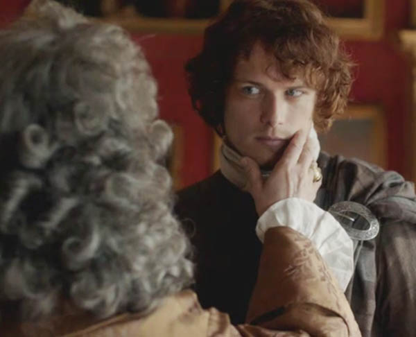
“You, of course, contain within you a sublime combination of the two!”

ep-110-Sandringham-03-KLS-edited

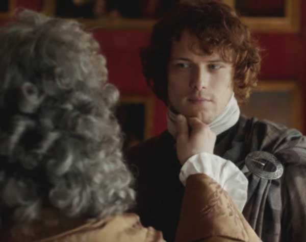
This smarmy old rascal is spot on: Jamie, you are indeed a sublime blend of beauty and belligerence! Best you look askance at the Duke and his roving fingers; Claire willna like him fondling yer chinny chin chin! Sandringham invokes the lyrics from “You Did It” (My Fair Lady): “Oozing charm from every pore. He oiled his way around the floor.” Grrrreasy!

Now for chin anatomy: More than 200 years ago, the German physician, naturalist, physiologist and anthropologist, Johann Friedrich Blumenbach (1752 – 1840) declared that the chin is a uniquely human feature. Nowadays, most naturalists agree that elephants (Photo A) and perhaps two other mammals have chins but few species other than humans can lay claim to this body part.
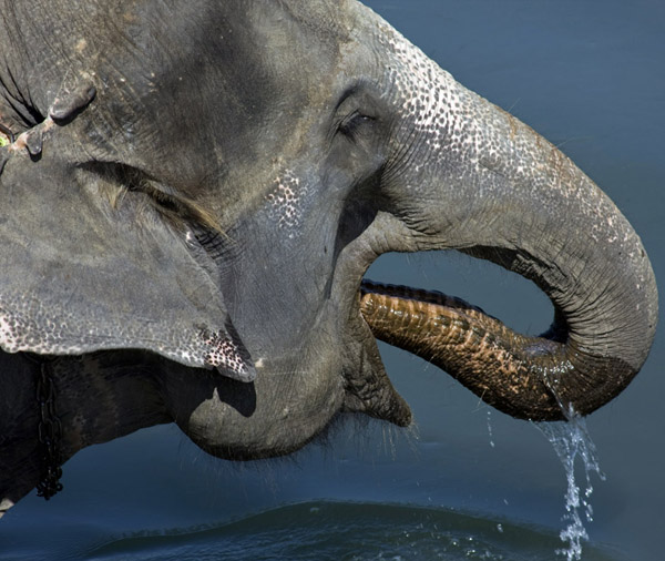
elephant KLS edited

Photo A

A few scholarly circles hotly debate, why do we have a chin and what is its purpose? One cool idea posits that the human chin emerged due to speech and mastication (chewing) patterns. Indeed, computer models show that mechanical stress relating to muscle pull could contribute to chin development.

Another proposed reason for the human chin is sexual dimorphism, the different appearance of a body part between the genders. Typically, female chins are smaller and rounder and male chins are bigger and squarer. Such differences, it is argued, contribute to attractiveness and augment mate selection. And, don’t our Claire and Jamie demonstrate chin sexual dimorphism to a T (Starz episode 101, Sassenach)?

ep 101 Jamie & Claire chin KLS edited

Alas, the June 2015 Smithsonian reports that the chin didn’t develop to make us swoon nor to accommodate speech or distribute the stress of chewing. Rather, newest thinking declares the chin is the by-product of a shrinking face: over the eons, the face has decreased in size and tilted inward which in effect pushes chin and jaw outward. As for me, I prefer the sexual rif, thank you very much!

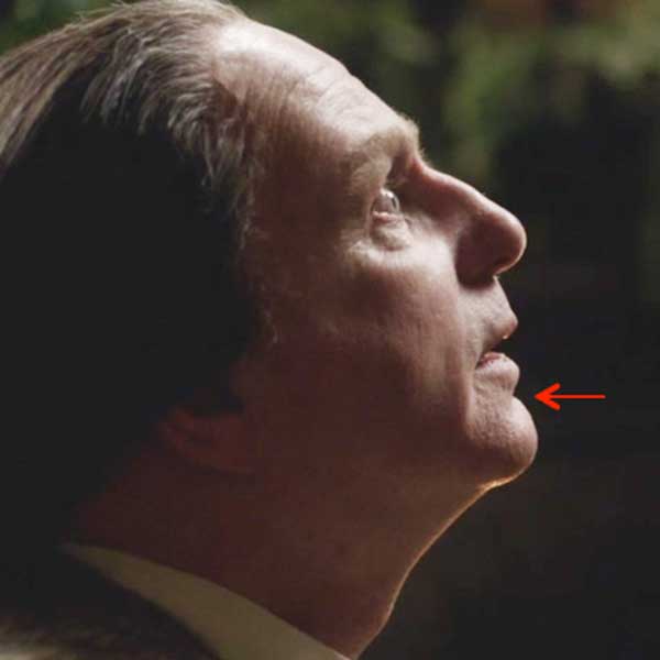
More chin anatomy: the mentus (Latin meaning chin) has several topographical features. First, the chin (Photo B –black arrow) and nose are typically the most forward projecting parts of the face. The chin has a bony base but the fleshy, moveable tip is the chin pad. From the point of the chin a pair of bony horizontal ridges project backward (Photo B – green arrow) each ending as a bony angle (Photo B – purple arrow). Between the lower lip and the chin is a horizontal skin groove, the mentolabial sulcus (Photo B – blue arrow).

Try this: What, there’s work to do already? Yep! Grip and wiggle your chin pad. Next, find your mentolabial sulcus, left and right horizontal bony ridges and bony angles. Very nice and good for you!
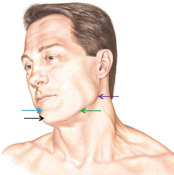
Figure0001-lower-jaw-KLS-edited

Photo B

The projecting point of the mandible (Latin meaning jawbone) provides the bony foundation for the chin. The mandible arises during fetal life where it develops as right and left halves joined in the midline by a thin plate of fibrocartilage (Anatomy Lesson #24). The paired mandibular halves persist until the second year of life when the joint ossifies into a vertical bony ridge, the mandibular symphysis (Photo C – drawing of newborn skull).
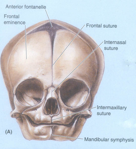
mandible 02 KLS edited

Photo C

During childhood, the chin persues the adult form as a midline triangular-shaped mental protuberance flanked on each side by a mental tubercle (Photo D). In the midline above the protuberance lies the unpaired bony ridge, the mandibular symphysis or symphysis menti (Photo D – black arrow).

Try this: Once again locate your mentolabial sulcus. Move one fingertip just below the sulcus and wiggle it back and forth. Do you feel a faint ridge? This is your symphysis menti. Move the finger downward to the bony tip. This is your mental protuberance. Now move your fingers to the left and right; do you feel a pair of small bony bumps? These are the mental tubercles. Well done!
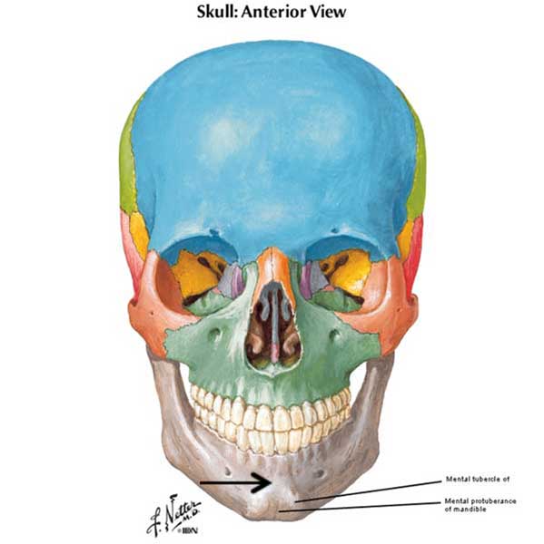
Figure0002A-lower-jaw-KLS-edited

Photo D

Let’s consider more about the mandible (Anatomy Lesson #11 and Anatomy Lesson #13), the parent bone of the mentus. The mandible (unpaired after two years of age) is the strongest, largest and lowest bone of the skull and is its only moveable bone. It has a U- or V- shaped body (Photo E) expressed as the lower bony ridges mentioned above. The body ends posteriorly as the bony angles of the mandible (Photo E – only left side labelled). Jutting upward and backward from each mandibular angle is a strong bar of bone, the ramus. In the midline are bony features of the chin as described above. Lastly, the mandible has an upper alveolar part (Photo E) that serves as a receptacle for 16 adult teeth which in the best case scenario includes: four front incisors, two canines (BJR’s dog teeth – black arrows), four premolars and six molars.

Try this: Palpate the body and angles of your mandible. Look in a mirror, open your mouth and find incisors, canines, premolars and molars (if present). If your mouth is too dark to clearly see the teeth, shine a flashlight into the mirror; the light reflects back into your mouth and nicely illuminates that inner sanctum!
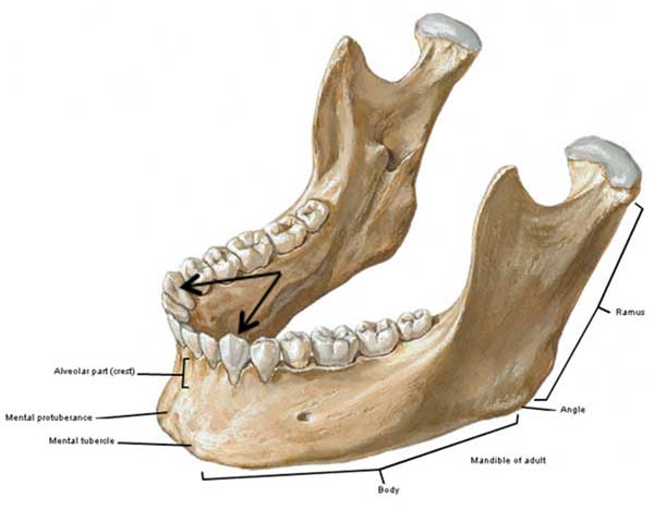
Figure0013A-mandible-KLS-edited

Photo E

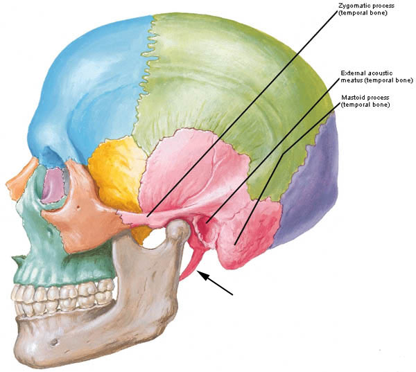
Each mandibular ramus ends in two bony projections: a condylar process the head of which articulates with a socket in the temporal bone at the TMJ (temporomandibular joint) and the sharp coronoid process (Photo F) onto which attaches a muscle of mastication (see below). On the inner surface, each ramus has an opening, the mandibular foramen, for passage of a nerve. On the outside of the chin are left and right mental foramina (pl.) which also transmit nerves.
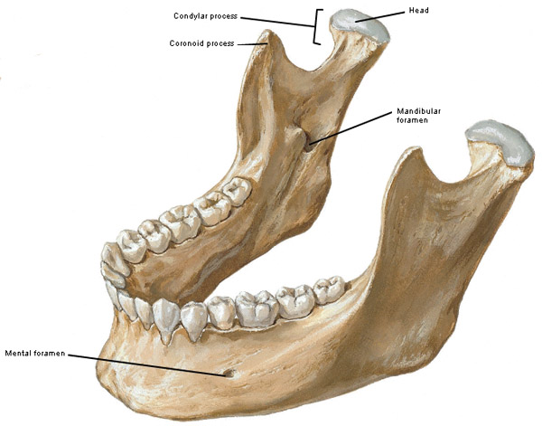
 

Figure0013A coronoid process KLS edited

Photo F

Warning: The next image (Photo G) shows a deep dissection of the head and neck. Please skip if you find such images challenging. Three important nerves on each side of the face are pertinent to our discussion. The paired mandibular nerves (branches of Cranial Nerve V) exit the skull deep to each cheek bone (zygomatic arch – Anatomy Lesson #9). Each mandibular nerve produces left and right inferior alveolar nerves or IANs that enter the mandible via the mandibular foramina (shown in Photo F) to supply sensation to the lower teeth. Ouch! Yes, IANs are the culprits that transmit tooth pain! Along the way, each IAN gives off a mental nerve which exits via its respective mental foramen and provides sensation to lower lips, gums and chin. Bet Jenny didna ken that a nerve was named after her kindly brown-haired laddie!

Try this: Straddle your mentus with thumb and forefinger. Place them about the same vertical level as your canine teeth. Press down gently until you feel slight hollows and a tingle. These are your mental nerves exiting the mental foramina.
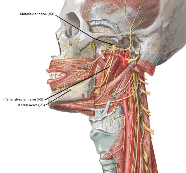
Figure0067A inferior alveolar nerve KLS edited

Photo G

A discussion of the chin is incomplete without considering associated muscles. A whopping 28 muscles attach to the mandible – yes, that’s 14 muscles per side! I won’t name them all. Muscles that attach to the mandible (see below) act on seven different head and neck regions. Suffice it to say that each side of the mandible has four muscles for chewing, one moves the cheek, three move the lower lip, one wrinkles neck skin, three move the tongue and two help us swallow. As if this weren’t enough, several of these muscles aid in speech by moving lips, tongue and hyoid bone (Anatomy Lesson #12). Whew! The mandible (and its chin) truly is a workhorse for head and neck muscles.

Three of the 14 muscle pairs mentioned above are easily demonstrated. Each mentalis muscle arises from the mental protuberance and inserts into the lower lip (Photo H). As they contract, the lip elevates and protrudes as in a pout; simultaneously, the skin of the chin wrinkles. They also add bulk to the chin pad. Each masseter muscle arises from the zygomatic arch (Photo H – black arrow) and inserts into body and angle of the mandible; contraction closes the jaw. The temporalis muscles are the third pair of muscles for today. These fan-shaped muscles arise from the sides of the skull and insert onto the coronoid processes (shown in Photo F) of the mandibular rami. Contraction closes and retrudes (pulls backward) the mandible.

Try this: Return to the mirror and wrinkle your chin-skin. Congrats! You just activated your mentalis muscles. Next, place your fingers in the hollow of each temple; close your teeth and retrude (pull back) the mandible. You should feel tension in each temporalis as they contract. Finally place fingertips just anterior to each mandibular angle. Bite down and feel the masseters tense as they close the mandible.
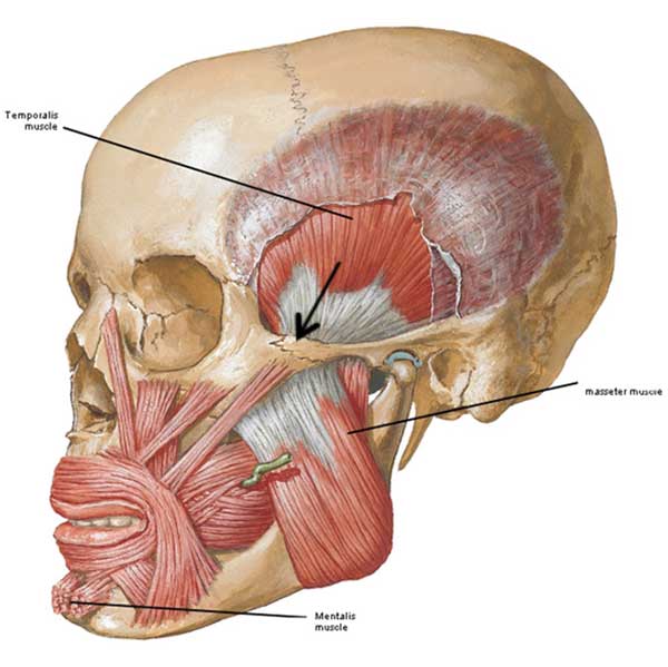
Figure0050A-mandibular-muscles-KLS-edited

Photo H

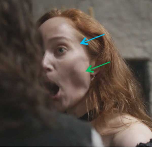
Are these muscles at work in Outlander? Oh, to be sure! Geillis offers us a wonderful visual as she interrogates Claire before sentencing at the witch’s trial (Starz episode 111, The Devil’s Mark)! Why are you here in Scotland (English lassie has no idea) and when will you stop lying (tell the truth, the whole truth and nothing but the truth!)? Here, Geillis opens her mandible using muscles of mastication (lateral pterygoids) that we have yet to learn and are not visible from the skin surface. With the mandible widely opened, the masseter is pulled taut (green arrow) and the temporalis is stretched creating a hollow at the temple (blue arrow). Try it yourself; it works!

ep 111 Geillis-masseter-02-KLS-edited

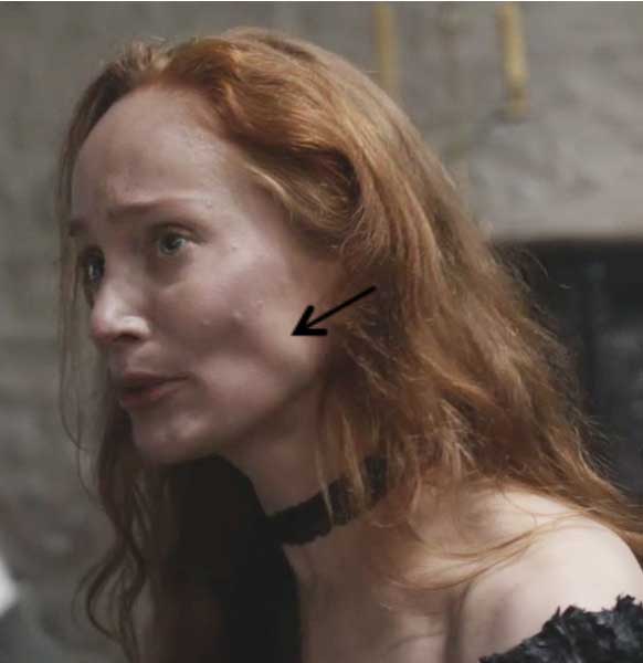
Next, Geillis closes her mandible using the powerful masseter muscles (black arrow). This wild wily witch needs some answers before she becomes kindling at her own personal bar-b-que!

ep-111-Geillis-masseter-KLS-edited

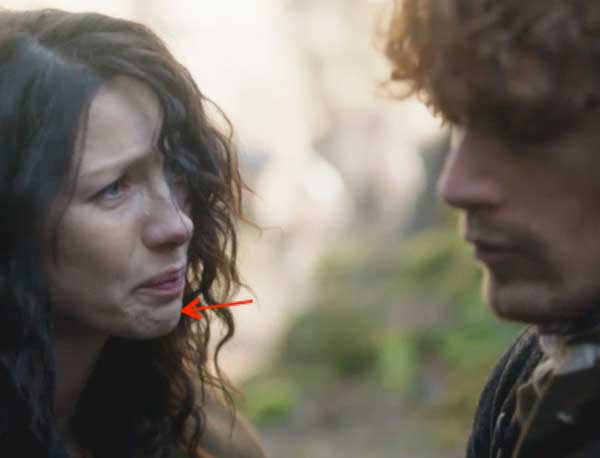
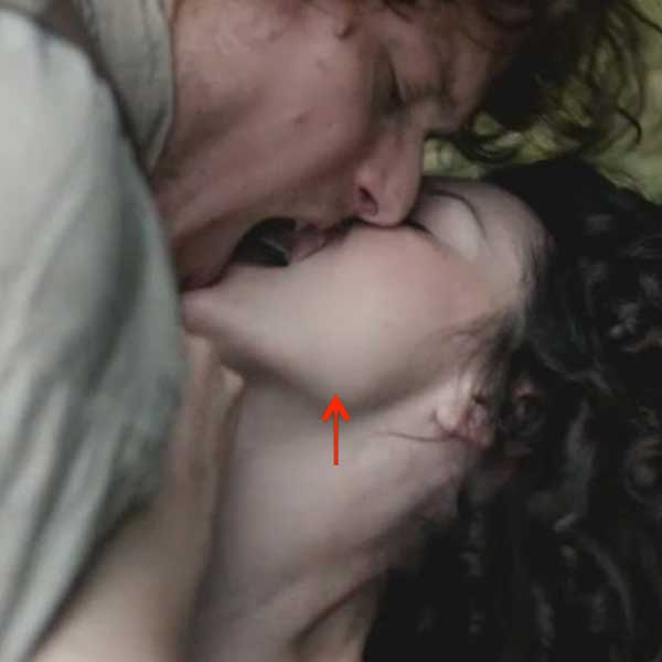
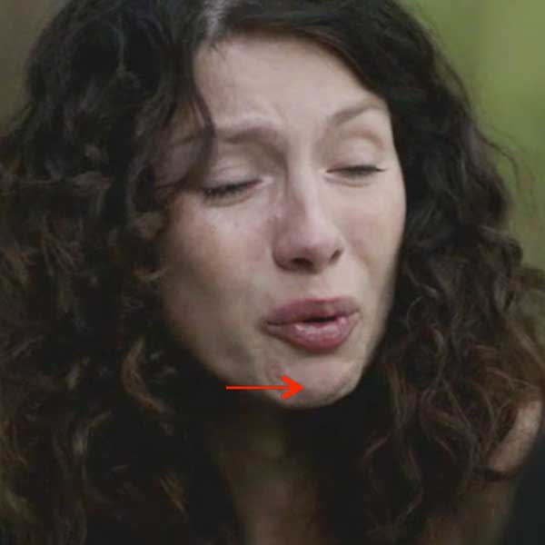
Mentalis muscles are at work “Down by the Riverside” as Sassynach and Big Red One go at it hammer and tongs! After Claire wounds an astonished Jamie with her sharp 20th century tongue, she feels verra sad (Starz episode 109, The Reckoning). This sassynach is the best chin-skin-wrinkler and lower-lip-pouter in the whole of Scotland! Yep, her mentalis muscles are working hard here!

ep-109-Claire-mentalis-01-KLS-edited

Finally, no chin discussion would be complete without considering chin shape: is a chin smooth, dimpled, creased, clefted or using urban-speak, the awful “butt chin” (who dreamed that one up)? All these chin shapes are considered normal variants but, interestingly, the cleft chin is most common in people of European descent.

Science teachers may use the cleft chin as an example of a dominant genetic trait. But like the earlobe (Anatomy Lesson #24), the broad range of chin types should not be observed if simple dominant-recessive inheritance is at work. Other possible explanations for chin shapes include variable gene penetrance but this is beyond our present discussion.

Humans have long favored chin dimples as a mark of beauty. In Persian literature, the chin dimple is a “well” into which a poor lover falls and becomes trapped! Because a forest of growth covers the chins of most Starz Highlanders (the lads, no the lassies!), assessing their chin dimples is a challenge.

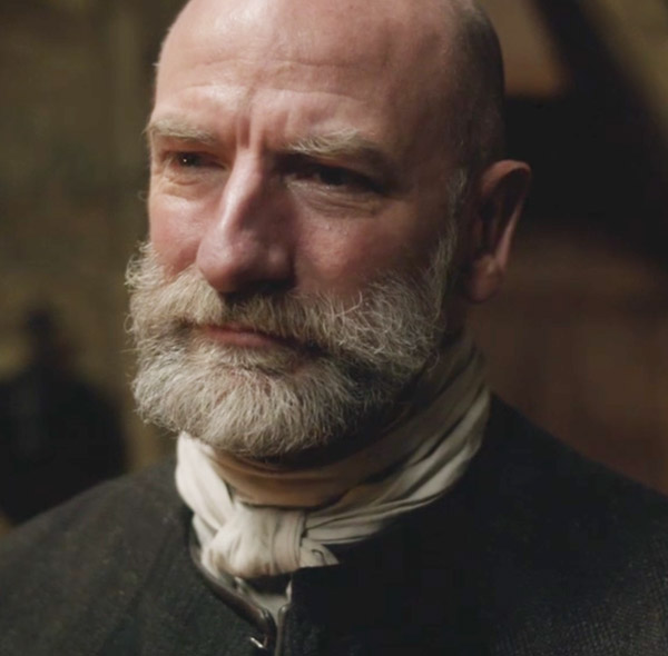
Consider dear Dougal whose chin and mandible are thickly furred. In honor of my good friend Jo Kc and the myriad of other Dougal fans, this image is for us! Here, big bad bro Colum abuses Dougal with names like “half-wit” and “numbskull” (Starz episode 110, By the Pricking of My Thumbs). Please employ your virtual imagination to identify as many chin and mandibular features as possible. Enjoy!

ep 110 Dougal chin KLS edited

Clean-shaven Highlanders are more rare than a wild haggis but here is that cutie, Willie; his is an excellent example of a smooth chin (Starz episode 114, The Search). Nice eyes, laddie!

ep 114 Willie chin KLS edited

Do ye like Claire’s wee but thoroughly charming chin dimple? Her chin enjoys one degree of separation from the smooth type. Here, she implores her husband: “Come back to me James Frasier” (Starz, episode 110, By the Pricking of My Thumbs). Oh, no! That clever, cunning Colum is sending her darling Jamie away!

ep 110 Claire chin KLS edited

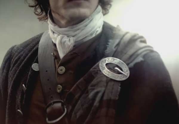
Moving two degrees of freedom from a smooth chin, Jamie’s awesome chin crease sends many a heart into cardiac arrest (Starz episode 113, The Watch)! Ye can see it well despite the bit o’ scruff that typically adorns his manly mentus. Herself writes about this in Outlander book:

“Good.” He loosened his grip and turned me to face him. At close range, I could see the bristle of auburn stubble on cheek and chin. I brushed my fingers across it; it was like the plush on an old- fashioned sofa, stiff and soft at the same time.”

Ummm, gulp!

ep 114 Jamie chin KLS edited

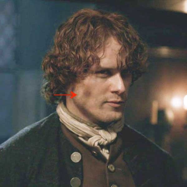
Oh, what? You canna see Jamie’s chin crease clearly enough? Okay, here is the only image of a clean-shaven Jamie I can find in the episodes (Starz opening credits). Do ye ken the crease now? Of course ye do. Won’t be sleeping tonight? Oooh, so sorry! Join the bazillions of fans who L-O-V-E Jamie’s chin!

into-Jamie-shaven-chin-KLS-edited

Okay folks let’s finish this lesson with a short pop quiz using matching questions. Here are six numbered structures followed by six lettered photos with arrows indicating the body part. Match the named structure with the body part. Answers appear at the end. Ready. Set. GO!

STRUCTURE:

  1. Mentolabial sulcus
  2. Angle of mandible
  3. Body of mandible
  4. Mentus
  5. Mentalis muscle
  6. Masseter muscle

photo-A

A

photo-B

B

photo-C

C

photo-D

D

photo-E

E

photo-F

F

ANSWERS:

1 = B   (Starz episode 110, By the Pricking of my Thumbs)

2 = C   (Starz episode 108, Both Sides Now)

3 = F   (Starz episode 110, By the Pricking of my Thumbs)

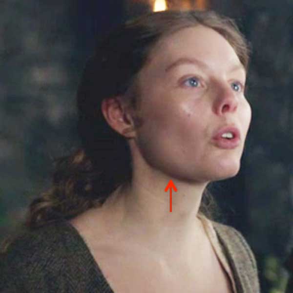
4 = E   (Starz episode 111, The Devil’s Mark)

5 = D   (Starz episode 111, The Devil’s Mark)

6 = A  (Starz episode 109, The Reckoning)

Hope you did well on the matching quiz. Let’s close this anatomy lesson with a big old chinny chin treat!

Ode to Jamie’s Chin

Jamie Fraser has a chin, a manly chin has he.

His tender fuzz will give you a buzz

And maybe two or three!

Dream of petting Jamie’s chin and leave a kiss or two.

It’ll grieve you much, but do not touch

Lest his wife come into view!

Claire Fraser is a lucky lass but a jealous wench is she.

Dinna touch his chin or she’ll slap your skin

into eternity!

 yum

(Starz episode 112, Lallybroch)

A Deeply Grateful,

Outlander Anatomist

Photo creds: Starz, Netter’s Atlas of Human Anatomy, 4th ed., Clinically Oriented Anatomy, 5th ed., www.en.wikipedia.org (elephant)

“If a Tree Falls – The Ear”

Summertime greetings to all Outlander anatomy students! Anatomy Lesson #24 covered the outer ear so today’s Anatomy Lesson #25 is the Ear – Part 2, or to be more exacting the middle and inner ears.

But first, what does a tree have to do with the ear? The title of this lesson derives from a 300 year old philosophical thought: “If a tree falls in a forest and no one is around to hear it, does it make a sound?” An 1884 issue of Scientific American correctly addressed this question. What do you think? Watch for the answer later on!

And to emphasize the importance of the ear in society, do you ken that the English language is replete with many idioms of all things ear? One website lists 120+ idioms including: a tin ear, all ears, music to the ears, up to the ears in, wet behind the ears, bend one’s ears, can’t make a silk purse out of a sow’s ear (who would try?), cute as a bugs ear (didn’t know bugs had ‘em), fall on deaf ears, in one ear and out the other, turn a deaf ear, and blow it out your ear (here’s to you, BJR!).

Now, onto the lesson. To review, in anatomy lesson #24 we learned that the human ear is divided into three ears: an outer (Photo A- green), a middle (Photo A- yellow) and an inner ear (Photo A- blue). That lesson dwelt almost exclusively on the outer ear. So, now we move to the middle and inner ears.

Ear-KLS-edited

Photo A

Let’s begin with the middle ears. Each middle ear is housed inside a cavity within one of our two temporal bones. The human skull includes 22 bones two of which are os temporale (Latin meaning temporal bone). The temporal bone is weirdly shaped (Photo B – pink bone). From a side view, it comprises most of the skull around the external acoustic meatus or EAM (Anatomy Lesson #24). The temporal bone also includes the zygomatic arch, part of the cheek bone (Anatomy Lesson # 8) and the gothic-looking styloid process marked by the black arrow (Anatomy Lesson #12).  Let’s add a new part of the temporal bone which is pertinent to today’s lesson, the rounded mastoid process.

Try this: Place your fingers behind the pinna of one ear and move downward until you feel a rounded mound of bone; this is your mastoid process. It typically lies just below the level of the EAM or ear hole as Claire called it (ha ha). Its outer layer is compact bone, but inside it is riddled with air-filled spaces. Well done folks!

Figure0004A-temporal-bone-KLS-edited

Photo B

A different image of the skull helps us appreciate the location of the middle ear. With the top of the skull and brain removed, Photo C shows the cranial base. The right side of the image shows the tortuous shape of the bony floor upon which the brain rests. Nerves and blood vessels pass through the many holes in the skull bones. The left side of the image is color coded so once again, the temporal bone is pink. See the bright green area? This is the location of the middle ear – it lies inside the petrous (Latin meaning stone-like) part of the temporal bone, one of the densest bones of the human body.

skull

Photo C

The middle ear is small but it contains a large number of components including tympanic cavity, inner leaflet of tympanic membrane, three bones, the opening of a throat tube, a posterior “attic door,” two wee windows, two tiny muscles, a nerve and a nerve plexus. Wow! That’s quite a list for such a small space! Let’s examine the components.

The tympanic cavity is an air-filled chamber (Photo D, yellow dashed line) inside the petrous temporal bone; its shape is so difficult to describe that many anatomists compare it to a small room with four walls, a roof and floor.  So let’s do that: the roof and floor are petrous temporal bone. The outer (lateral) wall is the tympanic membrane. The inner (medial) wall will be discussed shortly. The back wall has an “attic door” leading to the mastoid air cells (see below). The front wall receives the opening of the throat tube or pharyngotympanic or Eustachian tube (photo D) that extends between the back of the throat and the tympanic cavity.

Please understand this: normally, air pressure between the tympanic cavity and the EAM is equal. However, as we climb in altitude, air pressure becomes lower in the EAM than in the tympanic cavity which pushes the tympanic membrane outward causing discomfort, even pain. As we descend in altitude, air pressure is higher in the EAM than in the tympanic cavity which pushes the tympanic membrane inward, again causing pain. Air pressure equalizes on each side of the tympanic membrane when we open the Eustachian tubes: with altitude, air escapes from the tympanic cavity and with descent, air enters the tympanic cavity. Got it? Chewing and swallowing activates a pair of itsy, bitsy, teeny weeny, tensor veli palatini muscles that open the Eustachian tubes to equalize the air pressure. I won’t show an image of these wee muscles because it will clutter the lesson. Just know that they work well unless the Eustachian tubes are congested.

D.Figure0088D-tympanic-cavity-KLS-edite

Photo D

Remember the “attic door” in the back wall of the tympanic cavity? It has a longish Latin name, the aditus ad antrum, but the short of it is that the passageway leads to air cells which riddle the mastoid process (Photo E). These little spaces are thought to reduce the mass of the skull bones and provide physical protection.


Coronal-Section-of-the-Ear-Showing-the-Mastoid-Air-Cells KLS edited

Photo E

The middle ear bones bring us back to the anatomical rule of three! Three ossicles meaning tiny bones are the smallest bones of the human body. How small are they? Well, all three easily fit on a U.S. dime with plenty of wiggle room (Photo F)! There are three ossicles per middle ear and they are not included in the count of 22 skull bones.

ossicles 3 KLS edited

Photo F

The ossicles form a tortuous bridge spanning the tympanic cavity from outer to inner walls (Photo G). Each ossicle resembles the object for which it is named: the malleus (Latin meaning hammer) has a handle firmly attached to the inner leaflet of the tympanic membrane and a head that articulates (forms a joint with) the incus. The incus (Latin meaning anvil) articulates with the stapes (Latin meaning stirrup) and the stapes inserts into the oval window, an opening in the inner wall of the tympanic cavity. Although tiny, the joints or articulations between the ossicles are moveable.

Please understand this: imagination is sometimes required to relate the Latin names to their corresponding anatomical objects. However, ancient anatomists used objects found in nature to name the body parts. The names such as stapes or malleus seem quaint but I actually prefer them to the current naming trend which often includes meaningless words containing lots of “cs”, “xs” and “zs.”

ossicles KLS edited

Photo G

The inner wall of the tympanic membrane bears two holes piercing the temporal bone. One hole, the oval window, is plugged by the stapes (Photo H); the other hole or round window is closed by a membrane (Photo H – black arrow). The inner wall has several other features too, but these are beyond the scope of this lesson

middle-ear-KLS-edited

Photo H

Let’s stop for a short Clinical Correlation: The oral cavity and throat contain oodles of bacteria (Rupert and Angus consider germs in this hilarious video: Angus and Rupert Go Through The Stones). All is well unless they follow the Eustachian tube into the tympanic cavity where they can set up housekeeping to our detriment. Otitis media (Latin meaning inflammation of middle ear) is a rather common condition which typically includes bacterial (or viral) infections of the middle ear. Over 30 million doctor visits per year in the U.S. are due to otitis media. Symptoms include: pain, pulling at the pinna, irritability, sleeplessness, crying, etc. A trip to the doctor and an otoscope exam is in order (Photo I).

otoscope KLS edited

Photo I

Anatomy Lesson #24, showed the photo of a normal tympanic membrane (Photo J – left). But, with otitis media, the tympanic membrane is red, bulging because there is fluid in the middle ear and it hurts (Photo J – right)! Proper treatment is imperative.

ear-drums

Photo J

Unresolved otitis media can lead to mastoiditis (Latin meaning inflammation of mastoid bone). Now, dinna confuse mastoiditis with mastitis which is inflammation of the breast. Och, we are discussing ears, not mammary glands!

Here’s how mastoiditis works: recall that little attic door in the back wall of the middle ear that leads to the mastoid air cells (photo E)? Well, that door is a perfect conduit for bacteria to make their way from the middle ear into the mastoid air cells causing serious health complications in children and adults. Photo K (left side) shows bacterial invasion of the mastoid air cells and its presentation at the body surface (Photo K – right side). Signs include fever, redness, swelling, and tenderness behind the pinna which is pushed outward and forward.

photo-K

Photo K

A picture is worth a thousand words, so this photo shows a case of mastoiditis (photo L – left mastoid process). Ouch, that hurts!

Mastoiditis1 KLS edited

Photo L

Now back to anatomy. The inner ear is the last and by far the most complex of the three “ears”. At first blush it resembles a mutant squid or snail. It contains both bony and membranous elements. Bony parts include cochlea, vestibule and three (yes, three!)  semicircular canals (Photo M- tan structures) filled with fluid (perilymph). Membranous elements (Photo M – blue structures) are suspended within the bony parts; these are the cochlear duct, utricle, saccule and three semicircular ducts; these are surrounded by perilymph and are filled with endolymph. Only the cochlear duct is involved in hearing, the utricle, saccule and semicircular ducts are necessary for balance and equilibrium.

inner ear KLS edited

Photo M

The cochlea and its cochlear duct spiral 2.5 times much like a snail shell. Within each cochlear duct lies the organ of Corti, a strip of 15,000-18,000 specialized “hair” cells arranged in rows like soldiers. “Bristles” project from the surfaces of the “hair” cells although these are unlike the hairs of skin. When examined by scanning electron microscopy (Anatomy Lesson #6), the bristles resemble the pipes of an organ (Photo N – cat hair cells). The bristles are covered by a gelatinous membrane (not shown in Photo N). The organ of Corti is also our organ of hearing.

hair cells of cochlea KLS edited

Photo N

The following is a simplified version of how we hear, as the full vocabulary and structural and functional details would fill yet another anatomy lesson. Sound waves travelling through the air are gathered by the pinna and EAM; they strike the drum-like surface of the tympanic membrane pushing it inward with a thrust equal to the intensity of the sound. Ergo, loud noises push the eardrum inward more than soft sounds. The tympanic membrane vibrations are transferred through malleus, incus and stapes. With each vibration the stapes pushes inward at the oval window creating corresponding shockwaves through perilymph and endolymph of the cochlear duct (Photo O – black arrows). Movements of the fluids rub the bristles of the hair cells against the gelatinous membrane creating an excitation which is transfer to nerve cells forming the cochlear nerve (Photo O). The cochlear nerve joins with the vestibular nerve (see below) to form Cranial Nerve VIII (vestibulocochlear nerve). Impulses of each Cranial Nerve VIII follow auditory pathways into the brain. Hair cells near the base of the cochlea detect high-pitched sounds, such as ringing of a cell phone; those closer to the apex detect lower-pitched sounds, such as barking of a large dog.

Figure0087 inner ear KLS edited

Photo O

Electrical signals carried by the cochlear nerve make their way to the primary auditory cortex of the brain (Photo P – pink zone) where electrical signals are converted into “sounds” that we learn to recognize and understand.

In summary, outer, middle and inner ears work together to transfer sound waves through air (outer ear), solid (middle ear) and liquid (inner ear) where the good, good, good, good vibrations are converted into electrical signals that make their way to the brain for interpretation of sound. Go Beach boys!

250px-Brodmann_41_42 KLS edited

Photo P

Do you recall this lesson began with “If a tree falls in a forest and no one is around to hear it, does it make a sound?” Take a moment to think of the answer and then read on.

The answer to this philosophical riddle is that if a tree falls in the forest it creates sound waves but a receptor must be present to convert those sound waves into sound. If any creature is present with an organ than can perceive and interpret sound waves, then the falling tree does make a sound otherwise it only makes sound waves. Make sense? Good!

Now, every anatomy lesson must tie into all things Outlander and hearing is no exception. So next is a jolly good quote from Outlander book when Claire tells Jamie she is from a waaay different time zone (Starz episode 111, The Devil’s Mark). Well, Jamie knew there was something unique about this braw and bonny lassie!

“Do you know when I was born?” I asked, looking up. I knew my hair was wild and my eyes staring, and I didn’t care. “On the twentieth of October, in the Year of Our Lord nineteen hundred and eighteen. Do you hear me?” I demanded, for he was blinking at me unmoving, as though paying no attention to a word I said.

ep 111 he hears you 01 KLS edited

“I said nineteen eighteen! Nearly two hundred years from now! Do you hear?” I was shouting now, and he nodded slowly. “I hear,” he said softly.

Hmmm, Jamie is thinking: I ken now why the Sassynach didna take to my leather belt spankin’! It also ‘splains her twitchy-witchy “know how.”

ep 111 do you hear me 02 KLS edited

Now we must move on to how the inner ear provides balance and equilibrium. Our ability to balance is independent of outer ear, middle ear and cochlear parts of the inner ear but it is dependent on function of the three semicircular ducts (Photo Q – bone removed). The semicircular ducts contain endolymph and each bears an ampulla, a swelling at one end (Photo Q – red arrows). Each ampulla contains a patch of “hair cells” similar to those of the organ of Corti. Once again, the bristles are covered with a gelatinous membrane.

Anterior and posterior semicircular ducts are oriented vertically at right angles to each other. The lateral semicircular duct is slightly off the horizontal plane. Orientation of the ducts cause each duct to be stimulated by angular rotation of the head in a given plane.


Figure0090C-semicircular-ducts-KLS-edited


Photo Q

Turning your head from left to right sides (as in no) moves endolymph in the lateral semicircular duct. Nodding your head (as in yes) moves endolymph in the anterior semicircular duct. Moving your head to touch one shoulder or as in doing a cartwheel moves endolymph in the posterior semicircular duct. In aviation terms, the semicircular ducts are oriented such that they detect pitch, roll and yaw.

Here is how the semicircular ducts work: As the head moves in angular rotation, endolymph moves in the opposite direction bending the gelatinous membrane (cupula) and exciting the hair cells (Photo R). The hair cells transfer the signal to nerve cells of the vestibular nerve (part of Cranial Nerve VIII). The signal is carried to the brain which interprets it as angular motion of the head. The information can be used to activate various muscles to adjust the head and/or body positions.

ampullae KLS edited

Photo R

The final anatomical elements of the inner ear are utricle (Latin meaning leather bag) and saccule (Latin meaning money bag –  Dougal is into this one!) located between the semicircular duct and cochlear duct. These elements are also filled with endolymph (Photo S) and are designed to detect changes in linear acceleration or linear deceleration.

Figure0090C utricle and saccule KLS edited

Photo S

Utricle and saccule each contain a patch of hair cells with surface bristles (Photo T – bullfrog hair cell) covered by a gelatinous membrane (absent in Photo T). The hair cells of the utricle are oriented horizontally and those of the saccule are vertically oriented.

macula stereocilia KLS edited

Photo T

Seated atop the gelatinous membrane are wee otoliths (ear stones or ear rocks) made of calcium carbonate (Photo U); these add weight to the gelatinous membrane thus enhancing our sense of  gravitational pull.

otoliths KLS edited

Photo U

The utricle and saccule work this way: As our head undergoes linear acceleration or linear deceleration (forward, backward, upward, downward), endolymph, the gelatinous membrane and otoconia move in the opposite direction (Photo V). This bends bristles of the hair cells causing them to activate nerve cells of the vestibular nerve of Cranial Nerve VIII. The electrical impulses are carried to the brain. The utricle detects horizontal changes and the saccule detects vertical changes in linear movements of the head. This information arrives at the brain which then determines if and how much the head is tilted and if the body needs to be reoriented in space.

otoconia%20displacement KLS edited


Photo V

This brings us to the end of today’s important lesson but we must tie balance and equilibrium into Outlander. Here are a couple of great quotes from Outlander book. The book quote and the Starz images (Starz, episode 108, Both Sides Now) don’t quite match because the book scenarios weren’t filmed. Nevertheless, it is fun and you will get the idea. The first scene takes place as Frank and Claire descend from Craigh na Dun after watching the Druid’s dance. Frank, the soon-to-be Oxford professor, is so absorbed in thought he doesn’t watch where he plants his feet!

“He dropped into one of his scholarly trances … The trance was broken only when he stumbled unexpectedly over an obstacle near the bottom of the hill. He flung his arms out with a startled cry as his feet went out from under him and he rolled untidily down the last few feet of the path, fetching up in a clump of cow parsley… “Are you all right?” … “I think so.” He passed a hand dazedly over his brow, smoothing back the dark hair. “What did I trip over?” “This.” I held up a sardine tin…”

ep 108 Frank balance KLS edited

Compare and contrast Frank’s stumble-tumble with Jamie’s sure footedness (Outlander book). In this scene, it is nighttime and Jamie unerringly hauls Claire through the night:

“Jamie kept a tight hold on my arm, hauling me upright when I stumbled over rocks and plants. He himself walked as though the stubbled heath were a paved road in broad daylight. He has cat blood, I reflected sourly, no doubt that was how he managed to sneak up on me in the darkness.”

Yes, Jamie has grace, balance, and equilibrium (Starz episode 104, The Gathering). Nothing will trip-flip this kilt! Hang on tight, Claire! Snort!

ep 104 holding hands KLS edited

You may have seen this arabesque in the teaser but let’s appreciate it anyway. Jamie throws water on burning hay, a fire set by a Watch weasle! Our highlander fireman even makes tossing water a thing of beauty. Balance and equilibrium on one foot; 17th century ballet!

ep 112 Jamie balance 01 KLS edited

At last, the time has come to report the results of Lesson #24 and the Pinna Poll! Six images of pinnae from the Starz cast (episodes #103, #109, #115, #116) were shown and votes tallied. All the ear flaps are fabulous!

Not sure if you are surprised, but the winner is:…………..drum roll………………..Jamie! Congratulations Big Red! And it was an honest count, too; no stuffing of the ballot box!  Jamie garnered 38% of the votes while the remaining five pinnae were in almost a dead heat with a slight edge by bad boy BJR!

Great comments were shared by many readers including these: exquisite, lovely, charming, manly, rugged, beautiful, elfin-like, fawn-like, nibble-worthy, snuggle-worthy, elegant, awesome, symmetrical, balanced, proportionate and yum! Let’s thank our six wonderful characters and all of you for playing the Pinna Poll!

pinna-poll

That’s it for the ear. Complex and elegant! Oh, and a word to the wise: keep music turned down a notch or two. Over the years, hair cells of the organ of Corti become damaged by excessive noise. For years, the damage has been considered irreversible although a recent study shows some promise using a gene-activating drug regime. Stay tuned.

Closing with this fun poem from Mr. R’s World of Math and Science:

An ear splitting sound!

A crash and a boom!

Ringing so loudly,

It shook the whole room!

But I didn’t hear it,

I couldn’t at all,

My left and right ear,

Had gone to the mall!


My left and right ear,

My organs that hear,

Had gone to go shopping,

And that’s what I fear…

Without my 2 ears,

That spectacular pair,

I can’t hear sound waves,

Move through the air!


The deeply grateful,

Outlander Anatomist

Photo creds: Starz, Basic Histology, Junqueira & Carneiro, 11th ed., Concise Histology, Bloom & Fawcett, 2nd ed., Netter’s Atlas of Human Anatomy, 4th ed., Clinically Oriented Anatomy, 5th ed., www.aviationknowledge.wikidot.com (ampullae), www.clearwaterclinic.com (otoliths), www.fairview.org (mastoiditis), www.kids-ond.com (ossicles), www.mhhe.com (ampula hair cells), www.sciencepoems.net (Mr. R’s ear poem), www.student.com (ossicles with inner ear), www.teachmeanatomy.info (mastoid air cells), www.otopathologynetwork.org (ossicles on dime), www.wallpaper.com (fallen tree), www.wikipedia.org (mastoiditis with subperiosteal abscess)