Greetings everyone! Welcome to Anatomy Lesson #15: The Thoracic Cage. Now, don’t leave this lesson just because the title doesn’t include Jamie! He’s up next time. Promise.
This lesson follows the fascinating self-defense session from Starz episode 8, Both Sides Now, wherein Angus teaches Claire how to kill using the sgian dhu or hidden dagger. Is Angus’ lesson accurate and are his descriptions anatomically correct? Read on and find out. It will be interesting, I assure you.
A wee note of explanation: I analyze the images and self-defense lesson as an academic. If my comments seem cavalier, please know that the topic matter is difficult for me, too.
Now, knives flash everywhere in this episode. Mayhap RDM wrote it?
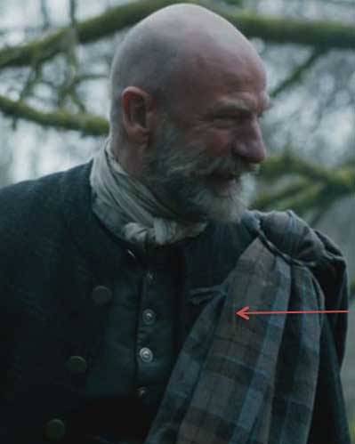
Knives are not my specialty (although I’m, um, good with a scalpel) but let’s start the topic with a brief intro to the sgian dhu. Apparently, 17th and 18th century Scots carried this dagger hidden in the sleeve or lining of a jacket. As a guest, however, proper courtesy and etiquette required that Highlanders remove the weapon from hiding and display it in a stocking top (Photo A – red arrow) as depicted in this elegant and manly portrait of Alasdair Ranaldson MacDonell of Glengarry (Henry Raeburn, artist – 1812).

Photo A
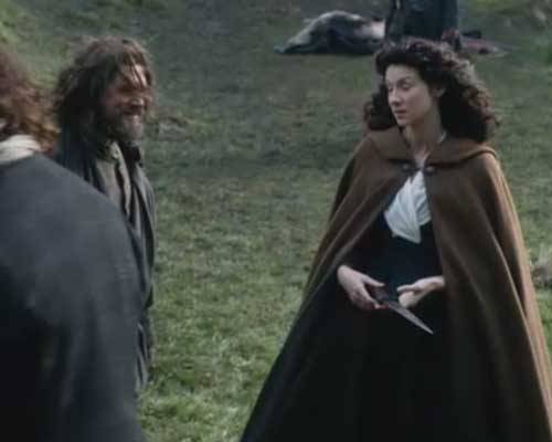
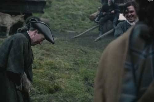
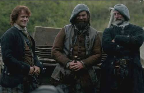
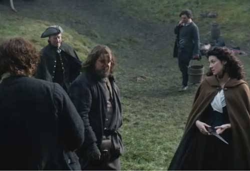
If ye recall, an important event leads to Claire’s sgian dhu lesson. Just blame it on the crouching Grants! Aye, those sneaky dudes were hidden in the rocks under veil of darkness with pillage on their minds. Springing from their hiding place, they attack the MacKenzie party and ruin Rupert’s mellifluous waterhorse tale; what a voice that man has!
Happily, Dougal and his fighting warriors quickly prevail and Uncle is so delighted that, wonder of all wonders, he hugs his nephew! I’ve burned this image on my retinas thinking it may be the only time we see Dougal show affection for the lad. Wait ‘til Jamie hears that his uncle has the hots for his new wife…probably no hugging after that.
Next morning the search is on for Jamie’s dirk, lost by Claire during the nighttime scuffle. Ah, gie it to my wife. But alas, the dirk willna work for the lass.
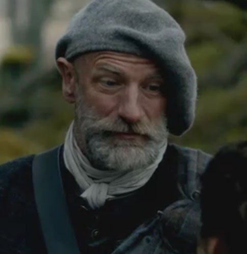
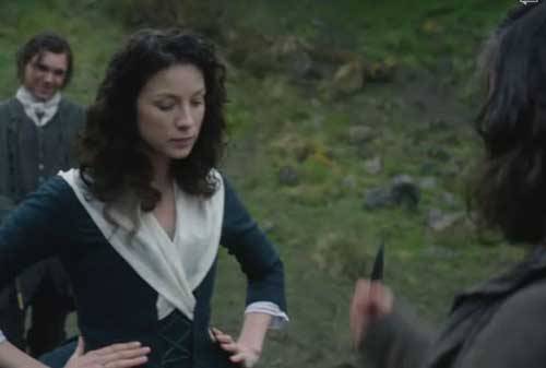
Ned Gowan thinks that someone should teach this leddy how to defend herself. Jamie agrees and war chief Dougal sits honing his dirk with a stone. Ever dapper in his wool bonnet and tidy beard, he intones that “the lass needs a sgian dhu.”Claire is – huh? – until her ruadh gallant translates: “sgian dhu, hidden dagger!”
So, does anyone have a sgian dhu for Claire? To be sure, that crusty solicitor Ned, does. A lot of folks keep theirs hidden in a stocking, but he keeps his “in another more private place”… ahem, who knew? Willie (what a cutie) thinks Ned’s hiding spot is verra clever. Careful Ned, ye need all those body parts and passions lest you lose face time with the strumpets!
Herself writes in the Outlanderbook:
“The tiny sgian dhu, the sock dagger, was deemed acceptable, and I was provided with one of those, a wicked-looking, needle-sharp piece of black iron about three inches long, with a short hilt.”
OK, she’s got a knife but who will give her a lesson? Turns out Angus is “a good man with a blade!” and he’s been waiting to teach Claire a thing or two. I recall him thrusting his dirk while watching Claire in the surgery at Castle Leoch (Starz episode 3, The Way Out) and trying to get a wee “keek at her breests” on her wedding night (Starz episode 7, The Wedding). Naughty Angus!
Herself records in the Outlander book (Here, Rupert is the teacher):
“So I was marched out into the center of a clearing and the lessons began… After a good deal of amiable discussion, they agreed that Rupert was likely the best among them at dirks, and he took over the lesson.”
Again from Herself’s own hand (Outlander book):
“He found a reasonably flat spot … in which to demonstrate the art of dagger-wielding … “Generally, ye want to use the underhand; overhand is only good when ye’re comin’
down on someone wi’ a considerable force from above.”
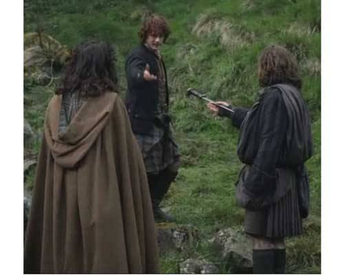
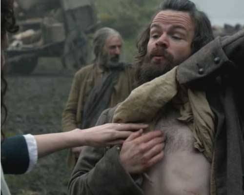
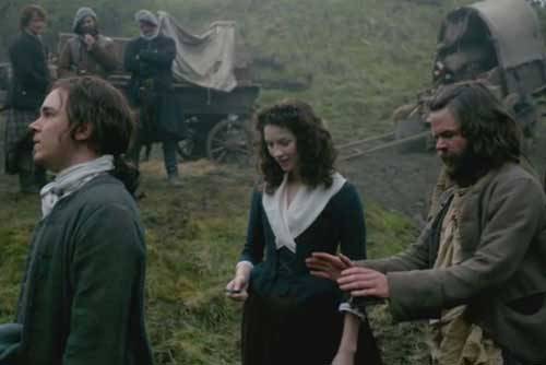
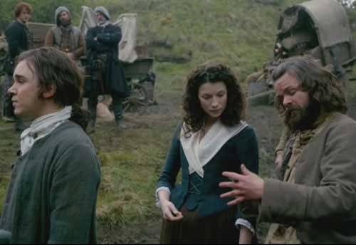
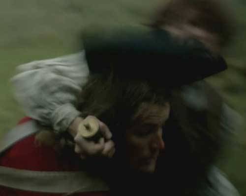
Angus demonstrates to dubious Claire several fierce upward jabs with the wee wicked weapon (dare ye to say that rapidly three times in a row).
“Aim straight up and as hard as you can into the heart” is Angus’ genteel advice. First try, Claire stabs upward near Angus’ breast bone and gets a warning! Noooo……. Dinna ye love the look on Angus’ face? He’s such an avid teach!
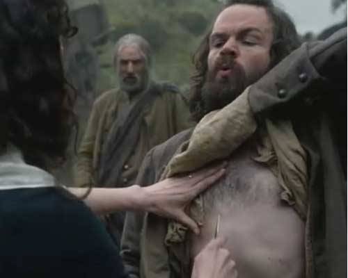
“Uh, oh, avoid the breast bone – you get your knife stuck in that soft part at the top and you’ll be without a knife.”
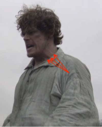
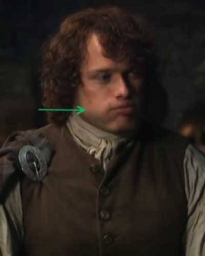
Whoa, there’s a tip-top issue here. Angus, did ye really say “at the top?” Ah, well, now, the adult human breastbone doesn’t have a soft spot at the top. After all, we aren’t chickens (well, maybe sometimes) …. I checked the CC script and sure enough the word is top. But, I think Angus says tip not top in which case, he is correct because the breast bone tip can be soft (see below). Mayhap he was misunderstood? Who cares? Anatomists to be sure. Otherwise, Angus’ anatomy lesson is flawless. Bravo professor Angus!
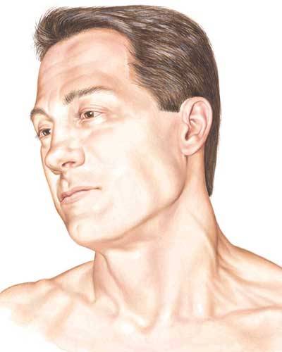
Lest Angus and Claire get too far ahead of us, let’s halt the lesson and start our own. Gruesome as it may seem, this lesson presents us with a fine opportunity to explore the thoracic cage and vital organs it protects. The thorax is the upper part of the trunk easily distinguished from abdomen because the former contains ribs. From the front, the thorax occupies the region shown in blue (Photo B) and is partially overlaid by chest and abdominal muscles.
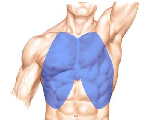
Photo B
The bony foundation of the thorax is the thoracic cage composed of thoracic spine (Anatomy Lesson #10), sternum, ribs and rib cartilages (Photo C). Let’s examine these structures.
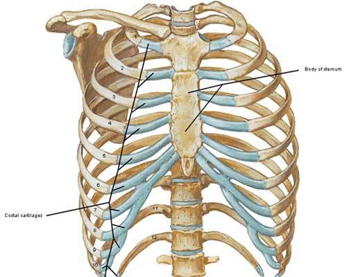
Photo C
Being verra imaginative men, early anatomists visualized the sternum as a sword and named its components accordingly. Can ye see the sword in Photo D? The sternum is divided into three parts:
- manubrium (grip of the sword – green),
- body (blade of the sword – turquoise)
- xyphoid process (tip of the sword – purple).
At birth, the sternum is typically six separate pieces of cartilage and bone. By 18-22 years, the segments ossify (become bony) into a single bone although the tip may remain soft (cartilaginous) until about age 40. So, yes, Claire’s knife could get stuck in the soft tip – not top!
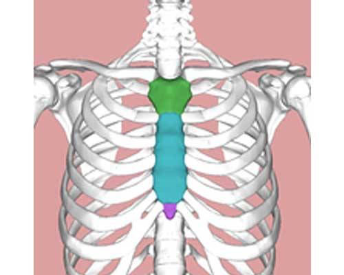
Photo D
The manubrium (Latin meaning handle) is fused with the body and articulates with costal cartilages of 1st and 2nd ribs (Photo E). It bears the midline jugular or suprasternal notch (Anatomy Lesson #12) and articulates with the clavicles (Anatomy Lesson #2) at paired sternoclavicular joints.
Try this: place a finger in your jugular notch and feel bony knobs at each side, the sternoclavicular joints. These are usually visible in a mirror; the thinner the person, the more prominent the joints. With a finger on one joint, lift, lower, pull forward, pull backward and rotate the same shoulder; feel the movement? This is truly amazing because the entire skeleton of each upper limb articulates with the thorax only at this single bony contact point!
The body or gladiolus (Latin meaning sword – as in gladiator) is the largest section of sternum; it is fused with manubrium, above and xyphoid process, below. It also articulates with costal cartilages
of ribs 2 – 7. Please palpate yours.
The xyphoid process (Greek meaning sword-like) is fused above with the body but terminates as a free end. Highly variable in shape, it may be curved, depressed, forked, deviated or pierced by a hole. Rarely it remains cartilaginous beyond 40 years.
Try this: suck in your breath as much as possible, hold your breath and lift the chest. Palpate the free tip of your xyphoid process – press gently as it may be tender.
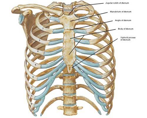
Photo E
Now, for the ribs: humans typically have 12 pairs of ribs (Photo F). Ribs 1-7 are true ribs because their costal cartilages directly join the sternum; ribs 8 -10 are false ribs because their costal cartilages join with costal cartilages of ribs above (Photo F – black arrows); ribs 11 and 12 are false and floating ribs because they do not join either costal cartilages or sternum. The joined costal cartilages of ribs 7 -10 form right and left costal margins.
Try this: holding your breath, run fingers along the free edge of the left ribs – this is your left costal margin.
Variations in rib count do occur: some individuals may have cervical or lumbar ribs or they may have only 11 pairs. Because of location, cervical ribs can compress arteries and nerves in the neck producing neurological and vascular symptoms. Otherwise, such variations are rarely a source of concern.
Between adjacent ribs are intercostal spaces, 11 pairs total (Photo F – red brackets). Although they appear large and empty in the image below, in life they are narrow and filled with intercostal muscles and fibrous tissue.
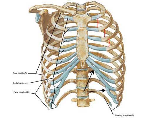
Photo F
Ribs vary in size and shape but each has a head (Photo G) which articulates with the bodies of two adjacent vertebrae and the intervening IV disc (Anatomy Lesson #10). Interestingly, if an individual rib is placed on a flat surface, it is seen as a twisted arch that will not lie flat. This twist affects the shape of thoracic cage and intercostal spaces.

Photo G
Another important feature of the rib cage is the manubriosternal joint also known as the sternal angle of Louis. Here, the union between manubrium and body creates a palpable bump (Photo H – side view) and is the level where the costal cartilages of the second ribs attach to the sternum. It is of clinical importance as various structures and anatomical boundaries occur at this joint.
Try this: place fingers in your jugular notch and work downward (about 2” or 5 cm) until you feel a bump; this is the sternal angle of Louis, a very useful landmark for health care workers to count ribs and determine locations of various thoracic structures. Men have more pronounced sternal angles than do women.
Occasionally, my acquaintances wish to wear heavy necklaces but cannot because of neck pain. I explain that they can likely wear heavy necklaces but only if the jewelry doesn’t fall below the sternal angle. If a heavy necklace is short, then clavicles and manubrium help support weight and relieve neck strain. Try it…
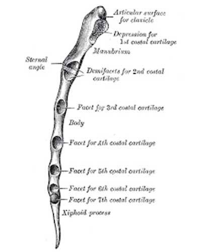
Photo H
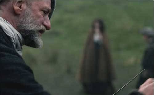
Now, back to Angus’ lesson… Next, he redirects Claire’s knife from his sternum to left rib cage. He tells her to stab straight up and into the heart. Having some acumen with a scalpel too, Claire “follows a man’s orders for once” (or wait, isn’t that twice?) with a thrust so convincing that Rupert laughs: “Oh, dinna kill him yet mistress. Wait ‘til the lesson is over!”
Now, is Angus correct: can a 3” blade reach the heart? Oh, aye – Angus scores again! The thoracic cage protects vital organs including heart, lungs and great vessels. In the following image (Photo I – anterior view) we see the heart in blue overlay with left and right borders shown in silhouette. Although the respiratory diaphragm divides thoracic and abdominal cavities, it rises surprisingly high under cover of the ribs. The heart sits atop the diaphragm and the abdominal liver is just below. Much of the heart lies protected behind the sternum but its point or apex reaches to the left (model’s left – not your left). A backward thrust with a sgian dhu pierces liver, too far left and it pierces lung. To reach the heart the blade must be thrust upward as hard as possible under the left ribs near the sternum so it pierces thoracic diaphragm and heart (yellow arrow).
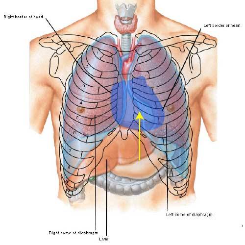
Photo I
Next, does the shape of the rib cage matter to a sgian dhu wielder? Weel, it should. Angus, for example, has a narrow rib cage and a narrow subcostal angle (Photo J – red lines) formed by union of costal cartilages and xyphoid process. The subcostal angle varies considerably from person to person. A typical angle is roughly 70° but some individuals have a wide rib cage and subcostal angle making a thrust to the heart easier to, erm, execute. In such people, the angle may exceed 100° allowing for a more successful stab.
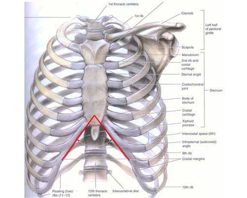
Photo J
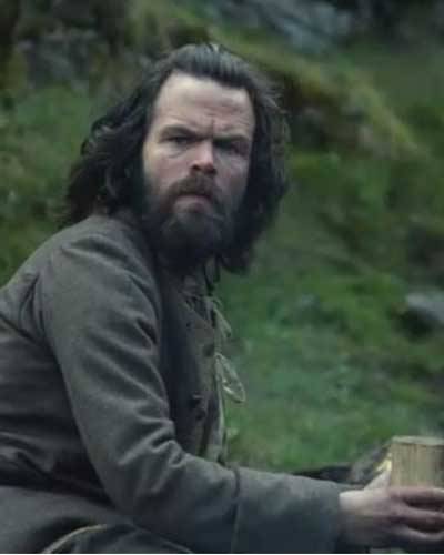
Taking the lesson very seriously, Angus next teaches Claire how to “kill from behind.” Sweet Willie is the model (Murtagh in Outlander book). Poor guy, he gets his share of abuse on this, his first rent trip.
And another fabulous Outlander quote from Herself (again, Rupert is teacher):
“I was timid and extremely clumsy at first, but Rupert was a good teacher, very patient and good about demonstrating moves, over and over.”
Aye, Angus is an excellent teacher as he patiently explains: “Now, this is the spot in the back. Either side will do. Now, you see all the ribs and such? Very difficult to hit anything vital when you stab in the back.”
He continues: “Slip the knife between the ribs, huh? That’s one thing but a lot harder than ye might think.” Now, is Angus correct about stabbing between the ribs, and if so, why? Stay with me now…
Yes, Angus is right! There are three reasons why stabbing between the ribs is difficult. As I explain, please recall that intercostal spaces are filled with intercostal muscles and fibrous tissue (Photo K).
- The intercostal spaces are not horizontal: most take a downward slope in the back and an upward slope in the front making it difficult to judge where to thrust a blade. Although a few medical papers report stab wounds between the ribs, these are often in the 1st or the 2nd intercostal spaces which are more horizontal than the rest.
- The intercostal spaces are narrow with an average width of about 2/3” (1.7 cm) so the chance of placing a knife in such curved, thin spaces is slim.
- Width of the intercostal spaces changes during each breath cycle so it presents a moving target.
Try this: place a finger in an intercostal space and breathe in as deeply as possible. Feel the change in width at deepest inspiration and again with complete expiration. During deep inspiration, the greatest width is discernible; with deep expiration, you’ll be lucky to feel an intercostal space at all. This reality greatly diminishes the chance of a successful stab into an intercostal space.
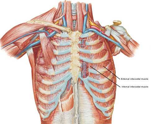
Photo K
Returning to the knife lesson, Angus instructs Claire to place her dagger “Here just under the last rib, you stab upward and into the kidney. Straight up – They’ll drop like a stone.” Whew, talk about a backstab! So, Nurse Claire delivers a well-placed thrust “straight up and in. See? Got it.”
So how did Claire do with her lesson? Was she a good student? Jamie’s proud of her knife skills and he can’t wait to get her alone so he can prove it; Murtagh’s thinking that mayhap Claire’s best weapon isn’t poison; and Dougal, well, that’s an admiring eye to be sure! Not sure what’s on yer mind Uncle, but ye best not share it wi’ Jamie!
So, is Angus right – can a 3” blade reach the kidneys from behind? Oh, aye, he scores again! Angus, you are on a roll, man!
Let’s see why. From behind, the thorax includes the area shown in blue (Photo L) and here, the underlying thoracic cage is formed of posterior ribs and thoracic vertebrae (Anatomy Lesson #10).

Photo L
In the back, latissimus dorsi and erector spinae muscles (Anatomy Lesson #10) cover the 11th and 12th ribs of the thoracic cage and deeper yet are the paired abdominal kidneys flanking the spine (Anatomy Lesson #8). A posterior view with muscles removed shows upper poles of right and left kidneys (Photo M- blue overlay) protected by 11th and/or 12th ribs. Note that the right kidney sits lower than the left making it more vulnerable to a knife thrust. This occurs because the right lobe of liver is considerably larger than the left lobe, displacing the right kidney downwards.
But, Angus is correct, either side will do: a 3” blade thrust under either 12th rib near the spine will surely strike a kidney and mayhap its renal artery. Ye may recall that the kidneys receive roughly 22% of the cardiac output (Anatomy Lesson #8)? This extremely high blood flow causes quick and profuse bleeding in the event of a significant laceration. Shock alone from such a wound could cause a person to “drop like a stone”. Although not always in the back, kidney stabs are fairly common injuries seen in urban ERs.
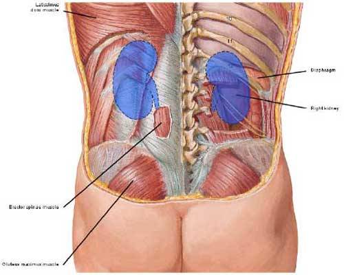
Photo M
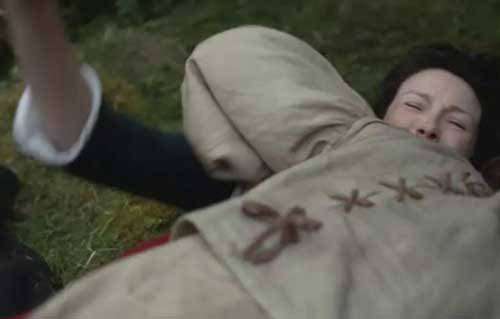
Harry a redcoat deserter soon provides Claire with a chance to apply her sgian dhu lesson. Talk about coitus interruptus! Those deserters sure know how to rain on a parade.Warning: If you dinna wish to see or read details about the stab wounds, please skip the next paragraph and three subsequent images.
Herself records the horrific, teeth-jarring attack in the Outlander book (Rupert and Murtaugh – not Angus and Willie):
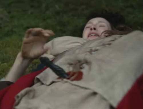
“I lay back, breathing heavily. I concentrated on my objective, trying to erase everything else from my mind. It would have to be in the back; the quarters were too close to try for the throat….I could see Rupert’s blunt finger stabbing at Murtagh’s ribs, and hear his voice, “Here, lass, up under the lowest ribs, close to the backbone. Stab hard, upward into the kidney, and he’ll drop like a stone……..I slipped my left arm around his neck to hold him close; holding the knife hand high, I plunged it in as hard as I could……… Unable to see, I had aimed too high, and the knife had skittered off a rib. I couldn’t let go now…”
“I stabbed again, with a desperate strength, and this time found the spot. Rupert had been right. Harry bucked in a hideous parody of the act of love, then collapsed without a sound in a limp heap on top of me, blood jetting in diminishing spurts from the wound in his back.”
BTW, happy is the anatomist to see the bright red color of arterial (not dark venous) blood from the stab wound in Harry’s back.
Now, its Jamie’s turn…… his dirk flashes and Arnold instantly joins his pernicious pal in death! Oh, aye, knives are everywhere in this episode!
From the Outlander book, Herself shares:
“Arnold’s attention had been distracted for an instant by the spectacle on the ground, and an instant was more than long enough for the maddened Scotsman he held at bay. By the time I had gathered my wits sufficiently….. Arnold had joined his companion in death, throat neatly cut from ear to ear by the sgian dhu that Jamie carried in his stocking.”
Whew! Jamie’s kill is so swift that I canna capture even one still image of the carnage!
And, not to be outdone by the other card-carrying knifers in episode 8, that crazy bastard, BJR, uses his own blade to harass and torment a terrified Claire. Not only does he destroy her pristine white fichu (how can that be after weeks of camping in mud and dirt?) but he cuts her lacings and tears her shift (aye, he’s a bodice ripper!). But, BJR isna really interested in women or their soft bodies; he wants intel and likes torture.
And, where is Claire’s hidden dagger when she needs it? Och! Safely hidden in the top of her lace-up leather boot whice she can’t reach (not bent over a desktop face-first with her skirts up)! Too bad Claire, otherwise ye could teach BJR your own wee dagger lesson! He’s earned it!
Pssst: just between you and me, I am fairly sure that Claire’s boots at Fort William are not the same ones she wears while climbing Craigh na Dun; the latter look a bit like UGGs! Mayhap there’s a Zappos near the fairy stones??
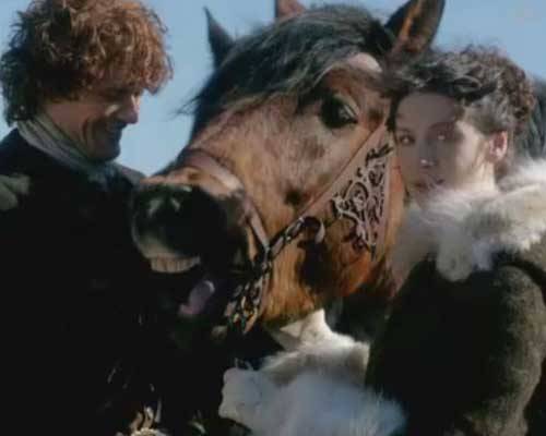
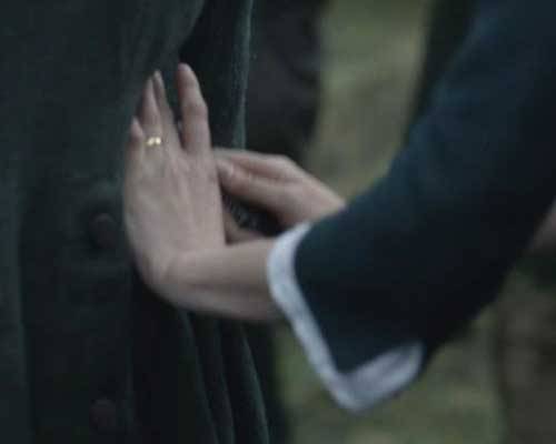
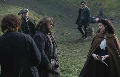
After all the blood and gore, let’s end this anatomy lesson on a happy note! Here’s the funny sequence between Jamie, Claire and Rupert over Jamie’s lost dirk after the crouching Grant’s attack. It happens so fast that ye may have missed the full effect. So here it is in slow-mo…
Ye recall Claire sighing that Jamie’s dirk is too long and heavy for her? With a huge, cheeky grin plastered on his face, Rupert chuckles “all the lasses say that to me.” Claire looks faintly disgusted. But lass, what’s wrong? Have ye never heard a man make a joke before? Aye, this time he’s the witty one!

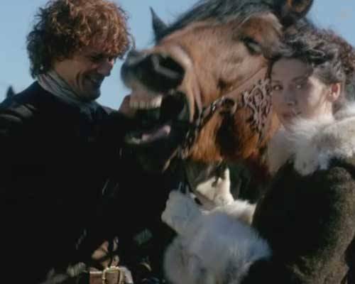
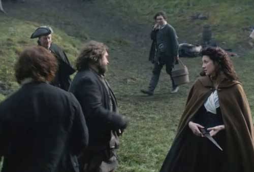
Oops! Not sure Jamie likes the double entendre… Suddenly, a wary Rupert isn’t grinning. I wish the camera had caught Jamie’s face as he swings toward the jokester.
Um, Rupert, ye best get while the getting’s good! Jamie doena like other men making ribald jokes to his wife. It’s one thing to pummel Jamie when he willna fight back, but ye likely won’t win if and when he decides ye are fish bait.
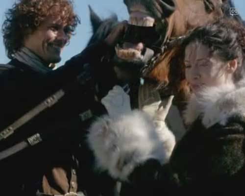
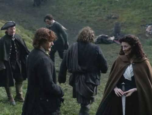
Aye, Rupert gets the message. Off he goes!
Claire finds the joke a wee bit amusing but Jamie isna smiling. Bye, bye, Rupert. For a big man, ye move pretty fast!
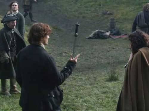
So, Jamie gets back his knife and rewards Claire by showing off. It’s not the first time we’ve seen him, erm, play with his blade. As Claire kens well, he’s darn good with a blade!
In closing, remember that the thoracic cage is a bony envelope formed of sternum, ribs, costal cartilages and thoracic vertebrae; it is engineered to protect vital organs including heart, lungs, great vessels and kidneys. Although vulnerable to trauma, a stab attack to the thoracic cage and its enclosed organs must be executed effectively to impose lethal harm. Mayhap Angus should teach the next Anatomy Lesson, you say? If I need guidance, I’ll ask. Snort!
A deeply grateful,
Outlander Anatomist
You can now follow me on Facebook and Twitter!
Photo creds: Starz, Gray’s Anatomy,
39th ed., Netter’s Atlas
of Human Anatomy, 4th ed., Clinically
Oriented Anatomy, 5th ed., Hollingshead’s
Textbook of Anatomy, 5th ed., www.Wikipedia.org