Fun Fact: Posterior Median Furrow

Anatomy Def: Midline vertical depression in the surface of the back (red arrows).

Outlander Def:  Deep, lovely groove of Jamie’s back!  

Get in the groove and Learn about Jamie’s back furrow by reading Anatomy Lesson #10, Jamie’s Back!

The posterior median furrow (PMF) literally means “back middle groove.” This vertical depression is most visible in thoracic and lumbar regions of the back. It tends to be shallow in the cervical region, diminishes over the sacrum and is continuous with the gluteal cleft (Aye, the sweet cheeks! <G>).

PMF is most visible in those with robust para-spinal (erector spinae) muscles. These muscles flank the vertebral column; the more developed the muscles, the deeper the PMF.

PMF is useful during physical exam because the vertebral spines can be palpated and counted in the groove. Specific vertebral spines mark levels for anesthesia delivery, location of abdominal organs, and vascular anatomy. For example, C3 marks the level of the hyoid bone (wink, wink to book readers).

More Fun Facts: English is replete with idioms about the back. These are  a few of the many:

  • Back away
  • Back me up
  • Back and forth
  • Back to square one
  • Back in the day
  • Back to the drawing board
  • Back down from
  • Back the wrong horse
  • Back to back
  • Back in circulation

Read about Jamie’s posterior median furrow in Outlander book. Yes, Herself describes it! 👏🏻👏🏻👏🏻

Compassionate Claire notes the groove as she describes Jamie’s badly damaged back: 

He sat patiently, not moving as I circled behind him and inspected his back…I was appalled…The scars covered his entire back from shoulders to waist. While many had faded to little more than thin white lines, the worst formed thick silver wedges, cutting across the smooth muscles. I thought with some regret that it must have been quite a beautiful back at one time. His skin was fair and fresh, and the lines of bone and muscle were still solid and graceful, the shoulders flat and square-set and the backbone a smooth, straight groove cut deep between the rounded columns of muscle that rose on either side of it…I tried not to imagine the muscular arms raised…ropes cutting into wrists, the coppery head pressed hard against the post in agony…Involuntarily, I reached out, as though I might heal him with a touch and erase the marks with my fingers. He sighed deeply, but didn’t move as I traced the deep scars, one by one, as though to show him the extent of the damage he couldn’t see.

Och! See Jamie’s PMF in Outlander, episode 105, Rent, after Uncle Dougal rends Jamie’s sark to expose BJR’s handiwork! 😱 Back off, Dougal!

The deeply grateful,

Outlander Anatomist

Follow me on:

Photo Credit: Sony/Starz

Anatomy Lesson #53: Dr. Abernathy Meets “Pretty Lady”

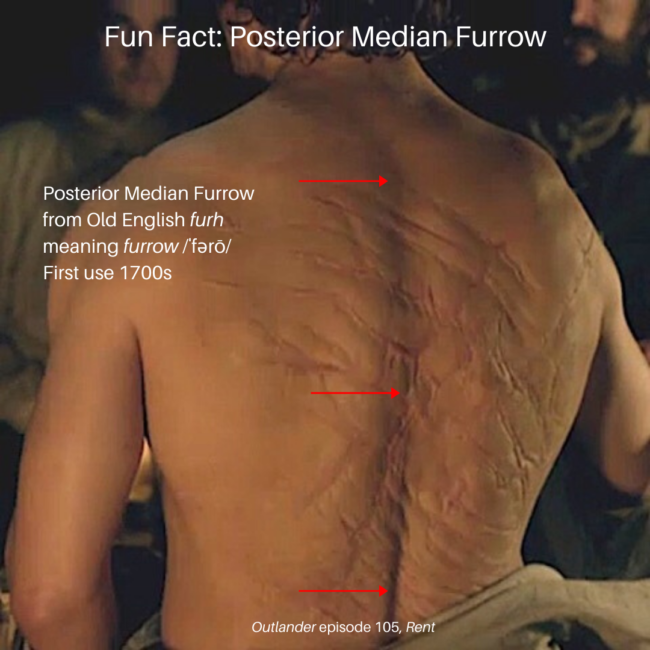
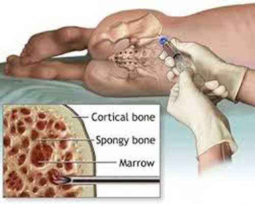
Greetings, anatomy students! Several of you have asked for an exploration of the “skeleton scene” shown in Outlander episode 305, Whiskey and Freedom. Although Anatomy Lesson #39, Dem Bones – Human Skeleton, examined the entire human skeleton, this lesson focuses on skeletal analysis performed by Dr. Abernathy with input from Dr. Claire. 

 The “skeleton scene” is a great adaptation of Chapter 20, “Diagnosis,” from Voyager book, by Diana Gabaldon. So, put on your forensic cap and let’s begin with a summary of that scene.

Here, the anthropology office at Harvard seeks Dr. Abernathy’s expertise to determine cause of death using skeletal remains. In the book, the skeleton arrives at his office in a box labelled “PICT-SWEET CORN.” I remember that brand!  

In the episode, bones are laid out on a desk top (Image A). From Voyager:

 Horace Thompson was probably someone from the coroner’s office, I thought. Sometimes they brought bodies to Joe that had been found in the countryside, badly deteriorated, for an expert opinion as to the cause of death. This one looked considerably deteriorated.“

…from the anthropology department at Harvard,” he said… “asked me would I have a look at this skeleton, to tell them what I could about it.”
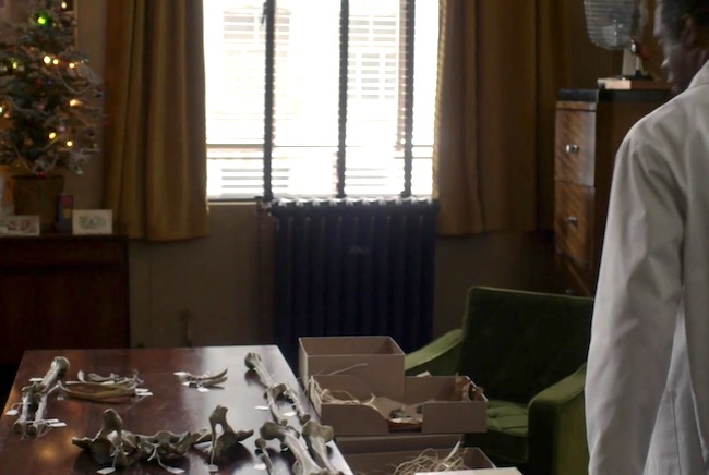
Image A

Dr. Abernathy reaches into the box and removes the skull (Image B).
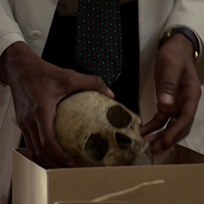
Image B

 Examining the skull, the good doctor concludes it belonged to a pretty lady who was mature and middle-aged (Image C). And, from Voyager:

As the saying goes, “Pretty is as pretty does.” The owner of this skull definitely did not do pretty things! 😳

“Oh, pretty,” he said in delight, turning the object gently to and fro. “Pretty” was not the first adjective that struck me; the skull was stained and greatly discolored, the bone gone a deep streaky brown. Joe carried it to the window and held it in the light, his thumbs gently stroking the small bony ridges over the eye sockets. “Pretty lady,” he said softly, … “Full-grown, mature. Maybe late forties, middle fifties.”
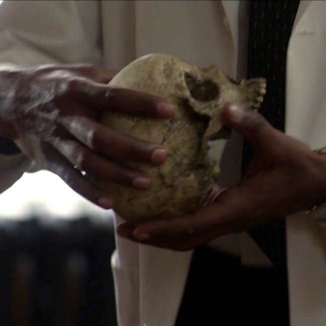
Image C

Clairvoyant Claire picks up the skull, which speaks to her (Image D).  Well, not really –  that would be weird – though Hallowe’en does draw nigh! Another quote from Voyager:

Then I held it close against my stomach, eyes closed, and felt the shifting sadness, filling the cavity of the skull like running water. And an odd faint sense—of surprise?

“Someone killed her,” I said. “She didn’t want to die.”
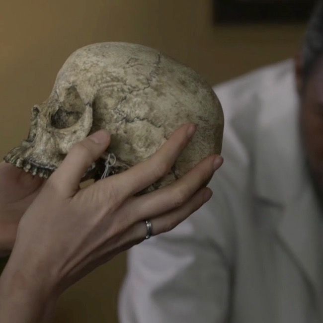
Image D

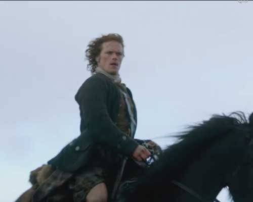
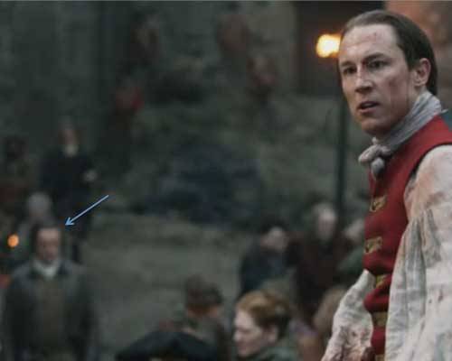
Dr. Abernathy fixes Dr. Claire with a gimlet eye. Lady Jane, have you lost your scalpels???  After all, how could she possibly conjure such details using a touchy-feely method of scientific inquiry? Well, the lass does harbor some awesome powers that seem to grow with time (Image E)? Again, from Voyager:

… “Where did you find her?” I asked….

“She’s from a cave in the Caribbean,” he said. “There were a lot of artifacts with her. We think she’s maybe between a hundred-fifty and two hundred years old.”
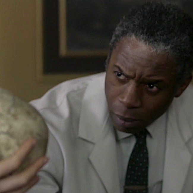
Image E

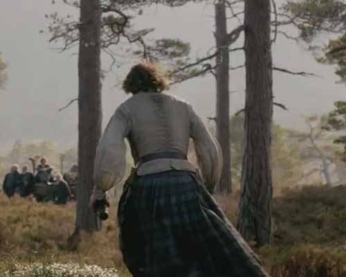
Next, Dr. Abernathy plucks two pieces of bone from the corny box (Image F). “You were right,” he says. Looking at them, he observes that the fracture plane runs through the centrum, although he doesn’t identify which bone has been fractured. 

Voyager book reveals the fractured bone as a vertebra (bone of spine) – more specifically, it is the axis, aka the second cervical vertebra (C2).

The wide body of the axis had a deep gouge; the posterior zygapophysis had broken clean off, and the fracture plane went completely through the centrum of the bone.

Joe’s finger moved over the line of the fracture plane. “See here? The bone’s not just cracked, it’s gone right there. Somebody tried to cut this lady’s head clean off. With a dull blade,” he concluded with relish.
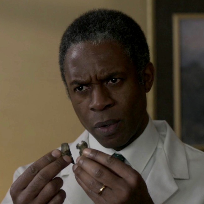
Image F

Then, Dr. Abernathy notes that although the burial site was a cave filled with slave artifacts, this lady was not a slave! He points to two leg bones (Image G), the tibia (Anatomy Lesson #9, The Gathering or Boar Gore) and the femur (Anatomy Lesson #7, Jamie’s Thighs or Ode to Joy!).  Ahhh, Claire sagely nods, “the crural index.” Back to Voyager, again:

“Not a slave,” he said… “No,” Joe said flatly. He tapped the long femur, where it rested on his desk. His fingernail clicked on the dry bone. “She wasn’t black.”

“Take a look at this,” Joe invited. “You can see the differences in a lot of bones, but especially in the leg bones. Blacks have a completely different femur-to-tibia ratio than whites do. And that lady”—he pointed to the skeleton on his desk—“ was white. Caucasian. No question about it.”

…“If you want to think blacks and whites are equal under the skin, be my guest, but it ain’t scientifically so.” He turned and pulled a book from the shelf behind him. Tables of Skeletal Variance, the title read.
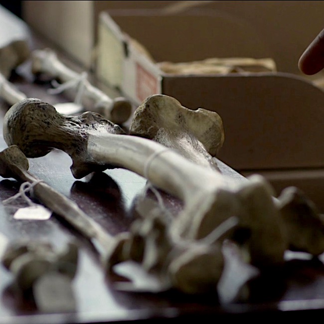
Image G

 OK, that pretty much summarizes the salient scientific points of this scene, although I see three issues that warrant comment:

      • Can bones reveal sex, age and/or beauty of the owner? 
      • Does the fractured bone match with “pretty lady’s” death? 
      • Can crural index determine race? 

Issue #1: Can bones reveal sex, beauty and/or age of the owner?

The answer is a qualified yes

When Claire arrives at the office, Dr. Abernathy has already laid out most of the skeleton and presumably, has already examined each of these bones. It’s pretty iffy to hazard a reliable guess with one measurement. But, if a series of measurements tend to fall within a given range, a forensic scientist can venture an educated guess. So, assuming he examined the skull and pelvis, then sex can be surmised with a resonable degree of confidence. Here’s why.

Sex & Skull: “Typical” female and male skulls exhibit differences (Image H). A female skull is usually smaller, with rounder orbits (red dot), prominent frontal eminences (yellow dot), smaller mandible (pink dot), less prominent temporal lines (green dot), smaller brow ridges (dark blue dot), and less pronounced glabella (turquoise dot), and the brow ridges are sharper. There are also other, more subtle differences, but understand that not all skulls show all structural differences, regardless of genetic sex.
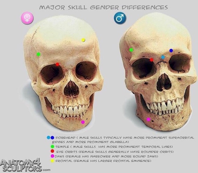
Image H

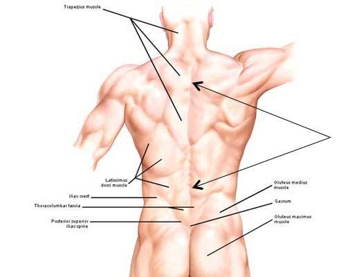
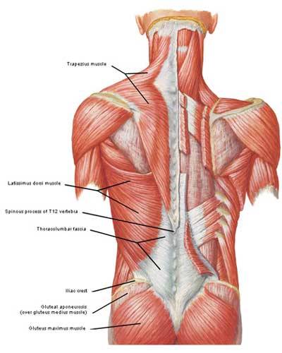
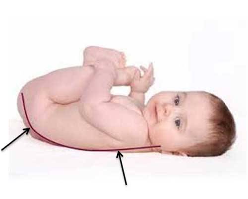
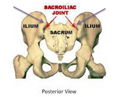
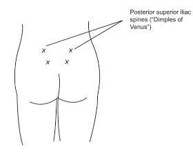
Sex & Pelvis: The bony pelvis also supplies important clues to sex. “Pretty lady’s” bony pelvis lies in three pieces (two hip bones plus sacrum). The pelvic width, shape of sacrum, sub-pubic angle and shape of obturator foramen (two front holes) are consistent with a female pelvis (Image I – obturator foramen not labeled).  Pelvic inlet and outlet are difficult to demonstrate in a dissembled bony pelvis but, assembled they would be similar to those shown in Image I, lower figure. Ergo, pretty lady’s pelvic features are consistent with those of a female.

Age:  Age can be estimated if cranial sutures (sites where cranial bones meet) are thin, bony ends are fused to bone shafts (growth plates are ossified), teeth are mature and bones are hard, along with the presence of wear-and-tear diseases, etc. So, yes, with careful analysis, general age can be estmated. Presumably, Dr. Abernathy considered these before Claire’s arrival. So far, so good!

Beauty:  As we all ken, beauty is in the eye of the beholder. Dr. Abernathy expresses an opinion when he dubs the owner a “pretty lady.” Of course, he cannot know how flesh draped those bones, but he considers the skull to be delicate and beautiful to his practiced eye. This is a subjective response on his part, but it is arguably consistent with the appearance of the skull which is delicate with good teeth. Nowadays, forensic scientists can reconstruct a face using computer programs, or the older clay sculpting technique

Thus, sex and age can be assessed with a fair degree of confidence if and only if multiple measurements and observations are considered, collectively. But, as beauty remains in the eye of the beholder, this issue receives a qualified yes.
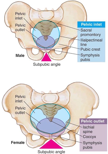
Image I

Issue #2: Does the fractured bone match with “pretty lady’s” death? 

Because I have some issues with this issue, the answer is mostly yes for the book, but no for the episode. Here’s why. 🤓

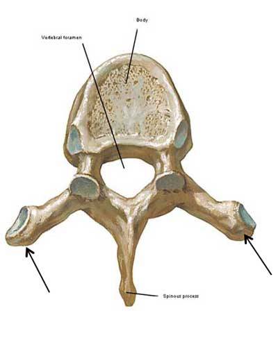
Dr. Abernathy holds up a wee bone, which is broken vertically into two nearly symmetrical pieces. Voyager identifies these fragments as belonging to the axis. The axis is one of seven neck bones (numbered 1-7 from skull downwards); it is also designated as C2, meaning it is second of the seven cervical vertebrae. The purpose of cervical vertebrae is to support the head and supply flexibility to the neck, augmenting movements of the head and shoulders.

Voyager book states:

The wide body of the axis had a deep gouge; the posterior zygapophysis had broken clean off, and the fracture plane went completely through the centrum of the bone.

Although the bone fragments of shown in episode 305 are supposed to represent a vertebra, the fragments do not  appear to be an axis. The axis is an atypical, weird-looking bone (Image J – gif):
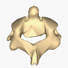
Image J

However, please appreciate that the axis is a splendid bone, allowing us to rotate our head from side-to-side in a “no-no” gesture (Anatomy Lesson #12 Claire’s Neckthe Ivory Tower!). 

Try this: Cervical vertebrae are buried rather deeply in the neck and difficult to demonstrate. But, if you able, sit up straight with chin level, place a finger in the groove of your neck just below the skull. You may feel a small bulge under your fingers. This is the spine of the axis. 

Let’s examine the parts of the axis (Image K – front view) to help us understand the quote from Voyager.

  • The axis has a robust body, located at its front surface.
  • Inside the body is a centrum, a remnant of embryonic development. You cannot see the centrum from the surface.
  • Dens is a tooth-like structure perched atop the body.
  • Superior articular facets (aka superior zygapophyseal processes) are flat surfaces where C2 forms joints with C1 above.
  • Inferior articular facets (aka inferior zygapophyseal processes) are flat surfaces where C2 forms joints with C3 below.
  • Joints are sites where movement occurs between bones – between vertebral facets these are called zygapophyseal joints.

To reiterate, Voyager book states:

The wide body of the axis had a deep gouge; the posterior zygapophysis had broken clean off, and the fracture plane went completely through the centrum of the bone.

Analysis: The book statement is a wee bit awkward because if the fracture plane passed through the centrum, it must necessarily cleave the body, so a deep gouge would not have been left to discern.

Also our vertebrae don’t have posterior zygapophyses – I suspect Diana intended inferior rather than posterior. Assuming this is correct, then: The dull blade cut horizontally (as in cutting off a head), breaking off the inferior zygapophyses (forming zygapophyseal joints with C3), and passing through the axial body and its centrum. Otherwise, the description makes sense.

One may also conclude, the blade must have passed through the lower part of the axis. Had the blade passed through its upper part, the stroke would have sheared off dens and superior zygapophyses but completely missed the centrum! Make sense? Yay! 👏🏻👏🏻👏🏻
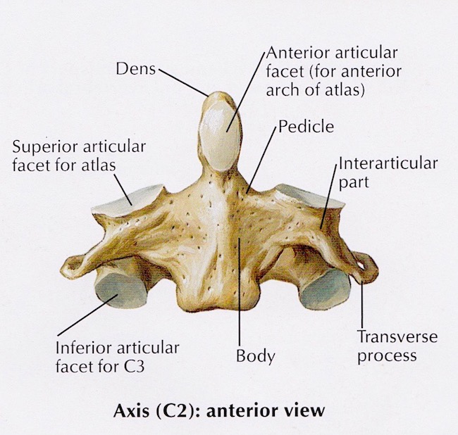
Image K

Conclusion: Dr. Abernathy holds two bony fragments of the fractured bone and I think, hum… that ring of bone appears to have been cleaved in halves by a vertical blow (Image L)! The only way a vertical fracture could occur is if the blade sliced downward, vertically cleaving the skull and its supporting cervical vertebrae. Gah! I think you will agree, this is pretty unlikely plus, the skull remains intact. So, no vertical swipe of the blade!

Ergo, although dramatic and interesting, the bone fragments do not reflect the axis damage as described in Voyager or by episodic Dr. Abernathy. Now, does all this keep me up at night? Hardly – I loved this scene despite its wee anatomical issues!

Image L

Issue #3: Can crural index determine race? 

The answer is a qualified no.

This topic is a super sticky-wicket but very important to consider, so bear with me.

Best start with a definition: the crural index, established in 1933, is the ratio of tibia-to-femur (not femur-to-tibia, as this yields different results). The formula to determine crural index is:

 (length of tibia x 100) / length of femur  OR  

(length of tibia/length of femur) x 100

Using this ratio, many studies showed that individuals of African descent had higher crural indices than those of European descent. Image M is a simplified, summary version of such findings.

In 1968, Dr. Abernathy was positive that a low crural index meant “pretty lady” was white….“No doubt about it.”

(BTW, I am pretty sure Claire pronounces this “cruel index;” but, it should be crural <kru-ral>, from the Latin crus meaning “leg”)
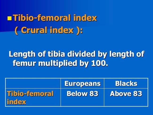
Image M

So, can race be determined from the crural index?

When I started graduate school in 1965, we were taught there were three human races: negroid, caucasoid, mongoloid. Fast forward 52 years and much has changed! Today, many biologists say race cannot be determined from bones because there is no such thing as race. These scientists posit that all living humans belong to one species (Image N): Homo sapiens sapiens (the second sapiens denotes the subspecies – that would be us).

Many designate the term ancestry, because race and even ethnicity have confusing connotations and definitions. Furthermore, they point out, more genetic variations can occur within “racial” groups than between them, meaning findings are limited by the sample studied. What a conundrum!

Just to clarify, some bony physical traits are characteristic of ancestry and can be traced to a particular global location. But, bear in mind, people of mixed ancestry may present features which do not fall neatly into any category. Also, humans are so similar that all bone morphologies are present in all  groups, just at varying rates. Despite such variations, skeletal analysis remains part and parcel of human identification especially when numerous skeletal measurements are obtained. Today, using calipers, x-rays, microscopy, DNA, and a mess of other tools, some of which were unavailable in 1968, forensic researchers can make reasoned guesses as to a person’s ancestry based on skeletal remains.

Summary: Nowadays, before a scientist suggests ancestry based on skeletal remains, (s)he makes multiple measurements, never relying on just one. And, prudent scientists avoid stating “we are sure” (even if they are). Instead, they posit, the data suggests or indicates or is consistent with or is likely. Verra prudent!

Hence the qualified “no” regarding the crural index; it is only one skeletal measurement and insufficient to make a judgement if a person was white or a not. However, Voyager accurately expresses views prevalent in the 1960’s. Make sense? Ta da!
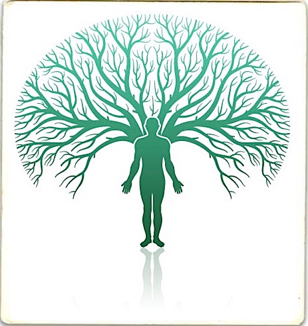
Image N 

Bottom line:

    • Today’s competent (my word) scientist cannot/will not declare ancestry using a single skeletal measurement such as crural index. 👎🏻
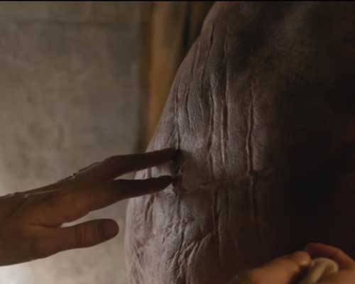
    • Cutting (hah) Dr. Abernathy some slack, his surety about the skeleton’s “race” was suitable and justifiable for his time (1968), but untenable for ours (Image O)!

Image O

So, this concludes a brief analysis of the “skeleton scene.”  Much more could be added, but would likely be too technical for most students. Hopefully, this summary was enlightening and will generate some thoughtful discussion and consideration in our ever expanding Outlander world. Buh-bye, pretty lady!

Ode to Pretty Lady

Your bones tell a tale.

Who are you?

Were you well?

Were you pretty?

Were you witty?

Were you sweet?

Did you cheat?

Were you bad?

Were you sad?

Or, were you mad?

Your bones tell a tale.

No Spoilers! Who are you “pretty lady?” Mayhap, we will find out during Outlander S.3!

The deeply grateful,

Outlander Anatomist

Follow me on:

Photo Credits: Sony/Starz (Images A-G, L, O), Clinically Oriented Anatomy by Moore and Dalley, 5th edition (Image K), www.pinterest.com (Image H), www.medical-dictionary.thefreedictionary.com (Image I), www.premiersourceshopping.com (Image O), www.slideshare.net (Image M), www.wikipedia.com (Image J – gif)

Anatomy Lesson 10: “Jamie’s Back!”

Anatomy Lesson #10: The Back.

Happy New Year to all of you! I’m a wee bit late posting this because I just returned from my trip to Fraser’s Ridge country. It was a verra beautiful region. I can see why Jamie and Claire made their home there.

Welcome to all newcomers and thanks to all those returning to learn about Anatomy Lesson #10: The Back. Because our red-haired hero is so utterly braw, I will use many images of Jamie to teach this verra important topic.

Claire first sees Jamie’s back in Starz episode 102, Castle Leoch, as she removes the blanket to clean his gunshot wound. Herself writes in Outlander book:

“I had finished the bandaging and stood quiet behind him, a hand resting on his good shoulder… His upper back was covered with a crisscross of faded white lines. He had been savagely flogged, and more than once. There were small lines of silvery scar tissue in some spots, where the welts had crossed, and irregular patches where several blows had struck the same spot, flaying off skin and gouging the muscle beneath.”

Then says Jamie:

“Lobsterbacks. Flogged me twice in the space of a week. They’d ha’ done it twice the same day, I expect, were they not afraid of killing me. No joy in flogging a dead man.”

image

I wish I could work off a blank canvas of Jamie’s back before it was scarred by that mad bastard Randall! Fact is we have only a brief and limited glimpse at Lallybroch (Starz episode 102, Castle Leoch) as Black Jack administers his first whipping of our brave hero. Ugh!

image

Now, take a wee gander at Jamie’s back after the first blow. The blue arrows mark welts or wheals! Our Starz Jamie has verra fair skin and these swellings appear to be real. Wheals are typically initiated by an allergic response or tough handling such as pressure, blows or friction. Mayhap Jamie was handled a wee bit roughly during this scene?

image

Back: Now, in anatomy, the human back is defined as the large area from the base of the head to the tail bone (Photo A). It is widest at the shoulders, narrows at the rib cage and ends as a “V” between the buttocks. The topographical (surface) anatomy of a well-muscled back displays a mess of lumps, bumps and valleys created by underlying muscles, etc.

The two black arrows in Photo A mark a prominent vertical midline groove, the posterior median furrow. You should also understand that for protection, back skin is thicker and has fewer nerve endings than, for example, belly or chest skin. It is also an area that we cannot typically see or touch without aid of mirrors, photos, or yoga practices. Namaste!

image

Photo A

Now, sans the skin, let’s take a peek at the superficial structures creating back contour (Photo B). The paired trapezius muscles (Anatomy Lesson #3) cover the upper back. You recall they take origin (begin) from bones of the skull and spine but they insert (end) on clavicle and scapula; acting together or separately, they these bones. Latissimus dorsi muscles cover the lower back. They also take origin from bones of the back but insert on and move the humeral bones (Anatomy lesson #2). Gluteus maximus and Gluteus medius (Anatomy lesson #1) muscles appear to be part of the back but they form most of the buttocks and belong to the lower limbs. The V-shaped depression at the base of the spine (white area) is the thoracolumbar fascia flanked by the gluteal muscles and iliac crests. The thoracolumbar fascia is a strong sheet of collagen that gives origin to back and abdominal muscles (cover in a later post). The iliac crest forms the top of each hip bone. The vertical midline groove (see Photo A) is created by the spine as it adheres to the dermis of the skin (Anatomy Lesson #5 & Anatomy Lesson #6).

image

Photo B

Removal of the superficial back muscles exposes the deeper erector spinae muscles (Photo C). They attach to the spine and ribs. In the physically fit, they are visible as thick longitudinal columns of muscle flanking each side of the spine. Their function is to arch or extend and rotate the spine (see below). Again, although seemingly part of the back, the buttocks and their gluteal muscles are actually parts of the lower limbs.

image

Photo C

The length of the back is determined by the spine or vertebral column which extends from the base of the skull to the tail bone (green in Photo D).

image

Photo D

The vertebral column forms the bony central axis of the back (Photo E). Five regions are identified from the cephalic end (Greek meaning head) to the caudal end (Latin meaning tail): cervical, thoracic, lumbar, sacral and coccygeal. There are 7 cervical (C 1-C7), 12 thoracic (T1-T12), 5 lumbar (L1-L5), 5 sacral (S1-S5) and 4 coccygeal (Co 1-Co4) for a total of 33 vertebrae (Photo E).

image

Photo E

A typical vertebra (some are atypical) shows the following main features (photo F):  a spinous process that projects posteriorly and provides ligament and muscle attachments; a cylindrical body that is flanked top and bottom by intervertebral (IV) discs; paired transverse processes (black arrows) that provide for muscle and ligament attachments; and a hole known as a vertebral foramen.

image

Photo F

The vertebrae of an intact spine are aligned and held in check by numerous powerful ligaments, connective tissue bands that link bone to bone (Photo G – don’t fash about their names).

image

Photo G

This arrangement makes the vertebral column strong, flexible and springy! It also allows for a considerable amount of movement including lateral (side bending) flexion, twisting (rotation), and anterior (forward) flexion (Photo H):

image

Photo H

And, for spinal extension (Photo I):

image

Photo I

Vertebral alignment also converts individual vertebral foramina into a vertebral canal that houses and protects the spinal cord. Photo J is a schematic of the spinal cord inside the vertebral canal (only a few  vertebrae are numbered). The colored coded regions give rise to spinal nerves issuing in pairs from cervical, thoracic, lumbar, sacral and coccygeal levels of the spinal cord; the nerves supply structures of the neck, trunk, upper limbs and lower limbs.

image

Photo J

The thoracic vertebrae are unusual in that they are directly anchored to the ribs and indirectly to the sternum (breast bone). This arrangement provides great stability and allows anterior flexion but little other movement (Photo K). Ribs in the image below are numbered but the thoracic vertebrae are not. Know that T1 vertebra articulates with the 1st pair of ribs and so on.

image

Photo K

Here’s an interestin’ tidbit: as bipedal humans, our spines transform during life. With infants, the entire spine is flexed anteriorly (Photo L – purple line).

image

Photo L

As we lift our heads, sit up and walk, four alternating curvatures develop that are readily apparent in the adult spine. The cervical and lumbar vertebrae curve posteriorly – the thoracic and sacral vertebrae curve anteriorly (Photo M – spine viewed from the left side).

image

Photo M

Take a deep breath and enjoy this image o’ Jamie’s back: ye can clearly see his thoracic and lumbar curvatures as well as the deep midline groove flanked by impressive columns of his erector spinae muscles (Starz episode 105, Rent). And, thank ye verra much but from now on, he’ll be mending his own shirt – since his tender Uncle Dougal keeps tearing it off in front of strangers! He doesn’t care if the lad wears rags from now on!

image

Now, Claire who makes love to Jamie with her eyes (and other stuff too) throughout all the Outlander books, observes that Jamie’s back is “as straight as an alder sapling.” She doesna mean that he lacks the four curvatures; she means that he holds his back upright and erect…good muscle tone, aye? Here’s a great example of Jamie’s back while playing with the fillies (Starz episode 2, Castle Leoch). Snort!

image

And, och, while giving the Watch the slip (Starz episode 5, Rent)!

image

And one of my personal favorites where Jamie strides away from Claire after telling her that it is he who is virgin (Starz episode 106 The Garrison Commander)! One of them better ken what they’re doing! That put a wee gleam in Claire’s eye!

image

Now back ta anatomy. Another interesting tidbit about the vertebral column: at birth, the wedge-shaped sacrum consists of five separate vertebrae. Fusion begins in adolescence and ends about 30+ years explaining why the adult sacrum is counted as five vertebrae. Also, the sides of the sacrum articulate (form a joint) with each hip bone at the sacroiliac (SI) joints (Photo N – red arrows) to create a rigid pelvis. The purple arrows mark bilateral bony elevations o’ the hip bones known as the posterior superior iliac spines (PSIS).

image

Photo N

The PSIS are also sites where the dermis adheres to underlying bone forming the so-called dimples of Venus (Photo O):

image

Photo O

Although the dimples appear in countless nude images, it turns out they aren’t just beauty marks; they are superficial and easily used for bone marrow aspirations (Photo P).

image

Photo P

Hold on to your chair! Claire shows her dimples of Venus in this scene from Starz episode 107, The Wedding (red arrows)! Here, Claire guides Jamie’s hands to her hips and he starts to put his right fingers over her left dimple!  Gasp, I think I just stopped breathing – need CPR STAT!

image

I cannot find any images of Jamie’s dimples of Venus but, aye, he’s got ‘em. It is just that they are absorbed into paired grooves where his lumbosacral fascia meets his very well-developed gluteus maximus and medius muscles (see Anatomy Lesson #1)! His butt muscles are so powerful that they create a groove on each side of the sacrum (Starz episode 105, Rent). Mmhm!

image

Now, this lesson must visit Jamie’s scourging by the Mad Man of Fort William! It breaks my heart to watch this episode (Starz episode 106, The Garrison Commander), but it is needful to understand. The harsh treatment of Jamie is not only a personal tragedy but is a harbinger of the approaching battle of Culloden and the subsequent crushing by the Redcoats of the Highlander way of life. You ken that Jamie had already received 100 lashes for trying to escape afore BJR decides he‘s due 100 more! The first strike of the whip causes the blood to flow freely because BJR is using the infamous Cat ‘o Nine Tails with lead weights at the tips! He means to bleed Jamie to the bone (You understand that if he had actually done so, Jamie would have been disabled or perhaps dead?)

image

Ye can see why Jamie dubs him that “mad bastard Randall.” Just take a keek at Randall’s face and eyes. This is hard work, right? And just in case you didn’t notice, the blurry figure in the background is Black Brian, Jamie’s father, so named because of the color of his hair whereas BJR is named for the color of his soul!

image

By the end, Jamie’s back looks like haggis. Little wonder he could barely sit astride a horse after his friends made shift to rescue him!

image

Herself provides another intimate appraisal of Jamie’s back in the Outlander book. Here, Jamie removes his shirt in Claire’s surgery after leaving Gwyllyn’s performance. The dressing is chaffing him and he asks her to remove it. Claire observes:

“He sat patiently, not moving as I circled behind him and inspected his back…I was appalled…The scars covered his entire back from shoulders to waist. While many had faded to little more than thin white lines, the worst formed thick silver wedges, cutting across the smooth muscles. I thought with some regret that it must have been quite a beautiful back at one time. His skin was fair and fresh, and the lines of bone and muscle were still solid and graceful, the shoulders flat and square-set and the backbone a smooth, straight groove cut deep between the rounded columns of muscle that rose on either side of it…I tried not to imagine the muscular arms raised…ropes cutting into wrists, the coppery head pressed hard against the post in agony…Involuntarily, I reached out, as though I might heal him with a touch and erase the marks with my fingers. He sighed deeply, but didn’t move as I traced the deep scars, one by one, as though to show him the extent of the damage he couldn’t see.“

image

Now, I must confess, this post wasn’t much fun because of its grim topic but history teaches us important lessons. During the Napoleonic Wars (1803-1815), soldiers in the British Army could receive up to 1,200 lashes, a number that could permanently disable or kill a man. The US followed a similar code and neither country abolished scourging until the late 19th century – a blot on both nations to be sure as scourging is intended to demean, humiliate and permanently scar its victims.

Flogging was also used during the US slavery period. The next photo taken at Baton Rouge in 1863 shows the back of Gordon (Photo Q), a scourged slave. His scars are pronounced because of keloid formation.

image

Photo Q

Lastly, let’s consider the importance of the back, the single largest canvas of the human body. It is the basis for many sayings focused on this area of vulnerability giving rise to idioms such as: “your back is against the wall”, “stab one in the back”, “friends have one’s back”, “watch one’s back” and “back off”. The back is also a symbol of strength and hard work, as in “back to work”, “strong back” and “put your back into it”. Indeed, the practice of showing explicitness on the back has been practiced for centuries through clothing, no clothing, art, tattoos and, in Jamie’s case, flogging.

The back can also be used to telegraph emotion. In the next image, Claire has just snubbed Jamie (Starz episode 105, Rent) because he said her face plainly showed that she was thinking of running like she tried at the gathering. Well, she did ask, but Jamie doesn’t like the brush off so he flaps his plaid over his back in annoyance. It’s very funny! See Claire “back” there? She just slipped into her tent. Darn woman…doesn’t know what’s billowing behind her back!

image

OK, that’s it for the back. Next time I will post on a lighter topic…like Claire, I promise!

The deeply grateful,

Outlander Anatomist

Follow me on:

Photo Credits: Netter’s Atlas of Human Anatomy 4th ed., Netter’s Atlas of Human Embryology, 1st edition, Hollingshead’s Textbook of Anatomy, 5th ed., Starz, ainsleyjason.wordpress.com, www.sportsmed.or.nz, www.Radiopaedia.org, www.wikihow.com, www.Wikipedia.org, www.massgeneral.org, www.faculty.washington.edu, www.wikipedia.com, www.momarock.wordpress.com, www.imueos.wordpress.com, www.forums.hardwarezone.com.sq, www.pedsoncologyeducation.com, www.stemcelldoc.wordpress.com, www.hercampus.com, www.prevention.com