In Voyager, third of the Outlander books, Herself writes about a character named Dr. Joe Abernathy. As he examines some human bones, he softly sings to himself:
“Oh, de headbone connected to de…neckbone…de neckbone connected to de…backbone…Now hear…de word…of de Lawd!”
This spiritual song, composed by African-American author and songwriter James Weldon Johnson (1871–1938), provides a fitting introduction for today’s Anatomy Lesson #12: The Neck.
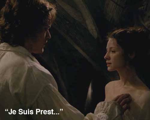
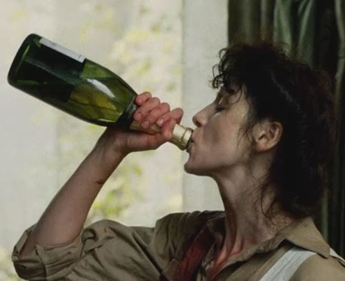
We need a model for our lesson on the neck and can ye think of a better one than bonny Claire? Here’s Claire drinking celebratory bubbly at the end of World War II; her ivory, blood-smeared neck rises from her torso as a long column of muscle, bone and sinew (Starz episode 101, Sassenach).
Definition: The neck is the body region between base of skull and top of sternum. The anatomical word for neck is cervix (Latin meaning neck), applying the neck region or neck of the uterus. For simplicity, I divide the neck into posterior and anterior compartments:
- Posterior neck contains cervical vertebrae muscles, nerves, ligaments, etc.
- Anterior neck compartment contains respiratory and gastrointestinal viscera (organs), important blood vessels, a bone and numerous muscles
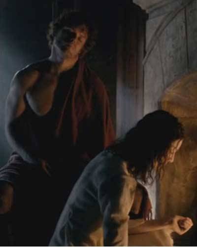
Image A shows the chin angled toward the right acromion (Anatomy Lesson #2), thus lengthening the left side and shortening the right.
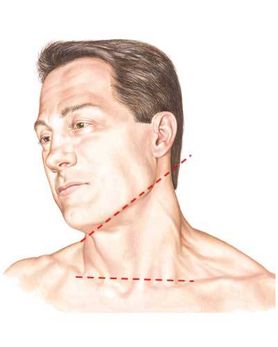
Image A
Posterior Neck: The posterior neck is supported by the cervical spine (see Anatomy Lesson #10 for thoracic, lumbar, sacral and coccygeal spine). The cervical vertebrae are numbered C1 – C7 and extend from base of skull to first thoracic (T1) vertebra (Image B – cervical spine, right side).
Image B illustrates C2 – T1 vertebrae. Anterior cylindrical bodies face right and posterior unpaired spinous processes point left. The spinous processes form midline knobs under the neck skin. These vertebrae form the cervical curvature, convex anteriorly and concave posteriorly, allowing for a springy and flexible neck that adjusts head position for optimal sight and hearing.
Although not shown in Image B, intervertebral (IV) discs separate vertebrae C2-C7.
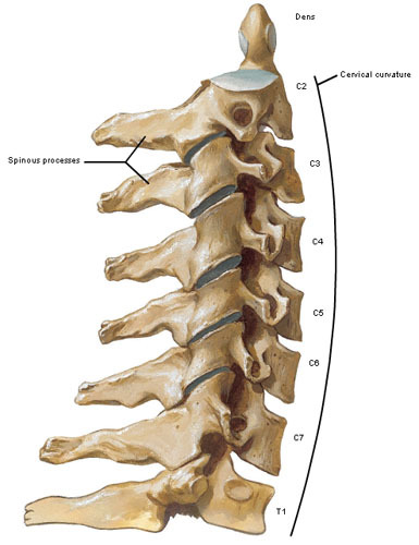
Image B
Ligaments: Viewed in situ (Latin meaning in position), several strong ligaments bind cervical vertebrae to each other and the skull base (Image C, right side). An unpaired ligamentum nuchae extends from skull base to C7 spine anchoring the cervical vertebrae and providing partial origin for the trapezius muscles (Anatomy Lesson #3).
In the neck, ligamentum nuchae covers the spinous processes and is aligned with the median furrow, a midline groove running the length of the back (Anatomy Lesson #10). Known as the paddywhack in sheep and cattle, ligamentum nuchae helps support head weight in four-legged animals. Dried paddywhack is sold as dog treats as suggested by the 19th century Welsh children’s song This Old Man:
“With a knick-knack paddywhack, give the dog a bone…”
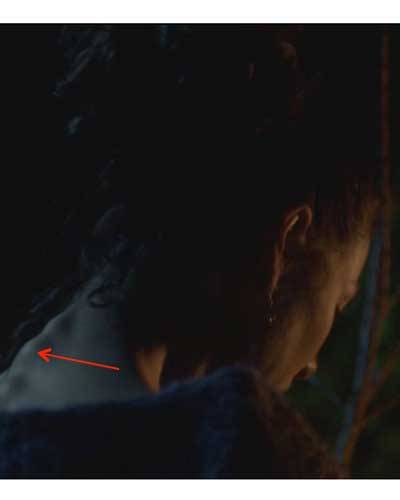
C7 bears a long spinous process, the vertebra prominens, because in most people it is most prominent.
Try This: Palpate your own C7 spine: Crop chin to chest and palpate the big bony knob of vertebra prominens at the neck base.
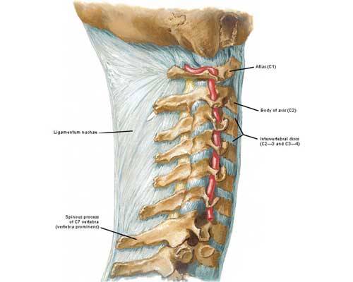
Image C
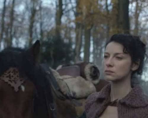
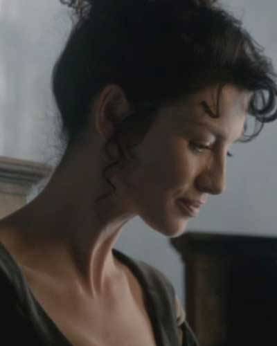
Claire’s slender, swan-like neck bears a visible vertebra prominens (red arrow) as she cries at Castle Leoch (Starz ep 103, The Way Out). She just teased Jamie in the dinner hall about kissing Laoghaire. She is envious of their intimacy and she misses her hubby!
C3 – C7 Vertebrae: Cervical vertebrae C3 – C7 are similar to thoracic vertebrae (Anatomy Lesson #10) except they are smaller and have paired transverse foramina (holes) to transmit blood vessels to and from the brain (Image D). The bodies are smaller and oval. Spinous processes project posteriorly and most are bifid – forked – just like Claire’s life line! Understand that the spinal cord passes vertically through the vertebral foramina (pl.).
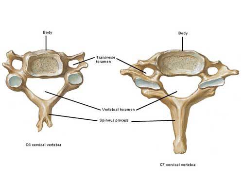
Image D
Atlas: C1 vertebra articulates with skull above and C2 vertebra below. C1 is also known as the atlas because it supports the head like Atlas of Greek mythology held up the celestial spheres. Its anatomy is also unique. Viewed from above (Image E), the odd atlas has neither body nor spinous process! Rather, it bears a small articular facet for the dens (see below) and a small posterior tubercle. Two peanut-shaped depressions (superior articular surfaces) articulate with two matching bulges (occipital condyles) of the skull base.
Try this: Nod your head up and down (as in “yes”). This sets up a rocking motion between atlas and occipital condyles of skull. No IV disc intervenes between C1 and occipital condyles base as it would be shredded with this motion.
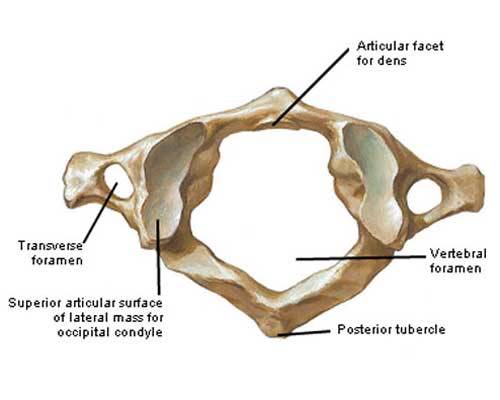
Image E
Axis: C2 vertebra, the axis, is also an odd vertebra (Image F). It articulates above with C1 and below with C3 vertebrae. It has an elongated body topped by the tooth-shaped dens (odontoid process). A strong ligament holds the dens against the articular facet of C1.
Wonder why C2 has a dens but C1 has no body? No one knows, but some biologists hold that the dens was once the body of C1 but became part of C2 during evolution.
Try this: Move your head from side-to-side (as in “no”) and understand that the dens acts as a pivot point around which C1 and the head rotates.
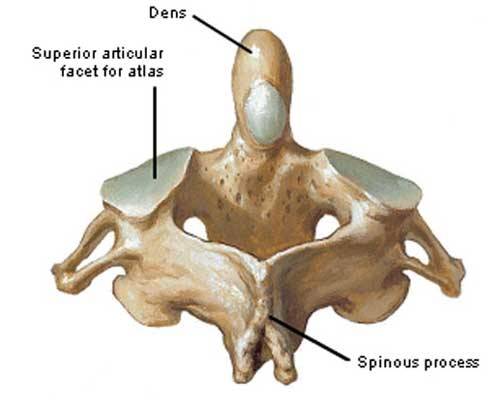
Image F
Try This: Insert a clean finger in your mouth and push it all the way to the back of your throat (stop if you start to gag…); the dens lies deep to your finger. For this reason, radiographs of the dens are taken via an open mouth. Image G (red arrow) is an x-ray of a normal dens; it projects upward like a large tooth (hence the alternative name, odontoid process). You can perhaps see the lower teeth embedded in the mandible?
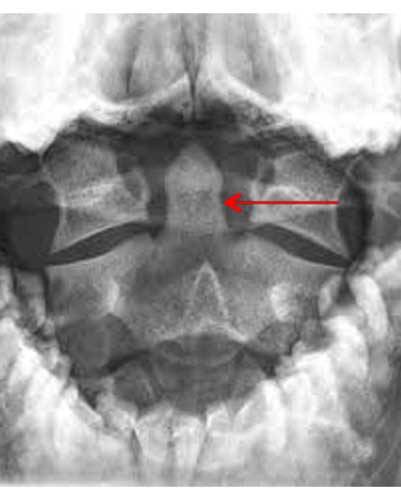
Image G
Dens and Hanging: The history of hanging is gruesome but pertinent. With the standard or long drop type of hanging, the condemned is suddenly dropped from a specified height. Weight of the plunging body plus a properly prepared and placed noose breaks the neck, meaning the dens snaps off C2 and is driven upward into the brainstem causing instantaneous death.
In The Fiery Cross, 5th book of the Outlander series, Herself explains:
“My horse moved suddenly, dodging past a group of men, and I saw them, three stick-figures, dangling broken in the tree’s deep shadow. The hammer struck one final blow, and my heart shattered like ice. Too late.”
The quote refers to the hanging of a character after sliding from the back of a horse. This type of hanging, known as short drop, results in a slow and agonizing death by strangulation because the body weight doesn’t drop far enough to break the dens! Had this character been hanged by long drop, the dens would likely have snapped from the body of C2 as shown in Image H (red arrow).
NOTE: I don’t have access to the medical history of the patient shown in Image H, so his/her fate is unknown. However, there are several types of dens fractures and treatment is possible.
I once had a medical student who fractured his dens in a skiing accident and survived! He was exceedingly careful when turning toward something or somebody: he rotated his upper torso and never rotated just his head for fear of dislodging the fractured dens.
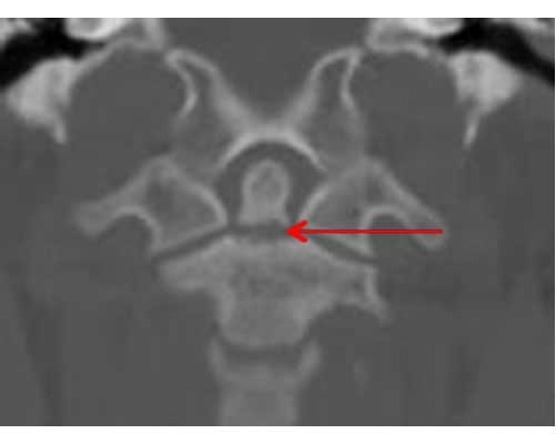
Image H
Muscles: Viewed from the back, the cervical spine is overlaid with layers of muscles in the back and at the sides.
The paired trapezii (Anatomy Lesson #2, Anatomy Lesson #3 & Anatomy Lesson #10) form the most superficial layer (Image I – left side). These help extend, rotate and flex the neck.
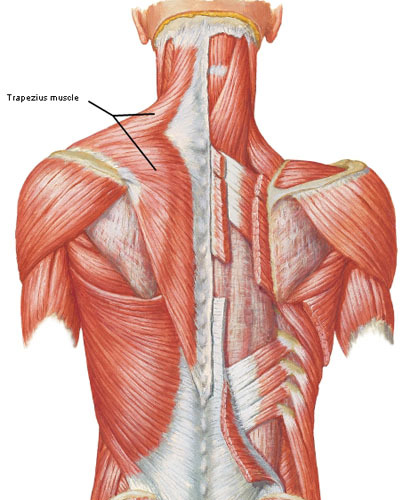
Image I
Two pairs of intermediate neck muscles lie deep to trapezius: splenius capitis and semispinalis capitis (Image J – left side); these also flex, arch and rotate the neck.
Believe it or not, there are even more deep muscles of posterior neck but time requires us to move on. This lesson is rapidly becoming as long as a master’s thesis!
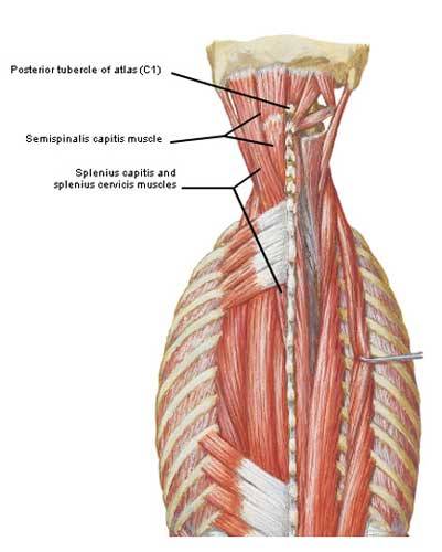
Image J
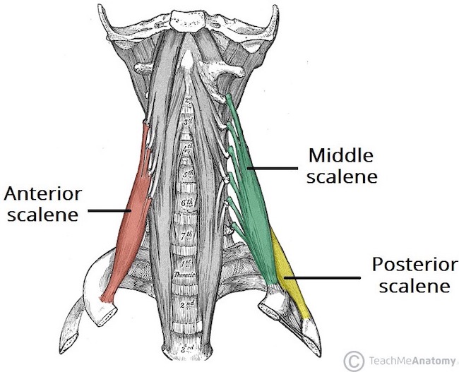
Three pair of muscles of the lateral neck are important. These are anterior, middle and posterior scalene muscles (Image K). These arise from the cervical vertebrae and insert on first or second ribs. Anterior and middle scalene lift the first rib and bend the neck towards the same side. Posterior scalene lifts the second rib and bends neck towards the same side. They all raise the ribs to increase the size of the thoracic cage and are thus secondary muscles of respiration.
Now, a question asked by many fans: why is Claire’s neck so long ( Starz episode 104, The Gathering)? It isn’t likely she has more than seven cervical vertebrae – an extremely rare variation! Two reasons likely account for her unusually elegant and swan-like neck.
First, the bodies of C2 – C7 vertebrae are probably a wee bit longer than most. Just a small additions to the lengths of each vertebrae and one’s overall neck length is enhanced!
Take for example the neck of an adult giraffe (Image L) which reaches 2 m (6.5’) in length even though this animal has only seven cervical vertebrae! Their neck length is achieved because each vertebral body can exceed 25.4 cm (10”) in height. Now, please dinna fash, I’m not suggesting Claire’s neck is like a giraffe’s! I use this extreme example to make the point.
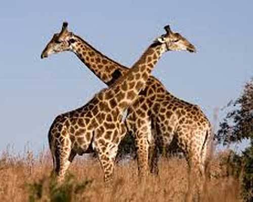
Image L
The second reason for Claire’s long neck is because her trapezius muscles allow for a definitive angle between their slope and the column of her neck. This gives a looong profile to her ivory tower.
Here she weeps as a sick BJR relates the horrific account of Jamie’s scourging (red arrows – Starz episode 106, The Garrison Commander). Beautiful photography as we even see the reflection of a tear fallen on her left breast.
Now, compare and contrast Claire’s trapezii with Jamie’s which are super well-developed! His amazing trapezii slope downward from head and cervical spine to clavicles and scapulae (Anatomy Lesson #2) filling in the aforementioned angles. This widens the neck and effectively causes it to look shorter, but no less appealing. Make sense?
Anterior Neck: This region provides passage for pharynx (back of the throat), larynx (voice box), trachea (wind pipe) and esophagus (tube to stomach), viscera that will be left for later anatomy lessons.
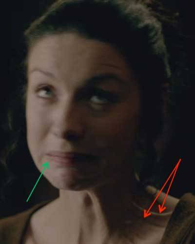
We will, however, consider two superficial structures of the anterior neck. First, do you recall the paired platysmas in Anatomy Lesson #11? Each is a thin, flat subcutaneous muscle that pulls down the lower lip (green arrow) and webs the neck skin (red arrows).
Here’s Claire with platysmata (pl.) contracted as she tells Jamie that she had a wee bit too much of Colum’s rhenish (Starz episode 103, The Way Out). Och, she’s had waaay too much!
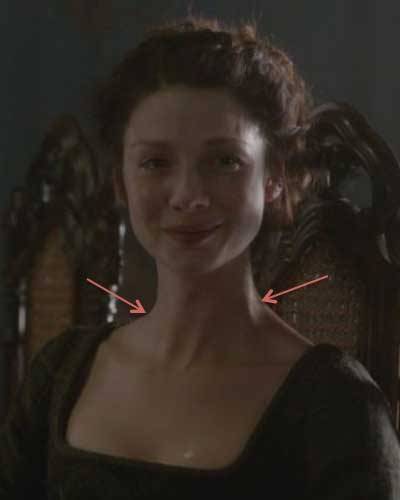
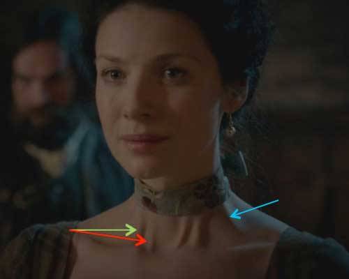
The second superficial structure of the anterior neck lies deep to each platysma; it is the external jugular vein. Each vein collects blood from its side of the face and empties it into a large vein at the base of the neck (Image M – black arrow).
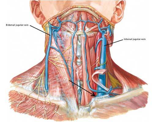
Image M
Claire’s right external jugular vein (red arrow) shows vey well in this scene where she verbally spars with dirty Dougal (Starz episode 102, Castle Leoch)! She’s clearly pissed and for good reason – Dougal has her watched day and night by those hooligans, Rupert and Angus, thinking she’s an English spy! There are other scenes where this vein is apparent in both Claire and Jamie. Hope you watch for it!
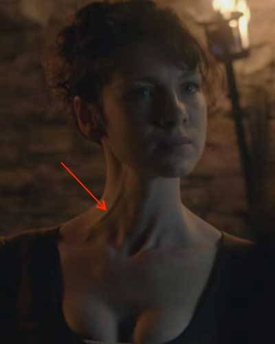
Next, an important pair of muscles provides contour to the anterior neck. To understand them, we must first learn their bony attachments. The adult sternum (breastbone) is divided into three parts; the top part is the manubrium (Latin meaning handle). The other two parts will be left for a later lesson. At the top of the manubrium is a divot known as the jugular notch or suprasternal notch (Image N). The clavicles (Anatomy Lesson #2 & Anatomy Lesson #3) articulate with the manubrium at paired sternoclavicular joints (black arrow – right side).
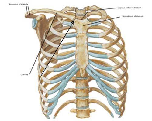
Image N
The next image clearly shows Claire’s jugular notch (red arrow) as she watches a tense exchange between Colum and the wee tailor (Starz episode 103, The Way Out) who unfortunately misjudges the length of the laird’s standard frock coat!
Try this: Place fingers at the top of your sternum and feel the bony indent; this is your jugular notch.
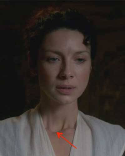
Sternocleidomastoid Muscles: The large paired SCM provide anterior neck contour (Image O – right side). Each SCM has two heads: the sternal head takes origin from the manubrium, the clavicular head from the clavicle. The heads merge into one belly that inserts on the mastoid process, part of the skull (Anatomy Lesson #11).
Try this: Place your fingers in the jugular notch – drag chin towards your chest and feel a stout tendon on each side – the sternal head of each SCM!
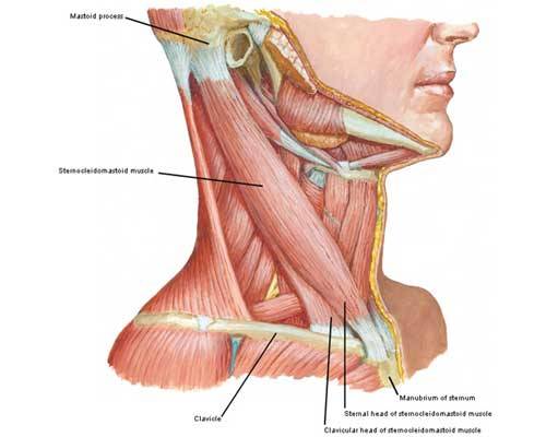
Image O
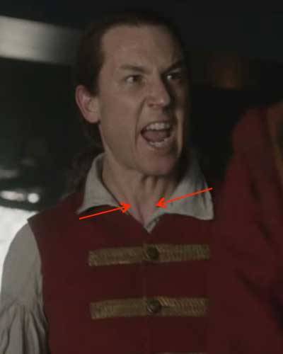
Although his collar is a bit in the way, you can clearly see the sternal heads of both SCMs in BJR’s neck (Starz episode 106, The Garrison Commander) as he bellows at corporal milk sop to “kick her!” Nice guy, eh? Wouldna want to meet him in a dark alley. Hey, wait, we did meet him in a dark alley in Starz episode 108, Both Sides Now…or that was his 6th times great grandson? Tcha, not much difference betwixt the two, at least in that episode!
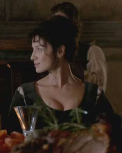
The clavicular head of each SCM is thicker and wider than the sternal head creating a mound easily viewed in this image of Claire getting liquored up by Colum (Starz episode 102, Castle Leoch). Yep, a servant has just delivered a jug of his rhenish! Do ye also see the sternal heads of each SCM?

Now, see Claire in her bonny ribbon with both SCM tendons tensed? She is sooo pleased and relieved by Jamie’s clever handling of his scheming uncles at the Gathering (Starz episode 104); he is her patient, after all!
A comment about Claire’s SCM. Some folks have more than two SCM heads on each side. In Claire’s case, the inner pair of tendons (red arrow – right side) belongs to the sternal heads; the next pair (green arrow – her right side) belongs to her first set of clavicular heads. The wide tendon (aqua arrow – her left side) belongs to her second set of clavicular heads.
This doesn’t alter function – in my view, it makes her long neck even more splendid. The tendons look very bonny on her ivory tower enclosed like it is by a pert and lovely ribbon (kudos to Terry and team for the gorgeous costuming!). No one wears a neck ribbon quite like Claire! It is the best except, mayhap, when Jamie is taking it off? Snort!
Try This: Check out your own SCMs in a mirror. Do you have more than two heads?
The SCM performs three movements for us (other neck muscles assist with these movements). First, both sternal heads contract to arch the neck and depress (lower) the chin. Here you see Claire (Starz episode 102, Castle Leoch) contracting both sternal heads as Colum grills her about the proper pronunciation of Beauchamp and her distant famly from Compiègne, France.
The second function of SCM is turning the chin to look over one shoulder. This occurs when the clavicular head of only one side contracts. Again, we see Claire smile at Colum at the supper table afore he starts her grilling (Starz episode 102, Castle Leoch). Now, her left SCM muscle tendons are clearly visible because they are stretched. But, in fact, it is her right SCM that contracts to move her chin toward her right shoulder.
Try it yourself! You might also re-watch this entire scene to appreciate the array of gracious neck movements performed by our braw heroine!
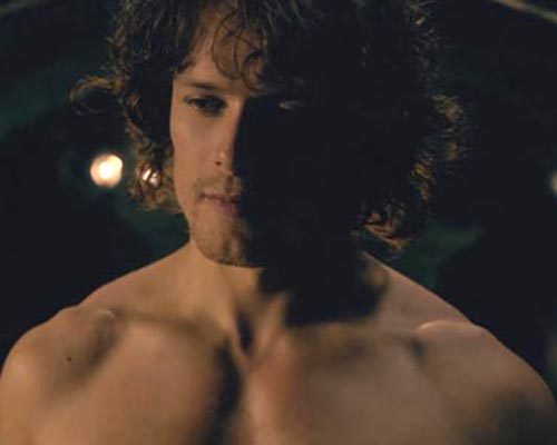
The third movement occurs when both clavicular and sternal heads of one SCM contract to pull the same ear toward the shoulder. Here is Jamie (Starz episode 102, Castle Leoch) having his gunshot wound cleaned. How many times have we seen guys perform this neck movement? It seems a universal guy thing, but, I didn’t ken it was done in 1743 by 23 y. o. Scottish virgin! Yup, that’s both heads of his left SCM in action.
Next, the anterior neck compartment contains an unpaired bone (Image P – viewed from above). The U-shaped Hyoid bone is Greek meaning shaped like the letter upsilon, the 20th letter of the Greek alphabet. In adults it measures roughly 2” (5 cm) side-to-side and front-to-back with a body and two cornua (Latin meaning horn). Unlike the other 205 human bones, the hyoid doesn’t articulate with any other bone!
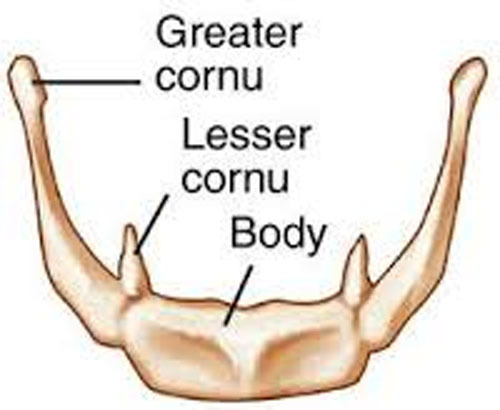
Image P
The hyoid bone sits in a mostly horizontal orientation about a finger’s breadth below the bottom of the mandible (Anatomy Lesson #11) and at the level of C3 vertebra (Image Q). The hyoid is suspended in the neck by two strong ligaments attached to our styloid processes, a pair of gothic-looking bony spikes. Aye, our skulls really do have these wicked-looking projections!
OK. Let’s stop for a funny story: once a student asked me to examine his plastic skull model for accuracy. It looked fine until I turned it over and noticed it had no styloid processes! I voiced the observation and he responded that he had broken them off because he thought them to be leftovers from the modelling process! We got a good belly laugh over that one!
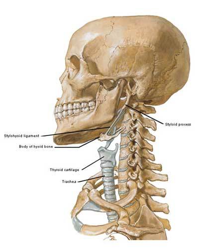
Image Q
Try this: Feel your own hyoid bone. Open thumb and index finger into a curved U and slip the opening over the front of your neck just below your jaw bone. Gently tighten your digits a wee bit and then cough or swallow. You should feel a hard bar move up and down on each side. This is the hyoid bone. If you push gently with your index finger your thumb feels the hyoid more easily on the contralateral (opposite) side. The hyoid is tucked so deeply under the mandible that it is rarely fractured except with compression during manual strangulation. Ugh!
Note: The hyoid bone appears in Diana’s 8th book Written in my Own Heart’s Blood but I will save that context for a later post. NO spoiler here!
A whopping nine pair of muscles attach to the hyoid. The thyroid cartilage (part of larynx) also hangs from it by a fibrous membrane. These muscles help control the tongue, floor of mouth, pharynx and larynx such that the hyoid plays important roles in breathing, speech, swallowing, chewing and coughing (Image R). Some muscles lift the hyoid forward or backward, others pull it down and forward or backward. Then there are combinations of movements between the muscles making for complex changes to throat, esophagus and airway. The arrows in Photo R indicate the directions the named muscle pull.
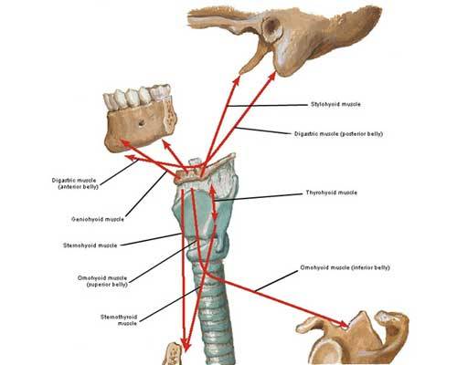
Image R
I won’t cover all nine pair of muscles attaching to the hyoid (only eight are listed in PhoImage R) because it is too much info. But, I will comment on the paired omohyoids. Each omohyoid (Image S – right muscle) has two bellies: an inferior belly arises from the scapula and ends at an intermediate tendon near the clavicle (Image S – black arrow) and a superior belly arises from the intermediate tendon and inserts on the hyoid. Although not shown in the image, the intermediate tendon is bound to the clavicle by fibrous tissue. As each omohyoid contracts, it pulls the hyoid bone downward and backward.
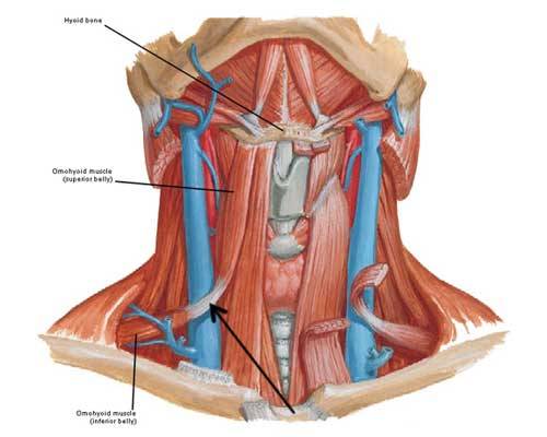
Image S
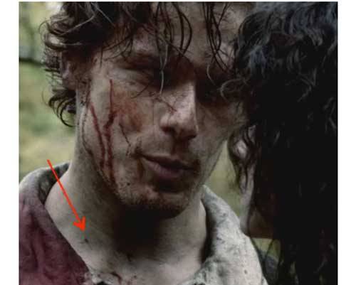
The omohyoid is rarely seen because it lies deeply. However, I saw this muscle in action during Starz episode 101, Sassenach. Now, I bet you watched this scene more than once but I encourage you to do it again. Here, Jamie has just chased Claire down (YES!). She spits “NO” ta his question: does she want him to pick her up and throw her over his shoulder (YES! erm…NO!). He says: “Well, then, I suppose that means yer comin’ wi’ me!” (oh YES, I mean NO!). Whew, gettin’ a wee bit dizzy here!
Now see the location of the red arrow on Jamie’s neck? Go back and watch this episode as he speaks the line about comin’ with him while keeping an eye peeled at the area indicated. Ye will see a bump rise and fall a couple of times. This is Jamie’s right omohyoid muscle contracting to depress his hyoid bone as he speaks. When I saw it, I nearly fell out of my chair! OMG it is bloody awesome! (Anatomists are a bit weird and easily entertained, ye ken?)
“Oh, de headbone connected to de…neckbone…
Let’s end this lesson with a final view of Claire’s magnificent ivory tower (not to mention Angus cleaning out a lug in the background)! Aye, she looks marvelous!
Before I close, I’d like to add a wee personal note at the request of a reader: I write my blog anonymously not because I have anything to hide but because I prefer to focus on Diana’s splendid books, the fabulous Starz Outlander series and fascinating human anatomy! I am a woman and a traditional one at that. If my blog encourages anyone, regardless of gender, to pursue an interest in science then I feel I have done my job. Namaste!
The deeply grateful,
Outlander Anatomist
Follow me on Facebook and twitter!
photo creds: Starz, Netter’s Atlas of Human
Anatomy, 4th ed., Clinically Oriented Anatomy,
5th ed., Hollingshead’s Textbook of
Anatomy, 5th ed., www.teachmeanatomy.info, www.radiopedia.org,www.wikipedia.org, www.commons.wikimedia.org, www.medical-dictionary.thefreedictionary.com,
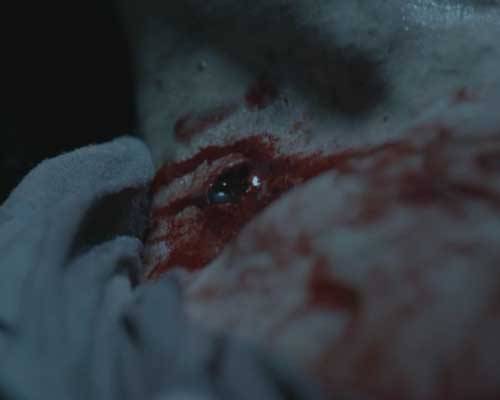
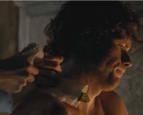
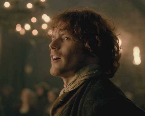
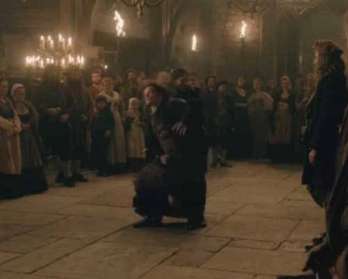
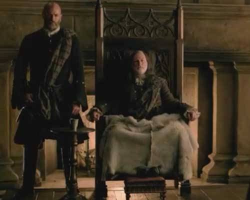
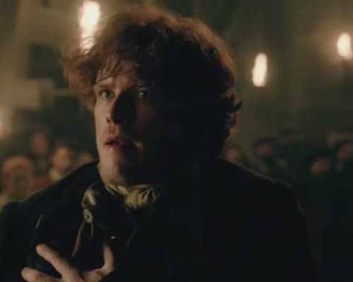
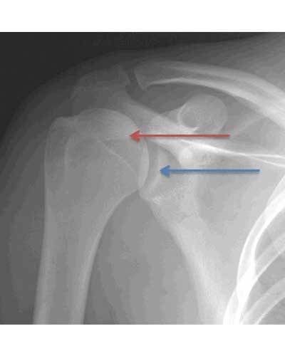
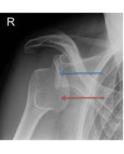
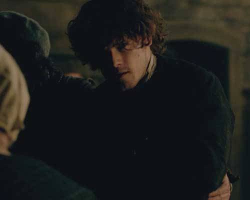
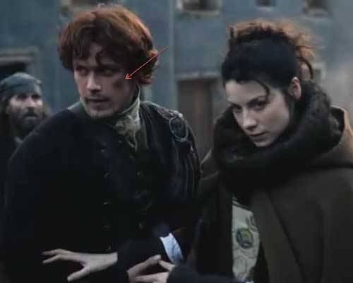
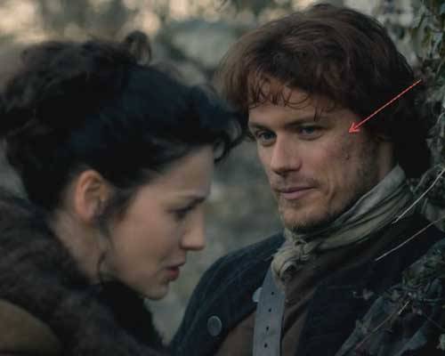
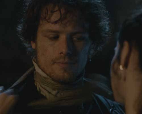
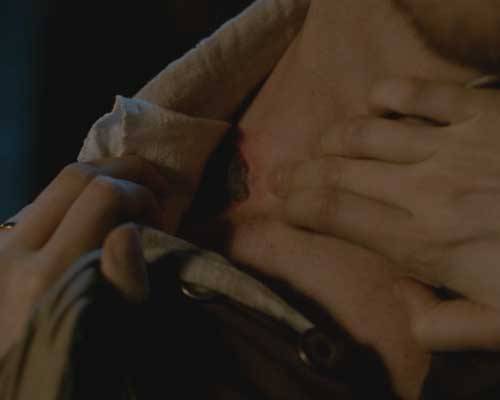

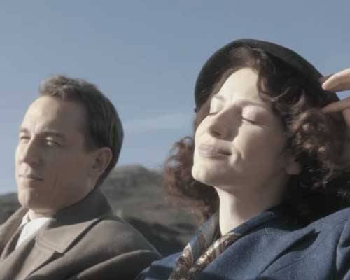
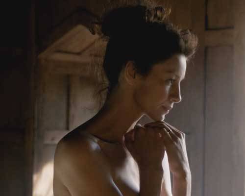
 Photo A
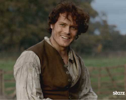
 Photo B
 Photo C

 Photo G
 Photo H
 Photo I