
Anatomy Lesson #7, Jamie’s Thighs, is a true ode to joy! I hope Ludwig won’t be offended by my reference to his 9th, but the title perfectly suits this lesson! Perfect timing with the upcoming US Thanksgiving Holiday. I’m down on my knees giving thanks because Jamie’s knees also appear in this lesson! 🙏🏻
Are you ready? Let’s go!
Just to prove my goodwill, take a gander at Jamie’s knees in the image below. May this tide you over as our lesson proceeds!
Definitions: Let’s begin with a few basic tidbits, otherwise much of this lesson might remain obscure!
- Joint – Site where bones meet and move relative to each other
- Muscle – Muscles attach to bones (some exceptions); these are voluntary meaning we can contract them at will – a.k.a. skeletal muscle
- Contraction – As muscles contract, a joint moves in various ways (Image A)
- Origin – Attachment site of muscle to bone – site moves little or not at all during contraction
- Insertion – Attachment site of muscle to bone – site moves during contraction
- Flexion – Muscle contraction closes (approximates) bones of a joint
- Extension – Muscle contraction opens (straightens) bones of a joint
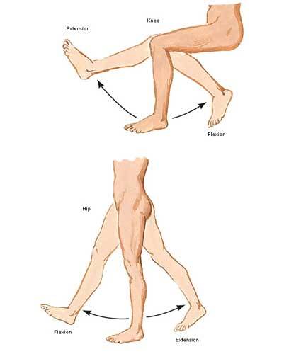
Image A
Lower limb: Our lower limbs are the body parts from hip joints to toes. Anatomists don’t use the terms upper and lower leg. Rather, the lower limb is divided into (Image B):
- Thigh – segment between hip and knee joints
- Leg – segment between knee and ankle joint
- Foot – segment beyond ankle joint
BTW, the dashed blue line in Image B represents a vertical midline through the body. A medial structure lies closer to this midline – lateral structure lies further from this midline. That’s the gist of it!
Try this: Locate your thigh, leg, foot and midline.
Test Q: Are your tiny toes medial or lateral to your middle toes?
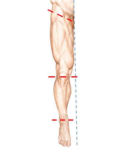
Image B
Test A: They are lateral. Great job!
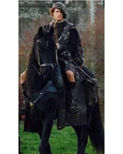
Outlander Time! The first time we rencounter Jamie’s thighs is in Outlander book where Claire straddles-his-saddle on the way to Castle Leoch. Herself writes:
My companion seemed to be having little trouble, in spite of being unable to use his right hand. I could feel his thighs behind mine, shifting and pressing occasionally to guide the horse. I clutched the edge of the short saddle in order to stay seated; I had been on horses before, but was by no means the horseman this Jamie was.
And yet another quote from Outlander book – this one from randy-dandy, Jamie:
But then that ride through the dark together….with that lovely broad arse wedged between my thighs…
Well! Starz Claire doesn’t sport a broad arse, but it surely is curvaceous and pert and we all ken where it was wedged during this ride! Mmphm!
Back to anatomy and more about bones!
Femur: The thigh contains one bone, the femur. I love superlatives, so here’s the first one for this lesson: the femur is the longest bone of the human body (Image C – front of right femur). The top of each femur ends in an angled neck and head that fits securely into the acetabulum, a deep socket of the hip bone Together, they form the acetabulo-femoral (hip) joint, a ball and socket joint.
Fun Facts: You may recall from Anatomy Lesson #2, When Claire Meets Jamie or How to Fall in Love While Reducing a Dislocated Shoulder Joint!: although the glenohumeral (shoulder) joint is very moveable it is also less stable due to a shallow ball and socket joint! Conversely, each hip joint is very stable but less moveable due to a deep ball and socket joint. The hip joint needs to be stable to support our weight against gravity. Very interesting!
At the knee, the femur ends in two sturdy knobs, medial and lateral condyles (Greek for knuckle); both knobs help form the knee joint (Image C).
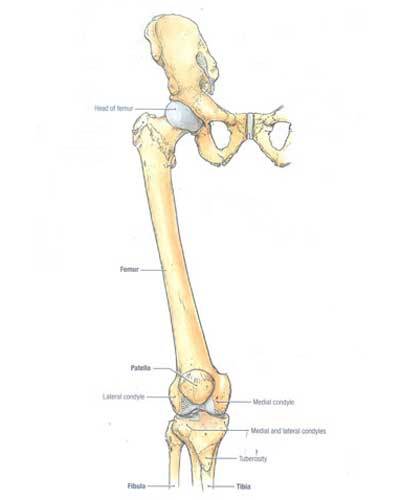
Image C
Tibia and Fibula: The leg contains two bones, tibia and fibula (Image D – front of right leg bones). The larger tibia is medial to the smaller, more lateral, fibula. The top of tibia ends in two flat surfaces, the medial and lateral condyles; lower down, it bears a midline knob, the tibial tuberosity. Only tibia helps form the knee joint; fibula plays no role.
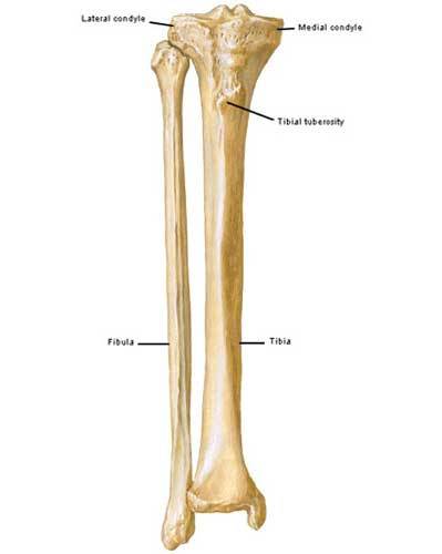
Image D
Patella: The small patella or knee cap is critical to knee anatomy. Patella is the largest sesamoid bone of the body (there are others) meaning it is enveloped in tendon. Its deep surface slides in a groove between medial and lateral femoral condyles as the knee joint extends and flexes (Image E – front of right knee joint).
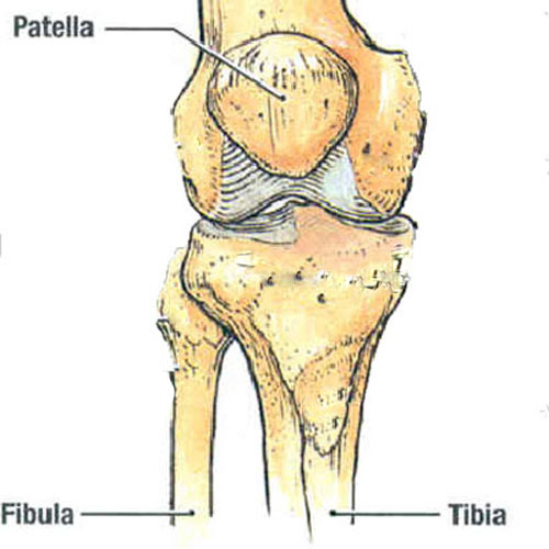
Image E
Thigh Muscles: Yes! Thigh muscles are massive because they support much of our weight and help maintain our bipedal stance against gravity.
There are 13 thigh muscles divided into three compartments. To keep this lesson shorter than a bloody master’s thesis, we will cover only quadraceps femoris (Latin meaning four-headed muscle of the femur) with a wee bit about iliopsoas muscle.
Quadraceps: The quads (as trainers call them) are four muscles at the front of each thigh:
-
- rectus femoris
- vastus lateralis
- vastus intermedius
- vastus medialis
Image F (front of right thigh) shows the three of the quad muscles. A fourth, vastus intermedius, is visible only after removal of rectus femoris (Image G).
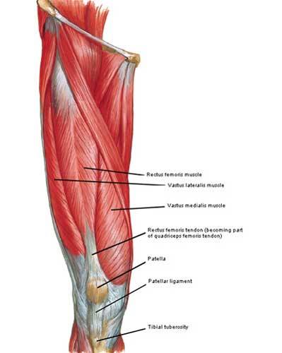
Image F
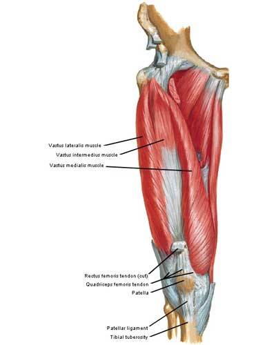
Image G
Origins: Each quad muscle has a different origin (Image H – red marks). Rectus femoris originates from hip bone and crosses both hip and knee joints . The three vasti (pl.) muscles take origin from different sites on the femur and cross only the knee joint.
Insertion: All four muscles fuse into a common quadraceps tendon that engulfs the patella and then continues as the patellar ligament to insert (Image H – blue mark) on the tibial tuberosity.
Actions: Acting together, all four heads of each quad extend the knee joint but only rectus femoris also flexes the hip joint.
Because quads are the only muscles that extend the knee joints, they are crucial for walking, running, jumping and squatting. Quads are also called anti-gravity muscles because they contract as we rise from a seated position or lower our bodies in reverse, holding our weight against gravity (think of thighs during snow skiing or Jamie lowering Claire onto the marriage bed. Yum!).
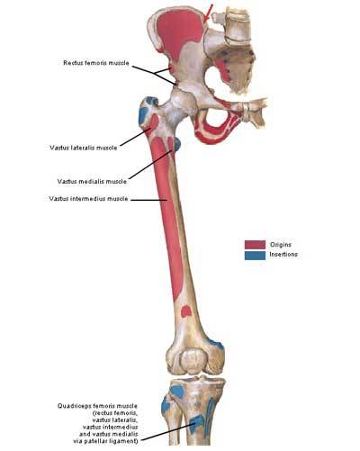
Image H
Iliopsoas: One last muscle… Although recti femoris (pl.) are decent hip flexors, iliopsoas muscles are the strongest. Like the quads, iliopsoas is a compound muscle formed by fusion of iliacus and psoas (pronounced soas) muscles.
Iliacus arises from the hip bone and psoas arises from the lumbar vertebrae (Image I – right front). They fuse into a single tendon that inserts into the femur. These muscles draw the femur closer to the torso, assisted by rectus femoris and a few other minor hip flexors. Or, with thighs held stationary, they draw torso closer to thighs (think sit-ups).
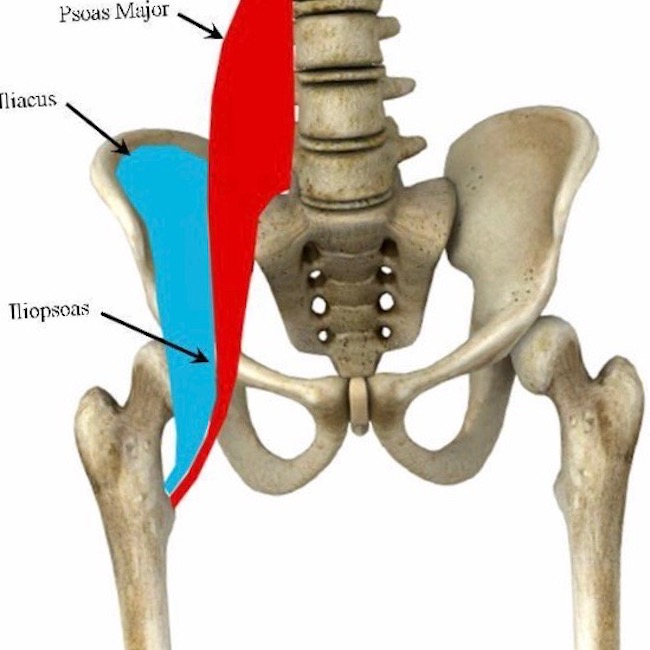
Image I
Try this: To test your quads, place back against a wall. Drop the tush while walking your legs away from the wall until you assume a squat position (Image J). Thighs and legs should be at 90° to each other (don’t drop lower – this is bad for knee joints!). Now hold your torso in place for 30 seconds and then gradually straighten (extend) the knee joints. If you feel wobbly, then you may need quad work as these muscles quickly lose mass and strength due to inactivity, sedentary jobs or aging. Quads can be strengthened by wall squats or by any exercise that adds resistence while extending the knee joint!
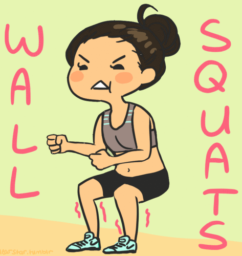
Image J
Knee Joint: Now on to the knee joint, largest joint of the human body (Image K – right side and front)! Here medial and lateral femoral condyles (knuckles) ride atop a flat plateau formed by the medial and lateral tibial condyles – no stable joint here!
All bony surfaces of knee joint are covered with articular cartilage, a bloodless, firm connective tissue that allows for smooth movement. This odd joint also has medial and a lateral meniscus (different type of cartilage) that create two shallow sockets atop the tibial plateau – one for each femoral condyle; these also act as shock absorbers.
As mentioned above, the patella glides between the femoral condyles during flexion and extension.
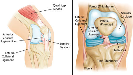
Image K
Knee Ligaments: The knee is stabilized by very strong ligaments designed to secure the tibia and femur (Image L – right knee in full flexion – patella absent):
- medial (tibial) collateral ligament
- lateral (fibular) collateral ligament
- anterior cruciate ligament
- posterior cruciate ligament (and, there are others!)
Overall, the knee joint is at risk due to our sports-crazy cultures and because supporting the body weight while in motion is a challenge for these relatively flat surfaces. Hence, the über-strong ligaments.
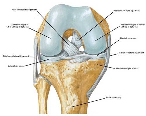
Image L
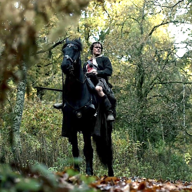
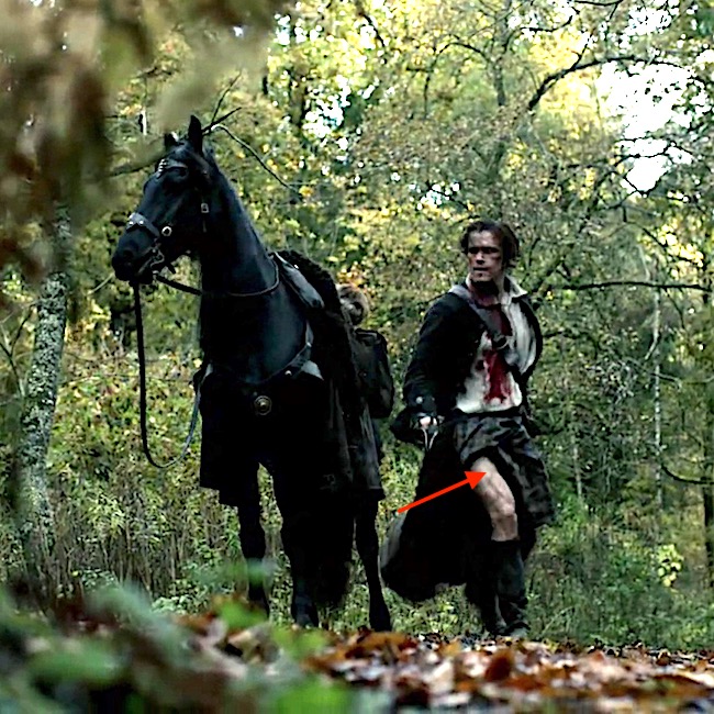
Now, for the fun stuff! I promised in Anatomy Lesson #3 that I would be returning to the scene where Jamie dismounts to grab Claire (Starz episode 101, Sassenach). Yep, that’s the one! Let’s relive that scene wherein we first spy Jamie’s thigh – aye, readers, there’s a first time for everything. Hah!
Wait fer it…………….”Lost yer way?”
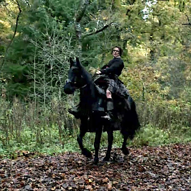
Wait fer it……….. Jamie’s shifts his weight. Um…..he looks a wee bit fashed!
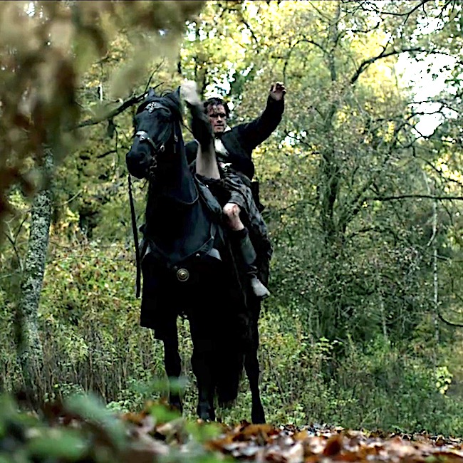
Wait fer it.…. With a high kick Jamie’s right thigh clears the steed’s neck. Yay, iliopsoas!
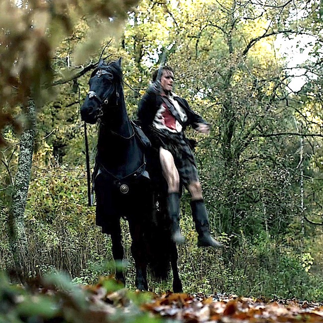
Wait is over! Jamie’s drops. Gad, this lad has loooooong legs!!!! Thank gawd that kilt didna do its job here! (That impressive thigh muscle is vastus lateralis!)
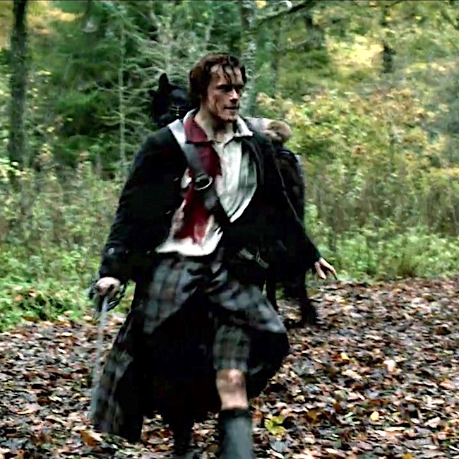
A firm landing and further evidence of those thighs and knees!
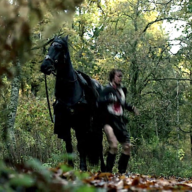
This just keeps getting better and better! Geez, Jamie is a perfect anatomical model!
Three of four quad heads are visible as he strides towards Claire. The red arrow points to the head of rectus femoris! (Claire! why aren’t ye laid out in a swoon?)
OK, Claire, now you are in fer it! Ye’re gonna get what ye deserve! Oops, sorry, wrong Starz episode. (Thinking 109, The Reckoning). Snort!
Okey dokey then, are you ready for a pop quiz? Let’s go for it!
Can you identify one of Jamie’s quads in the next image (Starz episode 7, The Wedding)?
Q: Name the major bulge (no, not that one! <G>) on the inside of his thigh (red arrow).
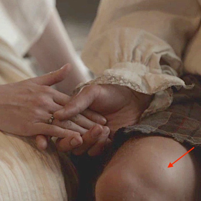
A: Right vastus medialis muscle. Good job!
Q: The light is dim, but name the quad at the red arrow.
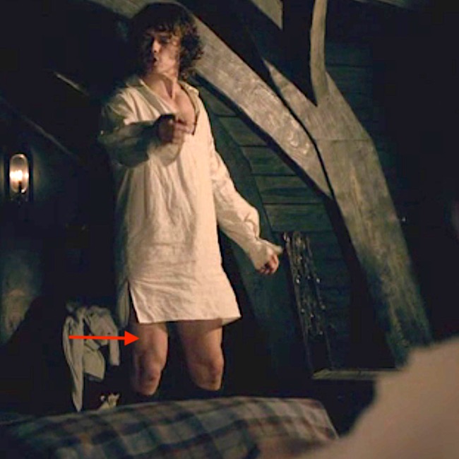
A: Left vastus lateralis muscle – just peeking out from under his wedding sark. He is sooo modest!
Back to anatomy!
Knees: Now for those gorgeous knees! I know gazillions of you have been awaiting another glimpse of Jamie’s knees! Sorry to mess them up with a quiver of arrows, but just so you ken:
- Blue arrow marks his lateral femoral condyle
- Red arrow is his medial femoral condyle
- Green arrow marks his patella
- Orange arrow is his tibial tuberosity.
So now you can name all the knuckles and knobs o’ Jamie’s knees!
Claire, lift your eyes lass! He squatted down to show off his gorgeous gams! He kens he is one damn fine-looking Scot – grubby sark or no!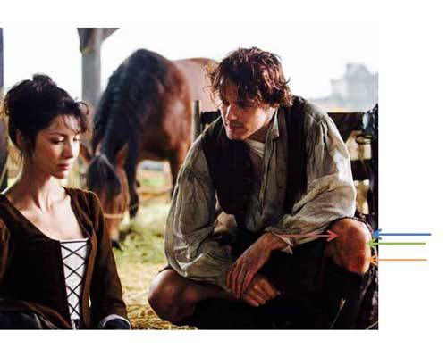
Almost done, so hang with me!
IT Band: Lastly, thigh muscles are wrapped in a strong sleeve of connective tissue, the fascia lata (Image M – right lower limb). Fascia lata thickens at the side to form the iliotibial (IT) tract or IT band (Anatomy Lesson #1, Jamie’s Tush).
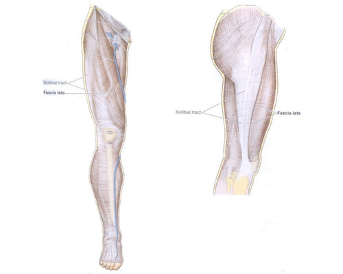
Image M
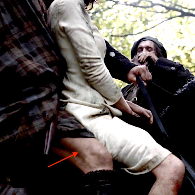
Now, for more applied anatomy! The red arrow marks a thick ridge of tissue at the back of Jamie’s thigh (Starz episode 101, Sassenach). Yep, that’s his right IT band! You can see it plain as Rupert swigging that raw whisky!
Fascia lata Function: You all should understand the importance of the fascia lata and IT band. The heart faces a long haul to pump venous blood against gravity from the foot back to the chest. Thigh muscle contractions help milk venous blood back toward the heart. Fascia lata aids this process by compressing contracting muscles against the deep thigh veins.
Danger! If thigh muscles get seriously injured, they swell and the inelastic fascia lata does not stretch to accommodate the swelling. When this happens, it is a medical emergency because swollen muscles restrict the return of venous blood to the heart and diminish the flow of oxygen-rich arterial blood to the lower limb – and, as you ken, tissue dies if deprived of oxygen!
This condition is known as compartment syndrome and the next photo (Image N) shows the result of medical intervention. This is the right thigh of a former students (he gave permission to post). After injuring his thigh muscles, the swelling threaten necrosis (death) of his thigh muscles so surgeons slit his fascia lata (near the IT band) to relieve pressure and re-establish blood flow. Now, when he contracts his right quads, they bulge through the slit in the fascia lata! An excellent visual of how the fascia lata compresses the thigh muscles!
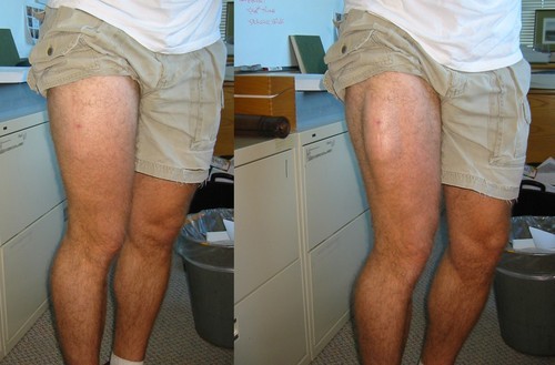
Image N
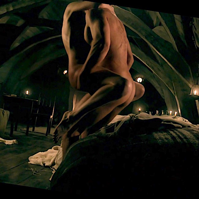
Okey dokey, with this example, we come to the end of our thigh and knee lesson! But, I’ll not leave you bereft. Here is one last image designed to increase your admiration and respect for Jamie’s anti-gravity quads and iliopsoas muscles!
In this, ahem, modest scene from Starz episode 7, The Wedding, Jamie bears Claire’s entire weight (9 stone or 126 lb – Herself records in Outlander). Then, he slowly lowers their combined weight (about 23 stone/322 lb?) to the bed afore he flips her over! That takes a whole lot of quad strength! Are you impressed? <G>
Ok, mukkers, that’s it for now. Please stay tuned for our next lesson. Still gobs of anatomy to cover as we work our way through the first eight episodes of Outlander!
The lessons are deliberately slow-paced to keep us occupied until Starz episode 109 makes its way into our hearts and minds!
Oh, almost forgot. You can now follow me on Facebook, Tumblr, IG and Twitter.
The deeply grateful,
Outlander Anatomist
Image credits: Starz, Grant’s Atlas of Anatomy, 10th ed., Netter’s Atlas of Human Anatomy, 4th ed., Clinically Oriented Anatomy, 5th ed, www.AuthenticFX.com, www.gouletballet.com, Wikipedia, OA archival photos, AAOC Website, Tumblr.com