Anatomy Lesson #40 “Snap, Crackle, Pop! or How Bones Heal”

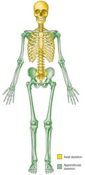
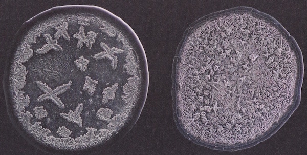
Welcome Outlander Anatomy readers! Our last lesson covered the human skeleton (Anatomy Lesson #39 “Dem Bones – The Human Skeleton”). Today’s Anatomy Lesson #40, “Snap, Crackle, Pop! or How Bones Heal” expands on bone anatomy but also addresses bone fractures and how they mend. As always, Starz Outlander images and quotes from Diana Gabaldon’s marvelous books are generously sprinkled throughout the lesson.

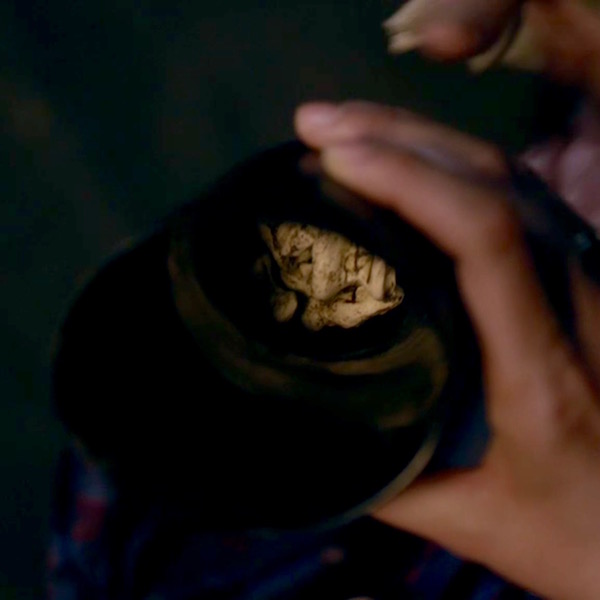
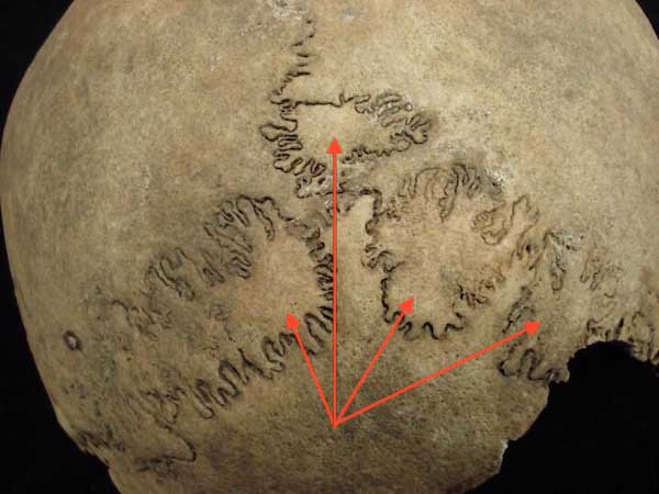
Last lesson began with spectacular images from Master Raymond’s hidden ossuary. This lesson, we start with images of a world famous ossuary located in Kutná Hora, Czech Republic. Known world-wide as the Bone Church, it is an ossuary (‘Kostnice’ in Czech) that houses the bones of an estimated 40,000 – 70,000 people (Image A).
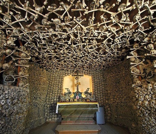
bone church 01 KLS edited

Image A

Here, bones are imaginatively and artistically arranged throughout the edifice. One of the most famous displays is a chandelier, purportedly containing every bone of the adult human body (Image B).

During the thirty years’ war, aristocracy of Central Europe wished to be buried in the hallowed graveyard at this site. As the number of burials outgrew available space, remains were exhumed and stored in the chapel, then assembled into artistic presentations.

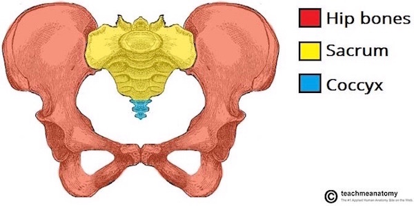
The ruffled collars beneath the candle-bearing skulls are hip bones! At the tip-top are inverted sacra (pl.). Dangling bones are humeri (pl.). Quite the room-lighter! Makes Master Raymond’s animal skulls seem almost quaint!
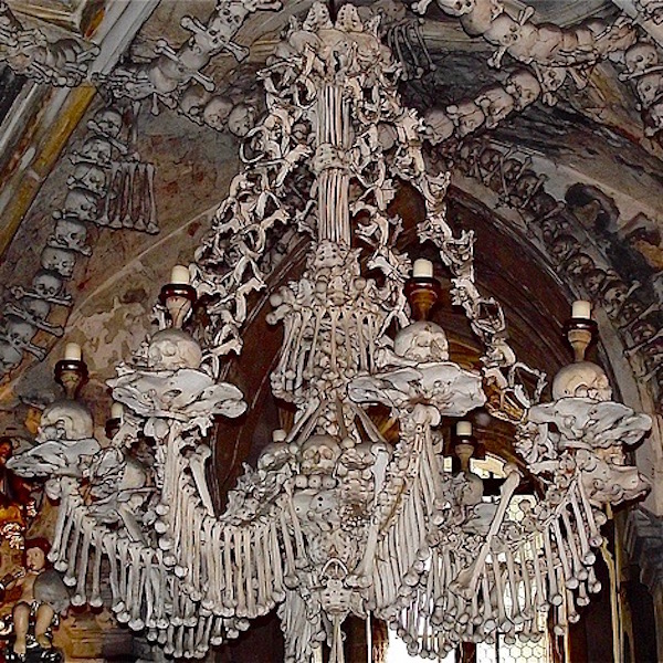
bone church 02 KLS edited

Image B

Surely, these images put us in the mood to learn more about bones, although this lesson covers  bones of the living. Today’s lesson will review and present more bone anatomy and types of bone cells. Then, we will cover events that ensue when bones go snap, crackle, or pop, as well as mechanisms of healing. Let’s go!

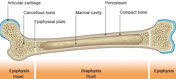
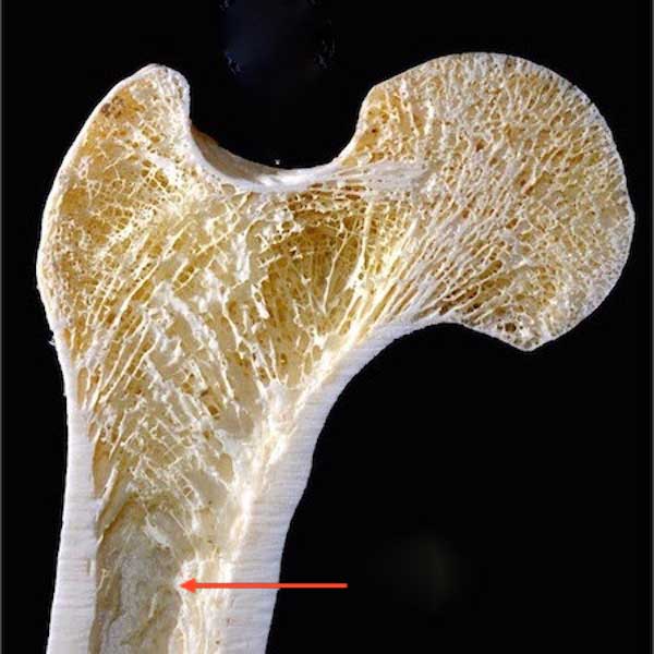
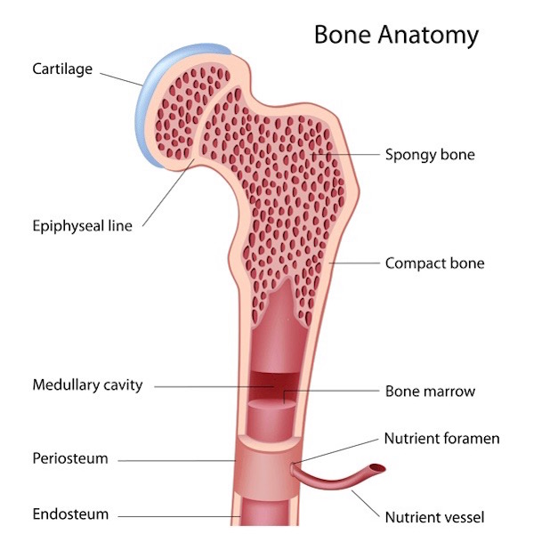
Review of Long Bone Anatomy: Lesson #39 presented long bone anatomy, but let’s take a moment to review. Once again, the femur, is our model (Image C). The shaft of a long bone is the diaphysis. The ends that form joints with other bones are the epiphyses (pl.); these are covered with articular cartilage (ball-shaped epiphysis is head of femur). The flared region between diaphysis and epiphysis is the metaphysis. Cortical bone forms a hard outer rind enclosing the marrow (medullary) cavity. Spongy bone fills epiphyses and metaphyses and is also scattered in the marrow cavity. Depending on the bone and age of the individual, the marrow cavity is filled with either fat cells or hematopoietic (blood-forming) cells, neither of which are elements of bone. Think of them as tenants not owners!
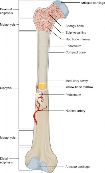
long bone parts 01 KLS edited

Image C

Periosteum and Endosteum: Periosteum is a thick fibrous layer covering the diaphyseal surface (Image D). Endosteum is a thin connective tissue layer lining the bone marrow cavity and covering all exposed surfaces of spongy bone. Both layers contain blood vessels and several types of bone cells as discussed below.
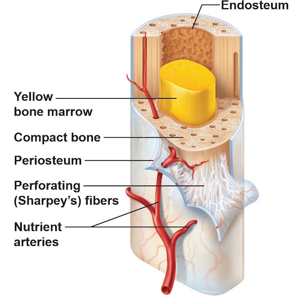
periosteum & endosteum KLS edited

Image D

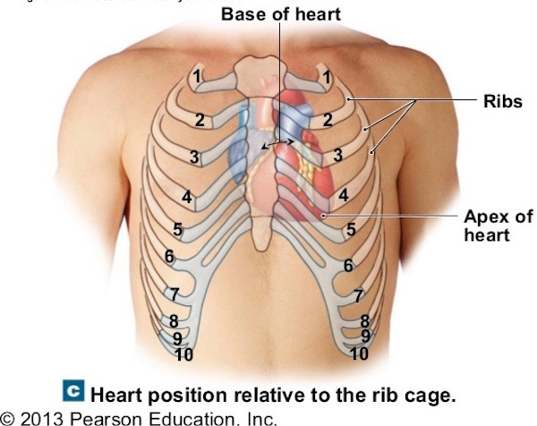
Vascular Supply: As we learned in Anatomy Lesson #39 “Dem Bones – The Human Skeleton,” bone is living tissue and thus requires a constant blood supply such that the human skeleton receives 5-10% of the entire cardiac output (amount of blood the heart pumps per minute).

To meet this demand, large bones receive several arteries (Image E). Periosteal arteries supply periosteum and outer compact bone. Epiphyseal arteries supply blood to epiphyses (pl.). Nutrient arteries supply inner cortical bone, endosteum, and help supply epiphyses and metaphyses.

These arteries access a bone via foramina (channels) that traverse compact bone to reach their respective turfs, then break into capillary beds where oxygen and nutrients are exchanged for carbon dioxide and waste products. Corresponding veins form and blood flows back to the heart. Round and round and round it goes…..
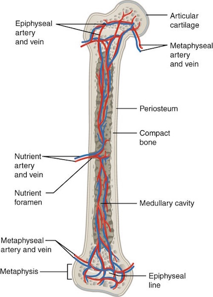
Nutrient artery femur KLS edited

Image E

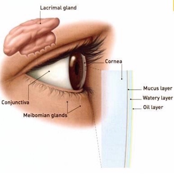
Nerve Supply: You may be surprised to learn that mineralized bone (organic matrix plus inorganic minerals – see Lesson #39) does not have pain receptors. Hum…if this is true, then why does a broken bone hurt so badly? Because periosteum and endosteum as well as articular surfaces are richly supplied with pain receptors (nociceptors). If these structures are compromised as with a fracture, then pain is severe!

Image F nicely illustrates nerve and blood vessel distribution in cortical bone and margin of the marrow cavity. Anatomy typically color codes arteries red, veins blue, and nerves yellow. The tiny yellow lines indicate that nerves follow blood vessels through cortical bone to the endosteum. Although not shown in Image D, nerves also follow periosteal arteries to innervate the periosteum.
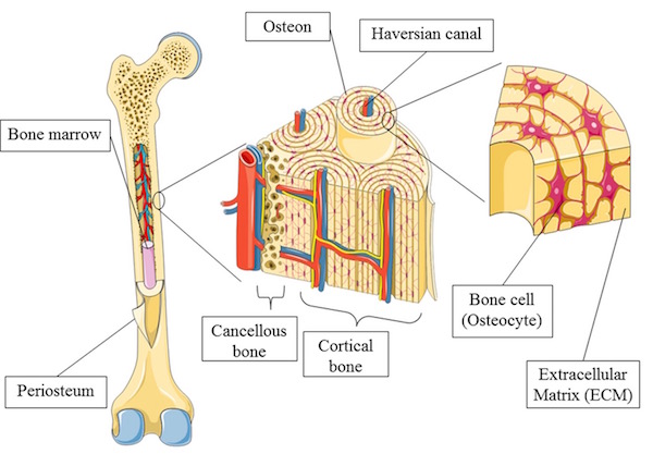
bone nerves KLS edited

Image F

Bone Cells: As mentioned above, bone contains several cell types including osteogenic cells, osteoblasts, osteocytes, and osteoclasts (Image G). These cells are located in periosteum, endosteum, or within mineralized bone. Again, although hematopoietic cells or fat cells fill spaces between spongy bone and within marrow cavities, these are not bone cells.

Osteogenic cells are stem or parent cells giving rise to osteoblasts (Image G); they reside in periosteum and endosteum.

Osteoblasts: From Greek osteo- meaning bone + blastanō meaning to germinate, osteoblasts produce collagen and other proteins as part of the organic matrix. These proteins are released into the extra-cellular environment where minerals deposit around them. Together, the organic matrix provides tensile strength and inorganic minerals provide compressive strength. As to their bulk, organic proteins represent 10% of bone mass with the remaining 90% being inorganic mineral. Osteoblasts reside in periosteum and endosteum.

Osteocytes: In the course of production and secretion of organic matrix and its subsequent mineralization, osteoblasts become “imprisoned” within bone and are renamed osteocytes. Located in compact and spongy bone, osteocytes help maintain the mineralized bone. Further, the mineralized matrix with imprisoned osteocytes is organized into cylinders of bone known as osteons (Haversian systems). A bone such as the femur contains millions of osteons but details about these units must await a future lesson.

Osteoclasts: Giant, multi-nucleated cells, the osteoclasts, are formed by the fusion of macrophages (Anatomy Lesson # 37, “Outlander Owies Part 3 – Mars and Scars”). Found in endosteum, their duty is to dissolve bone.
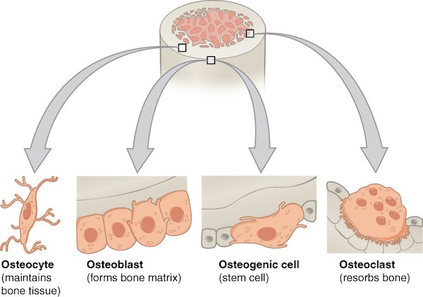
Print

Image G

As you might imagine, a good deal of coordination among the various bone cells is required to ensure homeostasis (balance) between bone production, maintenance, and resorption. For example, as a bone grows in circumference due to periosteal osteoblast activity, osteoclasts of the endosteum remove bone from the inside so the thickness remains fairly constant, a highly regulated process.

Image H shows in detail the distribution of bone cells in periosteum, endosteum and in compact bone.
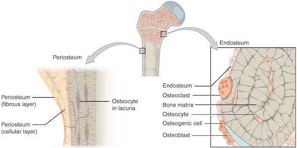
Print

Image H

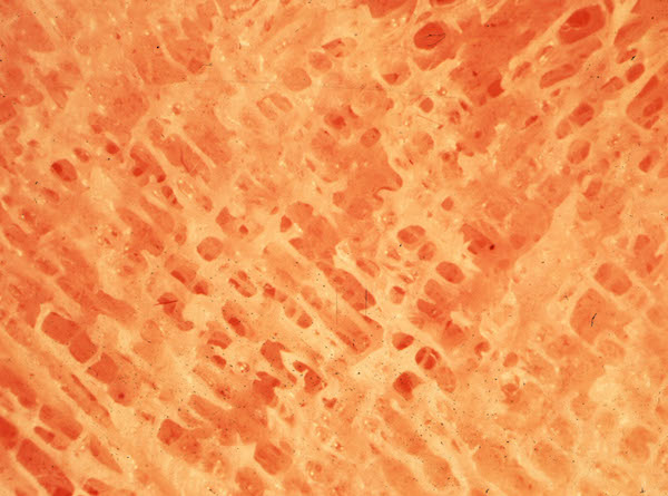
Next, let’s do something really daring and look at a microscopic image of bone cells as viewed through a light microscope (Anatomy Lesson #34 – “The Amazing Saga of Human Anatomy”). Please understand that microscopic anatomy, better known as histology (Greek histo- meaning “tissue” + ology meaning “to know”), is a challenge for most students. Watch this fun video by Harvard medical students as they lament about studying “histo!”

https://www.instagram.com/p/BEYogsIuTPz/

Ha ha! Very clever, medical students! But, you readers might respond the same way looking at a magnified image of a shard of spongy bone (Image I). This image is of a very thin slice of bone that has been stained blue and pink with H&E (hematoxylin = blue + eosin = pink). The so-called H&E stain is the most commonly-used histo stain in the US.

So, what do we see in Image I? The dense pink vertical band is a spicule of ossified spongy bone. The oval cells with pale nuclei along the left border of the shard are osteoblasts laying down a pale layer of unmineralized organic matrix (osteoid). The right border of the spicule show two large multi-nucleated osteoclasts busily dissolving bone – one such interface is a distinct divot indicated by the black arrowheads. Such divots are termed Howship’s lacunae (named for John Howship, a British anatomist). Several irregularly-shaped nuclei are scattered within the bony spicule – these are osteocytes encased in ossified bone.

That’s pretty much it! Being competent at recognizing cells and extracellular substance is based on pattern recognition. Looking at many examples of a given tissue is very useful in obtaining said competence. If you think this is impossible, understand that anatomical pathologists make their living looking at and diagnosing diseases from thousands of such tissue slides.
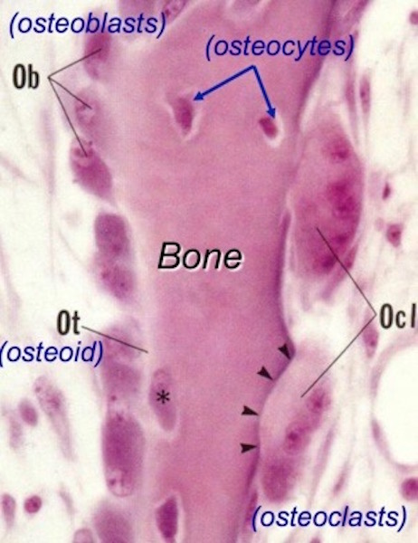
Osteoblast 02 KLS edited

Image I

Bone Fractures: Time for snap, crackle, and pop, and I dinna mean Rice Krispies! Compressive strength from mineral deposition and tensile strength from organic matrix give normal bones the ability to behave elastically. If trauma overcomes this elasticity, then bones fracture!

Bone fractures fall into two major categories: mechanical bone fractures caused by high force impact or stress, and pathological fractures caused by disease.

Pathological Fractures: Diana presents a fabulous case of fracture caused by disease in Colum. The Laird of clan MacKenzie is haunted by the aftermath of a pathologic fracture and accompanying deformities due to Toulouse-Lautrec syndrome (Starz episode 208, The Fox’s Lair).

Outlander book informs readers that at 18 y.o., Colum took a bad fall breaking the long bone (femur) of his thigh that subsequently mended poorly. Other skeletal anomalies quickly ensued. Diana explains in Dragonfly in Amber:

Legs crippled and twisted by a deforming disease, Colum no longer led his clan into battle…

…He glanced dispassionately down at the bowed and twisted legs. In a hundred years’ time, they would call this disease after its most famous sufferer—the Toulouse-Lautrec syndrome.

Read Anatomy Lesson #27, “Colum’s Legs and Other Things too!” for details about this rare syndrome (1.7 per million births). Of course, there are also many other diseases leading to pathological fractures. Nowadays, the most common in the US is osteoporosis.

Colum legs 01KLS edited

Mechanical fractures: High force impact or stress cause bones to snap. Different types of bones (e.g. short vs. long) often exhibit characteristic fractures. This lesson will consider fractures of long bones using the femur again as our example. Fractures are generalized into six different categories:

  • Closed fracture – bone is broken but overlying skin is unbroken
  • Open fracture – better known as a compound fracture, broken bone pierces the skin
  • Simple fracture – bone is broken but no other tissues damaged
  • Complex fracture – sharp fracture edges damage surrounding soft tissues
  • Complete fracture – bone breaks completely across the shaft
  • Incomplete fracture – bone is partly broken but remains in one piece

Then, fractures are named according to features of the break (Image J).

  • Normal – left image is the normal femur
  • Transverse – break crosses the shaft horizontally
  • Oblique – break crosses shaft at an angle
  • Spiral – torque on the broken halves twists these in opposite directions
  • Comminuated – bone is broken into four or more pieces (including main body of the bone)
  • Avulsion – a piece of bone is torn away usually by pull of a muscle tendon
  • Impacted – bone parts are driven together, also known as a buckle, or compression fracture
  • Fissure – crack along one side of the shaft
  • Greenstick – incomplete break through the shaft. Analogous to breaking a stick of green wood.
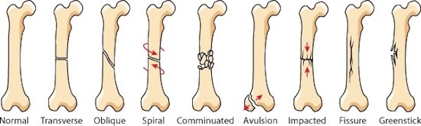
bone fractures 01 KLS edited

Image J

Fracture Healing: Fracture healing is the process by which the body repairs a broken bone and what a process it is! Several overlapping steps occur, different descriptors are used, and times may vary depending on the bone, but the general sequence is (Image K):

  1. Hematoma Formation (days 1-5): Bone fractures tear blood vessels, and periosteum and endosteum. Blood pours from the damaged vessels forming a hematoma (blood clot) between the fractured surfaces. Blood flow in intact vessels increases such that an army of white cells are delivered for defense and repair.
  1. Soft Callus Formation (days 3-40): Gradually, a bridge of cartilage and collagen replaces the hematoma and unites the broken ends. The cartilage bridge is made by chondroblasts (cartilage-generating cells) and is termed a soft callus.
  2. Hard Callus Formation (days 30-80): During this stage, cartilage of the soft callus is gradually replaced with new, woven bone (cartilage does not change into bone) and the site is renamed a hard callus. But, woven bone is immature and not as strong as mature compact bone. Mild exercise is often resumed during this phase.
  3. Bone Remodeling (days 80-100): If all goes well, the hard callus is gradually replaced with normal, mineralized compact bone that remodels to align with muscle pull and mechanical stress. Exercise promotes this process which is typically 80% complete within three months. But, depending on the bone and the fracture, step four can take up to 18 months.
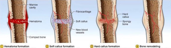
Fracture healing 01 KLS edited

Image K

Normal fracture healing requires the following:

  • Adequate blood supply: Oxygen and nutrients must be delivered to the repair site and carbon dioxide and waste products removed; these processes require blood vessels. Thus, re-vascularization occurs at the fracture site.
  • Fracture stabilization: Excessive movement of the fractured bones interrupts callus formation so fractures are immobilized with a variety of ingenious devices such as casts, rods, plates, screws, and external fixators.
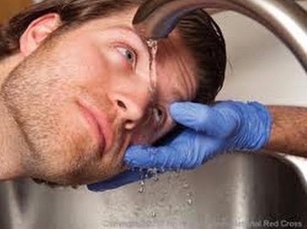
  • Adequate nutrition: On average, a fracture heals by 12  weeks. However, healing times depend on which bones are broken, severity of the breaks, age (younger folks usually heal faster), and nutrition. A vitamin sandwich (Image L) looks funny but animal and human studies show that fracture healing improves with supplements of Vitamins C, D, and E, as well as calcium and amino acids. Details of these experiments exceed our lesson goals, but understand that many average diets are marginal in these substances. So, be sure to consult with your physician about supplements while healing a bone fracture.
  • Rehabilitation: Immobilization of a fracture contributes to muscle wasting. Physical therapy can provide critical support in re-building muscle and encouraging proper bone alignment. This includes weight-bearing on a fracture after it has mostly healed in order to build bone strength.
  • No-No nicotine: Studies are pretty clear that nicotine in any form hinders the process of bone healing. Avoidance behavior!
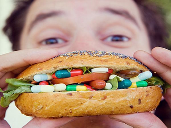
Vitamin sandwich KLS edited

Image L

Now for some real “fun.” With Starz images, let’s use applied anatomy to consider mechanical bone fractures and healing. Starz Outlander episodes offer two excellent examples of bone fractures, both are compound in type.

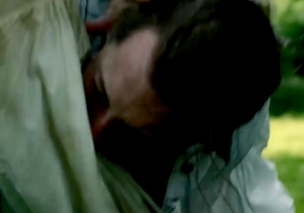
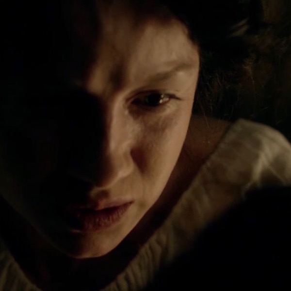
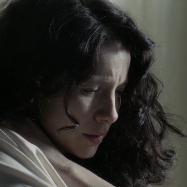
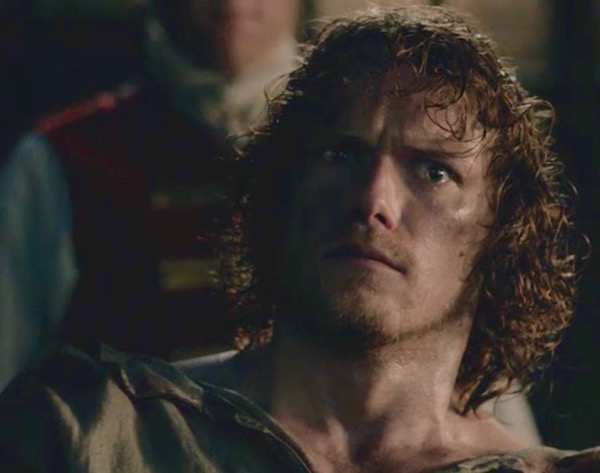
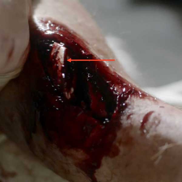
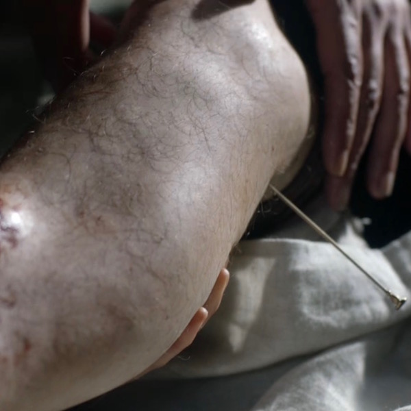
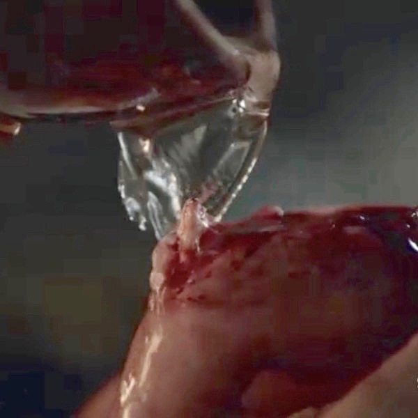
A workman is admitted to L’Hôpital des Anges in Paris (Starz episode 204, La Dame Blanche). He screams in agony because he has suffered a compound fracture of the right tibia (Anatomy Lesson #27, “Colum’s Legs and Other Things too!”). The skin is open and the upper part of the broken tibia is clearly visible at the red arrow.

This injury hurts like holy toothpicks because tears of periosteum and endosteum stimulate nociceptors (pain receptors). Then, muscles spasm as they attempt to stabilize bony fragments, upping the pain meter. And, adding insult to injury, a compound fracture rents the skin and we all ken how sensitive skin is to pain! All around, an agonizing situation.

ep-204-compound-fracture-05

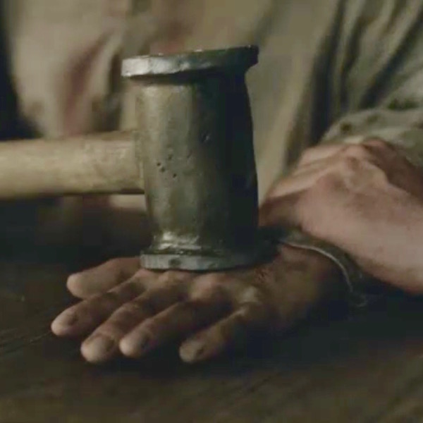
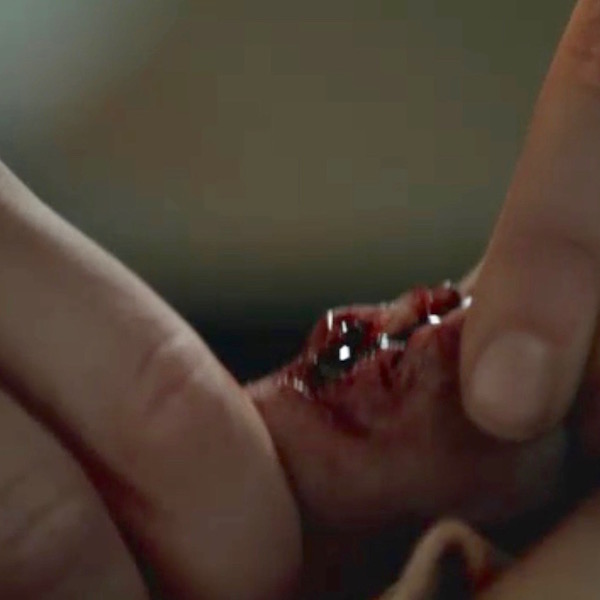
So, what does a good hangman do to ease his patient’s discomfort? Why, he hammers a nail into the side of the poor fellow’s knee, of course! What else? Presumably a type of primitive acupuncture, it momentarily halts the patient’s agony; mayhap from PTSD?

A quote from Dragonfly in Amber:

Monsieur Forez was at work today. The patient, a young workman, lay white-faced and gasping on a pallet. …The leg, though, was something else… Sharp bone fragments protruded through the skin … “Here, ma soeur ,” he directed, taking hold of the patient’s ankle. “Grasp it tightly just behind the heel… Monsieur Forez brought the point of the brass pin to bear …he drove the pin straight into the leg with one blow. The leg twitched violently, then seemed to relax into limpness.

ep 204 compound fracture 03 KLS edited

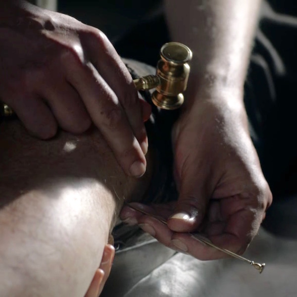
Monsieur Forez enlightens Claire regarding this startling exhibit of bedside manner (from Dragonfly in Amber):

“There is a large bundle of nerve endings there, Sister, what I have heard the anatomists call a plexus. If you are fortunate enough to pierce it directly, it numbs a great deal of the sensations in the lower extremity.”

Stop! I must pick at a wee bone: During the episode, Mr. Forez explains to Claire that anatomists describe a nerve at the inside of the knee which, if pierced, briefly anesthetizes the leg (Anatomy Lesson #27 – “Colum’s Legs and Other Things too!”). So, the human grease-guy whacks a 4” nail into the side of the workman’s knee for pain relief!

So this is what troubles me: the nail pierces skin near the saphenous nerve (a branch of the femoral) which, unfortunately, supplies only skin of the area and does nothing for bone pain. The correct nerve to pierce would be the tibial nerve, a branch of the sciatic; but, both of these nerves are in the back of the thigh! From the thigh, the tibial nerve sends fibers to the tibia in concert with its nutrient artery where it innervates endosteum and periosteum. Ergo, for this technique to work, the good Monsieur must pierce the tibial nerve in the back of the thigh.

I canna vouch for the efficacy of nail piercing, which seems a wee bit harsh, but I can question Forez’s anatomists and their data base. Soon after the shock of piercing subsides, the patient resumes screaming so I canna think it verra efficacious. Gah! Back to the dissection lab for those anatomists! But, I must say, the special effects are superb!

ep 204 compound fracture 04 KLS edited

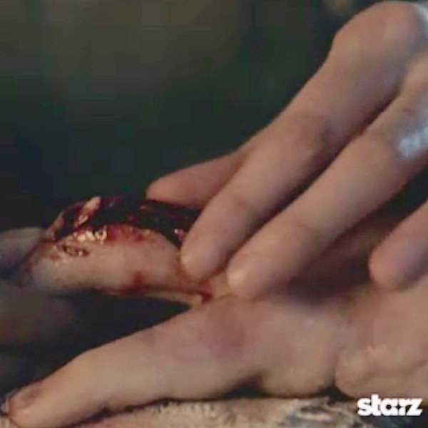
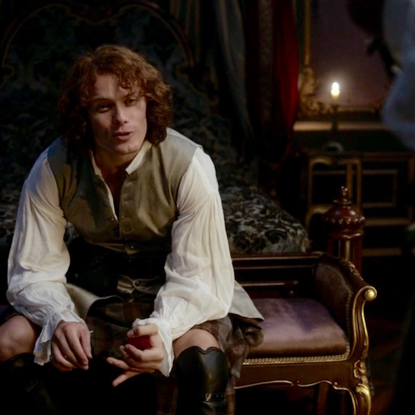
Next, we will consider another compound fracture, arguably the most infamous of the Starz series. The Wentworth Smack-Down delivered high force impact causing mechanical fractures of Jamie’s left hand bones (Starz episode 115, Wentworth Prison). Let’s do a walk through of these fractures.

ep 115 Mallet blow KLS edited

Heading to Outlander book, Diana enlightens us with these quotes:

Luckily the thumb had suffered least; only a simple fracture of the first joint. That would heal clean. The second knuckle on the fourth finger was completely gone; I felt only a pulpy grating of bone chips when I rolled it gently between my own thumb and forefinger…

The compound fracture of the middle finger was the worst to contemplate. The finger would have to be pulled straight, drawing the protruding bone back through the torn flesh. I had seen this done before—under general anesthesia, with the guidance of X rays.

We can easily deduce from Diana’s description that, at a minimum, Jamie’s hand suffered:

  • Simple fracture of the first phalanx of thumb.
  • Communiated fracture of the ring finger (4th finger in English system; 3rd in US system – see Anatomy Lesson #22, “Jamie’s Hand – Symbol of Sacrifice”) – proximal and middle phalanges smashed into bony bits.
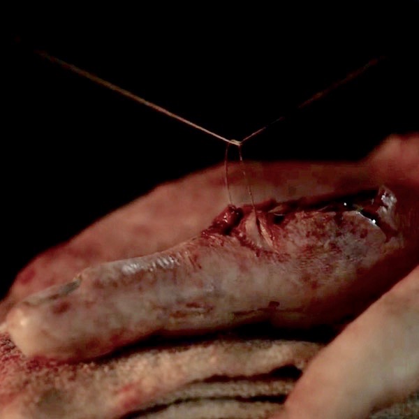
  • Compound fracture of middle finger – appears to be middle phalanx of the middle finger.

ep 116 Jamie hand 02 KLS edited

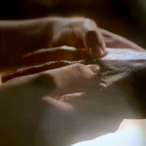
Like any combat nurse worth her salt, Claire cleanses Jamie’s wounds. She has no modern isotonic fluids or antibiotics, so she cleans his compound fracture with (no doubt) sterile water, perhaps containing a wee dram of alcohol for sterilization?

ep 116 Jamie hand 01 KLS edited

Next, Claire ponders the damage and then re-aligns and resets the bony fragments (Starz episode 116, To Ransom a Man’s Soul). And again from Outlander book:

I began to lose myself in the concentration of the job… deciding how best to draw the smashed bones back into alignment.

ep 116 Jamie hand 03 KLS edited

Now, as we learned above, a compound fracture tears surrounding flesh and skin. So Claire carefully closes the wounds with sutures (Starz S.2, introductory image) to exclude pathogens and reduce blood loss.

Opening creds Jamie hand 05 KLS edited

Earlier in this lesson, we read that movement of a new fracture interferes with callus formation. So, various devices are used to immobilize fractures as they heal.

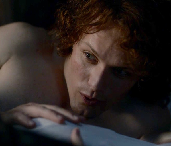
Ever resourceful Claire (or the monks?) devises an external fixator for Jamie’s smashed hand in the form if a very clever wire/leather, linen, and wood device! External fixation is a surgical treatment used to stabilize bone and soft tissues at a distance from the injury. So, check out the amazing invention (Starz episode 116, To Ransom A Man’s Soul) to immobilize Jamie’s hand! The middle finger is reset and stitched. The communiated and most severely wounded ring finger is stitched and splinted against the small finger. Brava, Madam Sassenach!

ep 116 Jamie hand 06 KLS edited

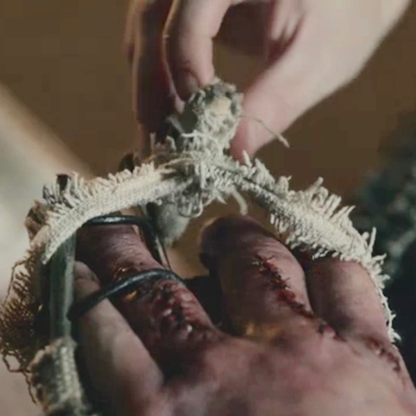
Jamie’s hand care is not yet complete! Claire provides physical therapy in the form a rag ball she made. Squeezing the ball helps Jamie regain hand mobility and strengthen the knitting bones as he and Murtagh plan how best to skewer BJR (Starz episode 203, Useful Occupations and Deceptions)!

In case you forgot from earlier in this lesson, exercising a repairing bony callus augments the ossification process, so Claire’s rehabilitation plan is spot on!

Two quotes from Diana’s Dragonfly in Amber:

It really wasn’t too bad; a couple of fingers set slightly askew, a thick scar down the length of the middle finger. The only major damage had been to the fourth finger, which stuck out stiffly, its second joint so badly crushed that the healing had fused two finger bones together. The hand had been broken in Wentworth Prison, less than four months ago, by Jack Randall.

He had regained an astounding degree of movement, I thought. He still carried the soft ball of rags I had made for him, squeezing it unobtrusively hundreds of times a day as he went about his business. And if the knitting bones hurt him, he never complained.

ep 203 Jamie hand KLS edited

And, pithy quotes from Diana’s Dragonfly in Amber remind us that months later, Jamie’s hand is still not whole (Starz Season 2 opening images), so clever caring Claire applies her personal version of massage therapy… a task that she and he undoubtedly relish! Hee, hee.

I crawled in beside him and took up his right hand, resuming my slow massage of his fingers and palm. He gave a long sigh, almost a groan, as I rubbed a thumb in firm circles over the pads at the base of his fingers.

Opening creds Jamie hand 07 KLS edited

Thus ends Outlander compound fractures and healing. Whether bone breaks or skeletons, people are endlessly fascinated by bones and the stories they tell. Book readers will recognize this poignant commentary from Dragonfly in Amber as Jamie and Claire find the bony inhabitants of an unknown cave in France.

He turned again then to the two skeletons, entwined at our feet. He crouched over them, tracing the line of the bones with a gentle finger, careful not to touch the ivory surface. “See how they lie,” he said. “They didna fall here, and no one laid out their bodies. They lay down themselves.” His hand glided above the long arm bones of the larger skeleton, a dark shadow fluttering like a large moth as it crossed the jackstraw pile of ribs.

Now, I have my own bone story to share. The following is a true event underscoring the amazing capacity of bones to heal. During WW II, my father was a welder of Liberty Ships in California. When I was 18 months of age, neighborhood kids accidentally ran over me and the trauma snapped off the head (ball-shaped epiphysis) of my right femur. Local physicians were unable to help so they sent us to the Treasure Island Naval Base where many of the best US physicians were stationed to assist with the war effort.

Those docs had never set a child’s femoral head before, but they courageously reset my leg, placed me in a 3/4 body cast, and prepared my parents for the bad news: the blood supply to the femoral head was likely severed, the femoral head would become necrotic (die), and the hip would freeze into an immobile joint. I would not walk normally, if at all.

Well, the good news is that my femoral head did not perish! Somehow and somewhere, an arterial supply survived. Although my colleagues and I share theories about which vessel might have remained intact, no one knows for sure.

Image M was taken in 1944 at Stinson Beach, CA, six months after the injury (like the artery, my appetite clearly remained intact <G>). Observe that the right leg is slightly rotated outward (externally), a classic stance following a femoral head fracture.

Today, the only residual is my right leg is 3/8” shorter than the left. Otherwise, nada! I am able to exercise vigorously and with no noticeable deficit. Blessings to those unknown healers who helped the wee bairn of a common laborer!
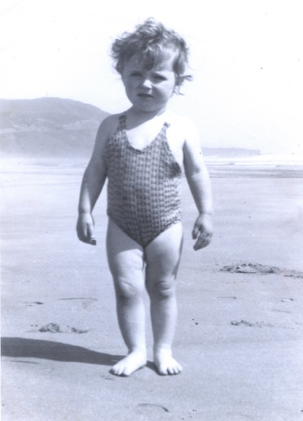
Karmen 1944 KLS edited

Image M

Our lesson on how fractures heal has come to an end. After two lessons about the skeleton, there remains much more to be studied, so we may revisit this topic at a later date.

A few last thoughts: Remember, bone is a dynamic tissue which constantly remodels itself throughout life. Individual bones are organs that collectively constitute the skeleton, our internal support system. Bone cells create, maintain, and destroy bone tissue to our benefit.

Bones have been prodded, examined, healed, revered, saved, feared, embellished, and cherished. Not all of this lesson is about happy stuff, so let’s end with this slightly irreverent image from the bow of the Titanic.
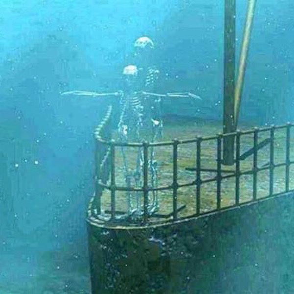
Titanic KLS edited

Image N

Dem bones, dem bones!

A deeply grateful,

Outlander Anatomist.

Photo creds: Starz, Outlander Anatomy archives (Image M), www.anatomy-medicine.com (Image F), www.askabiologist.edu (Image J), www.apps.carleton.edu (Image B) www.boneandspine.com (Image C), www.geneticliteracyproject.org (Image L), www.medcell.med.yale.edu (Image I), www.reddit.com (Image A), www.en.wikipedia.org (Image E; Image H), www.everythingfunny.org (Image N), www.commons.wikimedia.org (Image G) ,www.studyblue.com (Image D), www.ippe2ools.blogspot.com (Image K)

Anatomy Lesson #39 “Dem Bones – The Human Skeleton”

Hello, Outlander anatomy students, and welcome to today’s Anatomy Lesson #39, the Human Skeleton. This is a whopping subject so it will take two lesson to cover the bones!

Dem bones or dem dry bones refer to a spiritual song inspired by a vision recorded in the biblical Book of Ezekiel, 37:1-14. Ezekiel stands in a valley filled with dry human bones. Before his eyes and with the promise of hope, the bones join into human skeletons which become enshrouded with flesh. This wonderful spiritual has been rewritten for children:

Skeleton Lesson over! Naw, just kidding. The skeleton is a wee bit more complicated than the song lets on.

As if on cue, Starz Outlander team offers up a S.2 treasure trove of bone images just in time for our skeleton lesson.

Hum, look again, Claire, that cup isn’t half empty –  it’s half full (Starz episode 204, La Madame Blanche)! Thank you, Outlander!

Raymond bones 02

Claire glides into Master Raymond’s secret “little shop of horrors” overflowing with marvelous skulls from real and imagined beasties (Starz episode 204, La Madame Blanche). His wonderful ossuary includes a unicorn skull (lower right) embellished with head armor (chanfron for horses) complete with horn hole! A whimsical nod to the national animal of Scotland, no doubt. Love it!

Diana elaborates in Dragonfly in Amber.

Two walls of the hidden room were taken up by a honeycomb of shelves, each cell dustless and immaculate, each displaying the skull of a beast. …Tiny skulls, of bat, mouse and shrew, the bones transparent, little teeth spiked in pinpoints of carnivorous ferocity. …They had a certain appeal, so still and so beautiful, as though each object held still the essence of its owner, as if the lines of bone held the ghost of the flesh and fur that once they had borne.

Raymond bones 04

Claire examines an unusual skull, perhaps the remains of a horned carnivorous dinosaur such as carnotaurus (Latin meaning flesh bull). There are loads of horny creatures on Outlander. Ha, ha. Alas, such animals are no more declares Master Raymond. True, unless one takes a Spielberg detour to Isla Nublar!

Goddess of the Pen continues in DIA:

I reached out and touched one of the skulls, the bone not cold as I would have expected, but strangely inert, as though the vanished warmth, long gone, hovered not far off. ..“You see the teeth? An eater of fish, of meat”—a small finger traced the long, wicked curve of the canine… “Such beasts are no more, madonna.”

Claire & dino skull 02

Interested in old stuff, Master Raymond might like this bony specimen for his awesome collection (Image A), a 130,000 y.o. Neanderthal skull. Found in a limestone cavern near Altamura, Italy, the bones are shrouded with cave popcorn, limestone formations caused by splashes of mineral-rich water. Wondrous!

Neanderthal bones KLS edited

Image A

Master Raymond and his coy toys are endlessly fascinating, but it’s time to study the skeleton.

Gross anatomy (Anatomy Lesson #34, “History of Anatomy”) teaches bones and their relationships as they appear in the visible skeleton. We haven’t been exactly idle as prior anatomy lessons have discussed many individual bones. Some of these will be referenced in this lesson. Microscopic anatomy (Anatomy Lesson #34) studies bones as organs and tissues. Slightly different approaches and today’s lesson considers both.

Adult Human Skeleton: The word skeleton derives from the Greek skeletós, meaning “dried up”, because long after flesh has withered, the skeleton steadfastly remains (remains, get it? Hee hee). However, despite the definition, our skeletons are very much alive!

The skeleton forms the supporting structure of an organism. Some creatures, such as the Japanese beetle (Image B), have an exoskeleton (Greek exo- meaning outside), a stable outer shell to protect delicate innards, a type of organic armor. But exoskeletons present a couple of major disadvantages as grown and movement are limited.

Beetle KLS edited

Image B

We humans don’t walk around with our skeletons exposed – unless we suffered a Randall-scandal with BJ! Rather, humans enjoy an endoskeleton (Greek endo- meaning within); our skeleton lies inside the body, wrapped in flesh. The endoskeleton is a genuine boon because it gifts its owner with freer movement and growth potential.

The skeleton (Image C) is a composite of all bones in a human body. It is also heavy, accounting for 20% of our total body weight. The adult human skeleton includes 206 individually named bones whereas, the infant human skeleton contains about 300. The overall count drops during maturation as many bones fuse (e.g. skull bones), usually completing the process after three decades of life.

skeleton

Image C

Interestingly, the number of bones comprising the adult skeleton is always higher than 206, but, because some bones are not present in all people, are small, or are variable in number, they are excluded from the overall count. A couple of good examples follow.

Humans have small sesamoid bones (Latin meaning like a seed) housed within tendons of thumbs and great toes (Image D) where they influence the pull of muscles. These are not counted. The paired patellae (pl., knee caps) are the largest and best known sesamoid bones but, because of size and constancy, these are routinely included in the 206 count.

Sesamoid bones KLS edited

Image D

Another example of bones eliminated from the count are small, irregular wormian bones, which develop in sutures of the skull (Image E – red arrows). Although not rare, most skulls do not have them. Wormian bones can be markers of diseases such as brittle bone, but in normal individuals, their significance is unknown.

wormian

Image E

Back to the adult skeleton. The skeleton is divided into axial (Image F – yellow) and appendicular (Image F – green) parts. The axial skeleton includes bones of skull, vertebrae, sacrum, coccyx, ribs, and sternum, for a total of 80 bones. The appendicular skeleton houses bones of upper and lower limbs, including clavicle, scapula, and hip bones, for a total of 126.

Notice this, all bones of the adult appendicular skeleton are duplicated on each side of the body: two femurs, two humeri, (pl.) etc. However, the adult adult axial skeleton is variable: ribs and some facial bones are duplicated but all remaining axial bones are singular, lacking a counterpart. Three pairs of ear ossicles are good examples of duplicated skull bones (Anatomy Lesson #25, “If a Tree Falls – The Ear”).  Whereas, the frontal bone of the skull is unpaired (Anatomy Lesson #11, “Jamie’s Face” or “Ye do it Face to Face?”).

axial skeleton

Image F

Bones are Secure: Bones of the skeleton don’t swing in the breeze; they are joined by connective tissue elements. Unmoveable joints (think skull sutures) are united by strong layers of collagen. Moveable joints (think elbows – Anatomy Lesson #20, “Arms! Arms! Arms! – Redux”) are sites where adjacent bones move on each other. To be more precise, the articular cartilages of such bones move on each other.

I cringe when my yoga teachers mention the bad “bone-on-bone” plank position. They really don’t mean this because bone-on-bone means the articular cartilages are worn away ensuring arthritis as one’s constant companion!

Image G shows the moveable knee joint, one of the largest and most complex of the body. The femur (Anatomy Lesson #7, “Jamie’s Thighs” or “Ode to Joy!”) and tibia (ditto) are stabilized by four major ligaments, muscle tendons, joint capsule (collagen again), and shock-absorbing menisci (pl.). Similar anatomical elements (sans menisci) compliment all moveable joints.

BentKneeAnatomy

Image G

Bone Shape: Bones are classified into four categories based on shape: long, short, flat, and irregular (Image H). Most anatomists eliminate sesamoid and suture (wormian) bones from shape categories but all agree on the following four.

Long bones are longer than they are wide and are mostly confined to the appendicular skeleton where they engage in weight bearing and movement. Examples are femur, humerus, and phalanges. The femur (Anatomy Lesson #7, “Jamie’s Thighs” or “Ode to Joy!”) is the skeleton’s longest bone.

Short bones are as wide as they are long (cube-like), providing support and stability. Good examples are carpals of wrist (Anatomy Lesson #22, ”Jamie’s Hand – Symbol of Sacrifice”). The stapes (Anatomy Lesson #25, ”If a Tree Falls – The Ear”) of the middle ear is the skeleton’s shortest bone.

Flat bones are expanded into broad, flat planes. They protect underlying elements and/or provide wide muscle attachments. Good examples are bones of cranium, scapula, and sternum (Anatomy Lesson #15, “Crouching Grants – Hidden Dagger”).

Irregular bones have peculiar shapes; they provide protection and muscle attachment. Vertebrae and facial bones are good examples.

Now, the shape classification scheme is not without controversy as some bones cross boundaries, sharing elements of more than one category…. think of the flat and oddly-shaped scapula (Anatomy Lesson #2, “When Claire Meets Jamie” or “How to Fall in Love While Reducing a Dislocated Shoulder Joint!”).

Bone shapes

Image H

Bones as Organs: Now for some microscopic anatomy… an organ is a group of tissues that perform a function or group of functions. Like heart, lungs, liver, and brain, our bones are also living organs.

Bones are characterized by hard outer shells and “spongy interiors.” The outer shell is compact (cortical) bone; the interior is spongy (cancellous) bone, a distribution best illustrated using the femur (Image I). BTW, spongy bone is so named because it is riddled with holes, not because it is soft and pliable like the animal known as a sponge.

Parts of a Long Bone: Now for a crash course in bone anatomy. A long bone displays shaft (diaphysis), marrow cavity, and articular (epiphyses) ends (Image I). A connective tissue periosteum envelops the shaft and is richly endowed with pain fibers; anyone who has broken a bone kens this very well! Depending on the bone and age of its owner, the marrow cavity is filled with either fat (yellow marrow) or blood-forming tissue (red marrow). Both epiphyses are covered with smooth, firm articular cartilage (blue in Image I), which augments movement at the joints. Cancellous bone is abundant deep to the articular cartilage caps. Epiphyseal (growth) plates separate epiphyses (pl.) from shaft.

Growth plates are sites where long bones grow in length. Long bones continue lengthen until growth plates ossify, about two years after the onset of menstruation in girls and late teens for boys (there are variations). Whew, a packed mini-lesson!

Femur 01 KLS edited

Image I

Bone as Tissue: Named bones are organs, but bone is also classified as a type of connective tissue. Tissue is an aggregation of similar cells and extracellular material acting together to perform specific functions. Connective tissues range from fluids, such as lymph and blood, to semi-solids or solids such as cartilage and bone.

As a tissue, bone is a composite of organic proteins and inorganic bone mineral. The organic protein is collagen, the most ubiquitous structural protein of the human body. Collagen is abundant in bone where it acts as a scaffold for the deposition of minerals. Inorganic bone mineral is made of hydroxyapatite, tiny crystals of calcium, phosphate, and magnesium which endow bone with rigidity. Understand that normal deposition of bone minerals is a complex process involving several hormones (e.g. calcitonin), dietary calcium, phosphorus, and magnesium,, and sufficient Vitamin D (via sunlight and/or supplements)!

Tidy Test: This bitty bone test demonstrates the relative roles of organic and inorganic components of bone. Bake a raw chicken bone at low temperature for a few hours. Immerse another raw chicken bone in acid (vinegar works OK but something stronger is better) for many days. Baking destroys collagen (organic protein) leaving the bone brittle and friable; it readily snaps in two. Soaking removes inorganic minerals leaving a rubbery bone that can be easily bent. I don’t expect you to try this demonstration, but it has been done for many years in school science labs.

A dry femur (Image J), offers a superb example of how bony tissue is organized. Covering the surface is a rind of hard, cortical bone of varying thickness. The cartilage covering the epiphysis is absent. The interior of the epiphysis is filled with spongy bone, a lattice of thin, hard bony shards. Spaces in the marrow cavity (red arrow) and amid the spongy bone are filled with either blood-forming tissue or fat, depending on the bone and one’s age.

cortical-bone

Image J

Time for another outlandish image (Starz episode 204, La Madame Blanche). No, lass, dinna ask Master Raymond about future Frank! Dem bones, dem bones, goin’ talk about… you don’t want to know the answer!

Raymond bones 01

Which brings us to the very entertaining topic of sexual dimorphism. Don’t ask how I made that leap, it just seems to fit here! <G>

Sexual Dimorphism: Like many primates, the adult human skeleton exhibits sexual dimorphism; differences in form based on sex. Male skeletons are typically larger, heavier, and more massive than those of the female. There are also gender differences between some skull bones, canine teeth, and long bones (e.g. femur). But, the most reliable difference in discerning gender is via the bony pelvis. In 95% of cases, a skilled observer can assign an adult bony pelvis to the correct gender, although such differences are not evident before puberty.

To understand these sex-based differences, we must first consider anatomy of the bony pelvis (Image K), a ring formed by sacrum (yellow) and two hip bones (peachy-red). These three bones are held together by some of the body’s strongest ligaments. Why? Because, they bear the entire weight of torso, head and upper limbs, more than half our body weight!

BTW, I can immediately discern (with ~ 95% accuracy) that image F illustrates a female bony pelvis. You’ll understand how in a moment.

bony pelvis 01 KLS edited

Image K

Another consideration about the bony pelvis: at birth, each hip bone consists of three separate bones, ilium, ischium, and pubis, joined together by cartilage – one reason why there is righteous concern over young children doing rigorous gymnastics. By age 25, the three bones of each side, fuse and ossify into a single hip bone (Image L – right hip bone). From a lateral (side) view, the three bones meet at the acetabulum, the socket for the femoral head.

hip bone lateral KLS edited

Image L

Pubic bones join in the midline at the pubic symphysis (Image M). The ilia (pl.) form relatively immovable joints with the sacrum at the infamous sacroiliac (SI) joints (Image M – black arrows). The top opening of the bony pelvis is the pelvic inlet (Image M – dashed black oval). Flip the bony ring upside down and the bottom opening is the pelvic outlet (not shown, so use your imagination). The space beneath the pubic symphysis is the sub-pubic angle (Image M – red arrow).

Interestingly, if x-ray reveals a break in the bony pelvis, there will always be at least one or more additional breaks. Try breaking a round pretzel… one cannot break just one side… same with the bony pelvis.

bony-pelvis

Image M

Now that we understand the bony pelvis, back to sexual dimorphism… Table A summarizes typical differences between adult male and female bony pelves (pl.). Although there are always outliers, female pelves generally express traits that augment pregnancy and childbirth.

TABLE A

Males Female
Thick and heavy Thin and light
Narrower, taller pelvis Wider, shallower pelvis
Sub-pubic angle < 90° Sub-pubic angle > 90°
Pelvic inlet heart-shaped Pelvic inlet oval and rounded
Sacrum tilted forward Sacrum tilted backward
Small pelvic outlet Large pelvic outlet

Following puberty, the female bony pelvis grows becoming wider and shallower than the male pelvis (compare Table A with Image N). Let’s consider these differences.

The female sub-pubic angle is typically 90° or greater (Image N – green inverted V). The male sub-pubic angle is less than 90° (Image N – red inverted V). If one opens the space between thumb and index finger to a right or 90°angle; this is the typical extent of the female sub-pubic angle. If one spreads index and middle fingers as far apart as possible; this angle is less than 90° and is typical of the male sub-pubic angle. This is how anthropologists, forensic experts, and anatomists quickly identify the gender (historically, anatomists use the terms sex and gender interchangeably) of a bony pelvis.

Next, the female pelvic inlet is usually large and oval-shaped (Image N – top, right); the male pelvic inlet is small and heart-shaped (Image N – top, left).

Lastly, the female sacrum typically tilts backward such that the pelvic inlet of a standing woman faces mostly forward. Because the male sacrum tilts forward, the pelvic inlet faces mostly upwards.

Pop quiz! Return to Image F and determine if the yellow and green skeleton belongs to a female or a male. Answer follows Image N.

Pelves KLS edited

Image N

If you answered female, you are ready for a guest spot on one of those forensic TV shows. Congrats!

We now understand anatomy of the skeleton but what purpose does it serve? Well, it actually serves at whopping six purposes:

  • support
  • movement
  • protection
  • hematopoiesis
  • ion storage
  • hormone regulation

Support: Our flesh in the form of muscles, ligaments, tendons, blood and lymphatic vessels, nerves, and so forth, enshrouds the skeleton. If we did not have an endoskeleton for support and movement, we might look something like Mr. Blobfish (Image O), a deep sea fellow living off the coasts of Australia, Tasmania and New Zealand!

blobfish KLS edited

Image O

Movement: The body contains three different types of muscle: smooth, cardiac and skeletal. Skeletal muscles attach to bones via origins and insertions. As a skeletal muscle contracts, it moves the bone(s) to which it attaches. It goes without saying that movement allows us to negotiate our environment for survival.

We have over 700 named skeletal muscles accounting for over half our body weight! The foot of a running man from a 2008 Body World’s exhibit (Image P) shows 12 muscles (there are more that are not shown) of the right foot and leg engaged in lower limb locomotion… add the left leg and foot and the number doubles! Add muscles of thigh and buttocks and, the numbers continue to climb. What a wonder!

Running man KLS edited

Image P: Running man KLS edited

Protection: The skeleton offers sanctuary for vital organs such as brain and heart (Image Q). Seated in its bony thoracic cage (Anatomy Lesson #15, “Crouching Grants – Hidden Dagger”), all sides of the heart except its diaphragmatic surface are surrounded by bone. Ditto for trachea (most of it), lungs, bronchi, aorta, kidneys, brain, pituitary gland, eyes, tongue, etc., etc., etc. Our well-being has a vested interest in preserving vital organs from injury so surrounding them with bone is an ingenious devise.

Heart in rib cage KLS edited

Image Q

Hematopoiesis: Aaaah… What does this term mean? Hematopoiesis means the production of blood cells (Image R). Greatly simplifying a very complex process, circulating blood contains six classes of blood cells plus platelets  (Anatomy Lesson #37, “Outlander Owies! – Part 3,  Mars and Scars”), all of which arise in bone marrow. Side note: one class of blood cells (lymphocytes) also develop during immune responses in sites outside bone marrow.

Adding another layer of complexity, hematopoiesis is a tortuous process which varies throughout life (Image R). In utero, blood cells arise in the yolk sac (human embryos have one), liver, spleen, lymph nodes, and by the third trimester, in bone marrow. In children, the entire skeleton is engaged in hematopoiesis (it stops in the earlier organs). By adulthood, hematopoiesis confines itself to the ends of long bones and the axial skeleton. Sadly, in the aged, hematopoiesis declines even more, leaving elderly people challenged to produce enough blood cells for good health (exercise helps thwart this decline). Interestingly, blood-forming potential is retained such that under rare conditions, adult liver and spleen can resume hematopoiesis.

Hematopoiesis 01 KLS edited

Image R

Storage of Minerals: Remember calcium, phosphate, and magnesium that crystalize into bony hydroxyapatite deposits? As we know, these microscopic crystals form either compact bone or spongy bone (Image S – spongy bone). Either way, given the proper signals, the stored minerals can be mobilized from bone and released into the blood stream for other needs in the body. Thus, bones play an important role as storage depots for minerals.

bone marrow 04 KLS edited

Image S

Hormone Regulation: Last but never least, bone plays a hormonal role. Yes! Bone cells mostly in the medullary (marrow) cavity (Image T) produce two hormones. One, phosphatonin, targets the kidneys causing them to increase phosphate loss in urine thus regulating levels in the blood stream. A second, osteocalcin, stimulates pancreatic cells to release insulin and testicular (Leydig) cells to release testosterone. Ergo, bones are awesome, low-paid multi-taskers!

Femur 02 KLS edited

Image T

One last point before this lesson goes bye-bye. What about teeth? Where do they fit into the bony scheme? Most anatomists group teeth with the skeleton, in part because like the skeleton, they remain after all other tissues succumb. And, like bone, tooth enamel is made of specialized hydroxyapatite crystals, although harder, as enamel is the hardest substance in the body.

But, the real reason teeth are saved for the end of this lesson is because the devil made me do it! Yep, BJR is my “go to” guy (Starz episode 206, Best Laid Schemes)!

Moralizing Moment – S.2… Dueling with Jamie, the Snap-Dragon eschews codes duello as he sinks a Munch-Crunch into Jamie’s right arm (puir Jamie, he gets bitten a bunch in S.2)!

A duel bite? Shocking ! What “officer and gentleman” would disarm an opponent’s arm via a carnassal-chomp?

Where’s Murtagh, Jamie’s second, to demand the “field of honour” remains honorable? Sadly, Godfather is long gone – off to Portugal selling hijacked wine.

Jack bite 01

This is a perfect spot for Moralizing Moment – S.1. Something has been bugging me for months!

Some high level Outlander folks once opined that BJ has a “code of honor” because he kept his word, allowing Claire to escape Wentworth in exchange for Jamie’s  surrender. Och! I beg to differ. He is as despicable about that “promise” as about Battle-Bites.

I posit that Claire was “dishonorably discharged” from that hell-hole. Does shoving an unsuspecting lass down a 3-4 meter shaft seem honorable to you (Starz episode 115, Wentworth Prison)? Snort!

Claire fall 03

We have come to expect mind-boggling acts from Jack-the-Nipper – that fall could have broken Claire’s back. And, ugh, he pushes her into Wentworth’s garbage/dead body dump, where she finds Taran. We miss you big guy!

Holding a torch aloft, Jack-Jaw surveys his handiwork. Seeing Claire move, he can now “honorably” inform Jamie that his beloved has “left the building” (Starz episode 115, Wentworth Prison)! Ruadh, being a man of honor, honors his “end” of the bargain. Moral to the story: never dance with that dishonorable devil!

Claire fall 02

But, take comfort, budding anatomists……. Diana reminds us in a quote from Dragonfly in Amber that love redeems all, even dem bones (and teeth) – ! Yay, the skeleton scores!

” ’Blood of my blood, bone of my bone …’”  “I give ye my body, that we may be one,” he finished. “Aye, and I have kept that vow, Sassenach, and so have you.” He turned me slightly, and one hand cupped itself gently over the tiny swell of my stomach.

Whose your daddy (Starz, episode 206, Best Laid Schemes)? Hee hee!

Jamie love


“Let us rise up and be thankful. For if we didn’t learn a lot today, at least we learned a little… ”

-Buddha

Next lesson: how bones heal.

See you later, alligator….. that creature hanging in the apothecary shop. Oops! It’s an “after a while crocodile” dangling from Raymond’s rafters! Ta-ta!

alligator

A deeply grateful,

Outlander Anatomist

Photo creds: , Starz, Sony Pictures, Archival image from Outlander Anatomy collection (Image S), National Geographic Magazine, March 2016 (Image A), Body Worlds specimen (Image P), Frank H. Netter, 4th ed., Atlas of Human Anatomy (Image L; Image M), Kieth L. Moore and Arthur F. Dalley, 5th ed.,Clinically Oriented Anatomy (Image N), YouTube (dem bones), www.anthropology.si.edu (Image C), www.arbordoctor.net (Image B), www.archive.museumoflondon.org.uk (Image E), www.bbc.co.uk (Image I), www.en.wikipedia.org (Image H; Image R), www.highlands.edu (Image J), www.nim.nih.gov (Image T), www.skeletalsystemdev.weebly.com (Image F), www.slideshare.net (Image Q), www.teachmescience.info (Image K), www.theinertia.com (Image O), www.theknee.com (Image G), www.wikiradiography.net (Image D), Walt Disney’s Fantasia.

Anatomy Lesson #38: “Outlander Tears” or “Why, oh Why, do We Cry?”

Greetings anatomy students! Flurry over the Outlander premier has abated and S.2 is well underway so it is time for another anatomy lesson. Yay! The bell has rung and class is in session! This anatomy lessons turns our attention to the poorly understood act of emotional crying.

As always, we’ll use Starz images to set the stage for our lesson. Beginning with S.2 opening theme, we are greeted with excellent images of emotional weeping. Claire’s tears flow as she desperately pleads with Jamie after his torture at the hands of the Bloody Blackguard (Starz episode 116, To Ransom a Man’s Soul)!

Claire cry 04

Her tears flow almost unabated through the first half of Starz episode 201, Through a Glass, Darkly. The episode begins with Claire separated from Jamie and her pain is palpable as she shrieks her rage to the heavens (Starz episode 201, Through a Glass, Darkly)!

Claire cry 02

Her fury is quickly followed with helpless tears of loss and grief as she collapses amid the towering stone monoliths capping Craigh na Dun!

Claire cry 03

Wandering the road to Inverness, Claire meets a kindly Scotsman. She collapses in desperate sobbing after he reveals, yes indeed, the British won the Battle of Culloden! We empaths shed tears along with our beautiful, courageous heroine!

Claire cry 01

This quote from Diana’s Dragonfly in Amber book adds depth to Claire’s desolation:

I woke three times in the dark predawn. First in sorrow, then in joy, and at the last, in solitude. The tears of a bone-deep loss woke me slowly, bathing my face like the comforting touch of a damp cloth in soothing hands. I turned my face to the wet pillow and sailed a salty river into the caverns of grief remembered, into the subterranean depths of sleep.

Claire cry 05

Time to start this tearfilled lesson (sob) with a brief anatomical overview. Unless one suffers from an unfortunate condition such as dry eyes (keratoconjunctivitis sicca), tears flow under three different conditions: base-line tear production, non-emotional tearing, and emotional weeping. Regardless of the cause, all three types involve lacrimation (Latin, lacrima, meaning tear), the production of tears. So, sharpen your scalpels and let’s dissect each type of tear, one-at-a-time.

Base-line Tears: We learned in Anatomy Lesson #30, “Aye, Eye – The Eyes, Part 2,” each eyeball is equipped with a lacrimal apparatus which produces and drains tears. That apparatus includes the following features.

In the upper outer corner of each bony orbit (Anatomy Lesson #30, “Aye, Eye – The Eyes, Part 2,”) lies the small but powerful lacrimal gland (Image A). Most of the gland lies inside the bony orbit but a small part projects into the upper eyelid. Small ducts (channels) pierce the conjunctiva (transparent membrane) and convey the secretion onto the eyeball surface.

The lacrimal gland is designed to continuously secrete (discharge) the aqueous (watery) part of the tear film which bathes the surfaces of cornea and conjunctiva (Anatomy Lesson #30, “Aye, Eye – The Eyes, Part 2,” and Anatomy Lesson #31, Aye for an Eye – The Eye, Part 3). Although their roles differ, fibers from both sympathetic and parasympathetic divisions of the autonomic nervous system (the part we don’t control) supply the gland (see Anatomy Lesson #32, “A Real Eye Opener – The Eye, Part 4). Working together, parasympathetic fibers induce secretion and sympathetic fibers control blood flow.

The tri-laminar tear film (Image A) is composed of an inner mucous (adj.) layer produced by mucous cells of the conjunctiva, a middle aqueous layer produced by the lacrimal gland, and an outer oil layer produced by tarsal glands of the eyelids (Anatomy Lesson #29, “The Eyes Have It! – The Eyes, Part 1”). The tear film represents basal tearing and it is critical to the health of exposed eye surfaces.

tear film KLS edited

Image A

The tear film is continuously renewed and then drained by the following structures. Closure of the eyelids moves the tear film toward two small openings, the lacrimal puncta (pl.), at the nasal end of each eyelid (Image B – lacrimal apparatus of right eye). Each punctum (find yours) drains into one of two lacrimal canaliculi, tiny channels which empty into a reservoir, the lacrimal sac. From the lacrimal sac, tears traverse the nasolacrimal duct and drain into the ipsilateral (same side) nasal cavity. Remember, each eye has its own lacrimal apparatus.

The tear film doesn’t just give Jamie his sparkling bonny blue orbs; rather, it serves three important purposes:

  • Protects and lubricates exposed surfaces of the eyeball.
  • Washes away foreign particles.
  • Reduces the risk of eye infections (antibacterial).

Lacrimal apparatus KLS edited

Image B

Non-Emotional Tearing: Also known as reflex tearing, non-emotional tear production is a response to eye irritants. If the exposed eyeball is insulted, the lacrimal glands are stimulated to produce a flood of tears which overwhelms the lacrimal drainage system. Having no adequate outlet, they escape the eyelids and run down the face.

What are the causes of non-emotional tearing? Well, foreign bodies, objects that enter the eye from outside the body, are the most common cause. Although intuitively obvious, here are some important clues that a foreign body may have taken up residence in your eyeball:

  • Pressure
  • Discomfort or pain
  • Sensation of something “in the eye”
  • Extreme tearing (yes!)
  • Photophobia (pain or discomfort with light exposure)
  • Excessive blinking
  • Redness or bloodshot conjunctiva (image C)

red eye KLS edited

Image C

And just so you know, the following foreign objects are the most common causes of non-emotional tearing:

  • eyelashes
  • dried mucus
  • sawdust
  • dirt
  • sand
  • cosmetics
  • contact lenses
  • metal particles
  • glass shards

Those who read Anatomy Lesson #29, “The Eyes Have It! – The Eyes, Part 1” may recall examples of the now-popular eyelash jewelry, including among other delights, crystals glued to the eyelashes! Who thought that was a good idea? Well, if one is not verra careful, these fake jewels can wreak havoc as foreign bodies! Case in point, crystals float on the tear film overlying conjunctiva and cornea (Image D). These were likely applied to the upper surface of the lower eyelashes. The glue failed and the crystalline escapees drifted onto the eyeball surface.

Understand that if the conjunctiva is intact, objects deposited on the front of the eyeball cannot get lost behind it (Anatomy Lesson #30, “Aye, Eye – The Eyes, Part 2). They can, however, scratch the cornea and cause infection so foreign bodies in the eye should be resolved STAT!

foreign body 02 KLS edited

Image D

Then, there are really dangerous foreign bodies such as metal shards (Image E) that pierce the transparent conjunctiva and embed in the sclera (Anatomy Lesson #32, “A Real Eye Opener – The Eye, Part 4). Such objects can scratch the inner eyelids and cause infections. Och, I bet that hurts!

Protective eyewear can help prevent such injuries and should certainly be worn if working with:

  • saws, hammers, grinders, lawn mowers, and other power tools
  • dangerous or toxic chemicals

Foreign body 03 KLS edited

Image E

Non-emotional tearing also follows exposure to irritating substances such as the whiff of onions (Image F) or noxious agents such as insecticides, perfumes, detergents, smoke, dust, etc.

Chopping onions KLS edited

Image F

Well then, fair’s fair (Image G)! Too gruesome? If yes, why are ye watching/reading Outlander? Hee, hee.

Chopping human KLS edited

Image G

Speaking of noxious substances, if an across-the-counter product splashes into the eye, you can immediately relieve discomfort and assist tear formation by washing the eyeball. Yes, you read it correctly! Every science laboratory worth its salt sports an eye wash station as part of its safety equipment. Since few homes have such an apparatus, you can use a sink faucet or garden hose. Run a stream of cool water (NOT hot!) from the tap or hose. Open the irritated eye and turn head so the affected eye is DOWN. Let the water run across the eyeball from the nose towards the ear (Image H). Do NOT run water from the ear towards nose! Why? Because the irritant will flow into the lacrimal drainage structures and nose. Voila, now the problem is compounded!

Flush the eye for 15 minutes! Yes your water bill will skyrocket, but the irritant must be thoroughly diluted. Depending on the substance (think acids, lye, etc.), hie to the nearest urgent care facility or call EMT/fire department. In the meantime, wash!

eye washing KLS edited

Image H

Finally, if something splashes into both eyes, use a garden hose or a faucet spray nozzle. Look down and widely opening both eyes, allow water to flush them simultaneously. Do not turn the head to either side.

The following is an informative and well-done Youtube video that includes images of how to deal with splashes involving both eyes. Note that although it starts with showing a one eye splash, later it demos both eyes involvement.

Emotional Tearing: Now, we get to the nitty-gritty! Also known as psychic tearing, this is the same as crying, the shedding of tears trigger by emotions. Crying synonyms include weeping, wailing, sobbing, whimpering, squalling, mewling, and bawling. Humans cry if we feel grief, stress, sadness, happy, overwhelmed, pleasure, anger, and suffering. But, the bottom line is, scientists understand very little about why emotions provoke human crying!

Again, Outlander S.2 comes to our rescue: Rage, grief, and despair fuel Claire’s tears in Starz episode 201, Through a Glass, DarklyBut, in the same episode, Frank shares with us fine examples of emotional tears!

Tears of gratitude – Claire has finally returned to him. He doesn’t care who she bedded during her Highland gad-about, nothing could ever change the way he feels about her! No way, Jose!

Frank tears 01

Tears of wonder – After Claire reveals that she has a “bun in the oven”, Jack, oops, I mean Frank, is thrilled and responds with more pop up tears. Oh, my, he is delighted,  desperately happy and “over the moon”! Ah, erm…wait! How could such a miracle happen because his doctor told him that …

Frank tears 02

Tears of fury – What? That red-haired bastard (weil, son of a bastard) knocked up his beloved Claire? Who the hey does that guy think he is? And, presto, just like that, Franks’s tears of joy turn into rivulets of rage! So much for his “nothing you could ever do” speech.

Recall not-her-name Sally and her alley cat friends (Starz episode 108, Both sides Now)? Frank came very close to sharing the same black-jack knuckle sandwich with Claire! Fist of fury!

In rapid succession, Frank delivers a wallop of emotion-ladened tears just in time for this lesson. TY, Frank. Much obliged!

Frank tears 03

Back to our lesson: many of us ken that newborns wail without tears. Their nascent lacrimal glands produce just enough baseline tears to moisten their eyeballs. Somewhere between 1-3 months the lacrimal glands develop enough to shed tear droplets in response to physical discomfort (Image I).

Around puberty, tears from emotional pain usually overtake those from physical discomfort. Gradually, with age and experience, people add moral crying in response to acts of courage and self-sacrifice or to symbols such as the flag of one’s country or to the sound of bagpipes (my personal favorite)!

emotional crying

Image I

Now, scientists do have insights about how tears and emotions are linked. Emotional crying is a complex secretomotor phenomenon characterized by the shedding of tears sans ocular irritation. The lacrimal gland is linked to the limbic system (Image J), part of the brain that processes emotions. The limbic system (waaaay too complex for this lesson) is hard-wired into the autonomic nervous system/ANS (the part you cannot voluntarily control). With the proper emotional trigger, the limbic system stimulates the ANS to release the neurotransmitter, acetylcholine, a wee molecule which then stimulates the lacrimal gland to shed emotional tears. Ergo, emotions interpreted by the limbic system activate the ANS which releases acetylcholine which turns on the water taps! Voila, we sob with feeling! Pretty remarkable.

Limbic System KLS edited

Image J

What we don’t understand is why do strong emotions cause us to cry? What is the purpose of emotional tears? Well, there are lots of ideas, one dating back more than 2,000 years.

Greek philosopher and scientist, Aristotle, posited that tears are waste products like urine and therefore, discarded by the body (Image K): “That they are of one nature is plain to the taste.”

Take comfort, Jamie! Claire isna the only healer to taste urine (deferred to a future lesson). Truth be told, dedicated physicians used to routinely taste their patient’s urine. Talk about the call of duty. Pitooey!

Aristotle KLS edited

Image K

Here’s a good one…in the 1940s, American psychoanalyst and physician, Phyllis Greenacre, proposed that female weeping is a sign of penis envy and the way a woman can imitate a man urinating. A hypothesis to which I offer this scientific response: Snort!

Others have proposed that tears, like urine production, cleanse the blood. However, the average cry yields only about 20 tear droplets or the equivalent of 1 ml, a totally inadequate amount of fluid loss to alter blood composition.

Some suggest that crying purges the body of harmful chemicals produced under duress. Forty years ago, a biochemist found that emotional tears were richer in protein than non-emotional tears. Unfortunately, others failed to replicate his findings so they lost momentum. A basic tenet of scientific research is that independent laboratories must be able to replicate another’s results…. an excellent check and balance system.

However, later studies have shown that emotional tears contain elevated levels of prolactin (cuddle hormone), adrenocorticotropic hormone (induces adrenal cortex to release the stress hormone, cortisol), and leucine enkephalin (a natural painkiller). So, there may be merit to the idea of emotional purging. Verra complex, are our emotional tears!

Along a similar vein, Darwin proposed that in addition to lubricating the eyeballs, tears “serve as a relief to suffering,” and the idea that crying is cathartic remains viable but unproven. General wisdom suggests that emotional crying does make one feel better. But, why? Well, if misery is short lived, our mood may lighten by the time we finish a good cry. Or, in the midst of despair, something wonderful might happen to completely alter one’s mood such that eyes spout tears of joy!

Intriguingly, some researchers consider emotional crying as a social signal that a person needs nurturing; a sort of primal “shoutout” for help. One interesting study showed pictures of tearful faces to subjects. Within 50 milliseconds (.05 sec.) test subjects reported a boost in feelings of empathy and friendship towards people shown in such images. This very interesting hypothesis awaits further investigation.

Here’s some Outlander proof that social signals work: a mess of fans were ready, willing, and able to comfort that ginger-haired laddie as he wept at Wentworth Prison (Starz episode 115, Wentworth Prison)! Yep!

Jamie tears 02

Here’s another fascinating aspect of emotional tearing. Boys and girls cry with equal frequency until puberty when something complex happens. In Western cultures, boys are conditioned to restrain tears such that women cry twice (one study says 5x) as frequently as men. Biology may also play a role as male puberty is marked by increased testosterone production and some studies hint that this male hormone helps suppress emotional tearing.

Even more interesting, in some social settings such as sports, male displays of feeling such as hugging, cheering, and crying are OK; perhaps because people expect emotions to run high at sporting events. Consider Mario Balotelli, a world class footballer (soccer in US) for team Italy.  At the 2012 Euro final, Spain defeated Italy and Mario wept with deep regret (Image L). Apparently, this emotional display did nothing to hinder his career and may have helped it.

Finally, it seems that emotional crying in men can be downright desirable. Studies show that if powerful men display controlled weeping in response to sad or challenging situations, they are perceived as more competent than men who do not. Consider the lion-hearted WW II British PM, Winston Churchill, who has been dubbed the most tearful politician of all time!

mario balotelli tears KLS edited

Image L

Another consideration: scientists agree that animals shed tears to protect their eyes but also posit that humans are the only animals that cry based on feeling. Now, I ken that this supposition is bound to rouse some mighty powerful responses from readers who swear their pets display emotions (I’m pretty sure mine do)!

Charles Darwin wrote in The Expression of the Emotions in Man and Animals that keepers of Indian elephants at the London Zoo claim their charges shed tears of sorrow (Image M). And, social media is rife with anecdotal reports that, indeed, animals do demonstrate sadness.

However, science can barely evaluate human emotions much less interpret the emotional status of animals. Understand that this scientific position makes psychic weeping in animals neither true nor false, just not provable at this point in time.

elephant tears

Image M

As if this isn’t enough of teary stuff to contemplate, photographer Maurice Mikkers has recently photographed evaporated human tears and found that no two are alike in salt and mineral deposition (Image N)! ”Every tear is as unique as a snowflake,” writes Maurice. The significance of this fascinating finding awaits further studies.

Dried tears 02 KLS edited

Image N

So, one may safely surmise that the scientific jury is still out on why humans engage in emotional crying. Love it or despise it, crying appears to be a complex, multifactorial response which is crucial to our well-being! Emotional tears are potent symbols of who we are as individuals and as members of the collective whole (such as Outlander fans), celebrating our deepest connections to the world.

Sniff! Now that Outlander has returned to Starz, where is my box of tissues? Awaiting new episodes, but thus far, my favorite emotional tears are those shed by Jamie after Claire declares, “take me home to Lallybroch” (Starz episode 111, The Devil’s Mark). Wake up lad! She’s baaaaack!

Diana’s words from Outlander book!

He slept on his back, as he usually did, hands crossed on his stomach, mouth slightly open. The last rays of daylight from the window behind me limned his face like a metal mask; the silver tracks of dried tears glinted on golden skin, and the copper stubble of his beard gleamed dully…I kissed his cheek, damp and salty.

Jamie tears 01

I close this lesson with an amateur’s haiku poem in honor of Jamie’s emotional weeping:

Ode to Jamie’s Tear

Single tear slips free

Silent, salty and serene…

Pledge of endless love

A deeply grateful,

Outlander Anatomist

Photo creds: Starz, Maurice Mikkers – photographer of human tears (Image N – 27 Feb. 2016, New Scientist), www.dailybhaskar.com (Image D), www.emedicine.medscape.com (Image E), www.en.wikipedia.org (lmage J), www.evolutionaryparenting.com (Image I), www.hubpages.com (Image C), www.huffingtonpost.com (Image M), www.huntingtoneyecare.com (Image A), www.pickchur.com (Image G), www.inthestands.co.uk (Image L), www.studyblue.com (Image B), www.thekoreanforeigner.blogspot.com (Image F), www.vikasacharya.wordpress.com (Image K), www.youtube.com (Image H)