Anatomy Lesson: The Eyes of Aunt Jocasta

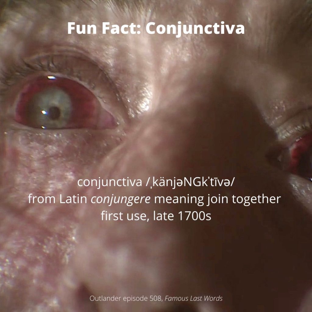
Anatomy Def:  Jocasta’s Blindness – painful pressure in the eye

Outlander Def: Jocasta’s Joy – pain relieved by smoking hemp flowers

Greetings, anatomy students!

Let’s learn about the disease that robbed Jocasta Cameron of her eyesight: Glaucoma. Yep, that’s the one. Glaucoma (glaw-koh-muh), is a word derived from the Greek glaukommatos meaning “gray-eyed.”

But, first, a wee divergence for a quick anatomy lesson of the eye which, in my opinion, is the body’s most anatomically elegant structure. This brief review is necessary to glean any useful understanding about this disease. 

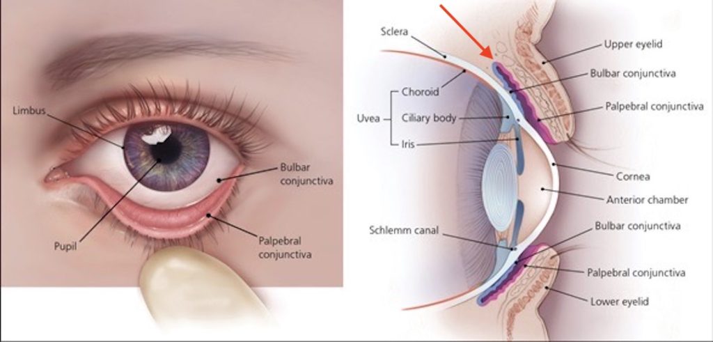
Eyeball: The next image shows a horizontal-section of the right eyeball (globe) as seen from above. The diagram shows in succession (from L to R) the cornea, iris, and lens; the opening in the middle of the iris is the pupil (red arrow). 

Two Chambers: The eyeball contains two important spaces (peachy-pink in the image, below). A large area behind the lens is the vitreous chamber – it is shaped like a sphere. In front of the lens is the smaller anterior chamber – it is shaped like a disc. The pupil supplies an open route between the two spaces.

Optic Nerve: At the back of the globe is the large optic nerve, an extension of the brain into the the eyeball. It is formed by filaments from cells of the retina. When photons (packets) of light excite retinal cells, they send signals via the optic nerve to the brain where it interprets the signals as the object viewed. 

Vitreous Chamber: The vitreous chamber is filled with the vitreous body, a jelly-like material that becomes more fluid with age. 

Anterior Chamber: The anterior chamber is filled with a watery substance known as aqueous humor.

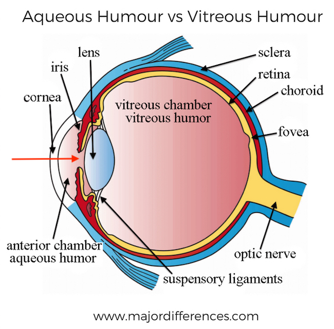
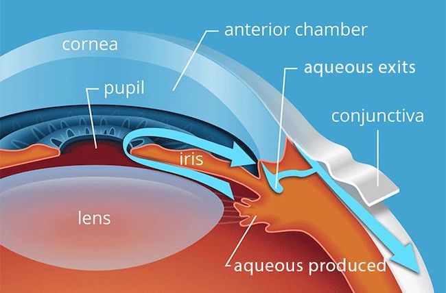
Aqueous Humor: Aqueous humor is similar to blood plasma; it fills the anterior chamber (next image). Aqueous humor is produced 24 x 7 –  continuously made by tissues near the lens and constantly drained by tissues near the iris.  

Blue arrows show the direction of flow of the aqueous humor from its production site, through the pupil into the anterior chamber, and then into nearby exits to be carried away.

A very quick lesson, indeed!

Learn more about the eye in not one, but five anatomy lessons! 

Q:  Why so many lessons, prof?  

A: Because the eye is exceedingly complex! 🤓

The lessons are:

*Anatomy Lesson #29: The Eyes Have It!

*Anatomy Lesson #30: Aye, Eye, The Eyes!

*Anatomy Lesson #31: An Aye for an Eye!

*Anatomy Lesson #32: A Real Eye Opener!

*Anatomy Lesson #33: Eyemax!

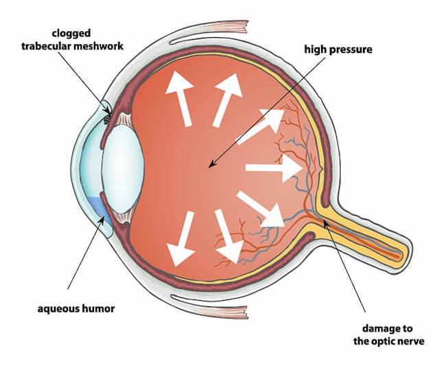
Glaucoma: When a person suffers from glaucoma, aqueous humor is either overproduced or the production is normal but it does not drain properly, or both may be culprits.  If the fluid cannot drain, not only does it fill the anterior chamber, it also backs into the vitreous chamber. 

Symptoms: So, what happens when aqueous humor builds up? Well, several things, usually over time.

    • Eye pain or pressure
    • Headaches
    • Rainbow-colored halos around lights
    • Nausea and vomiting
    • Red eyes
    • Loss of side (peripheral) vision
    • Low vision, blurred vision, narrowed vision (tunnel vision)
    • Blind spots
    • Blindness
  • Not-So-Fun-Fact: Glaucoma is the leading cause of blindness in people over 60.

Comparison:  Let’s  compare a normal eye (panel A) with a glaucomatous eye (Panel B) so we understand how things work.

    • Panel A shows normal flow and removal of aqueous humor
    • Panel B shows aqueous humor accumulating in anterior chamber and in vitreous chamber because there is too much or it does not drain properly.

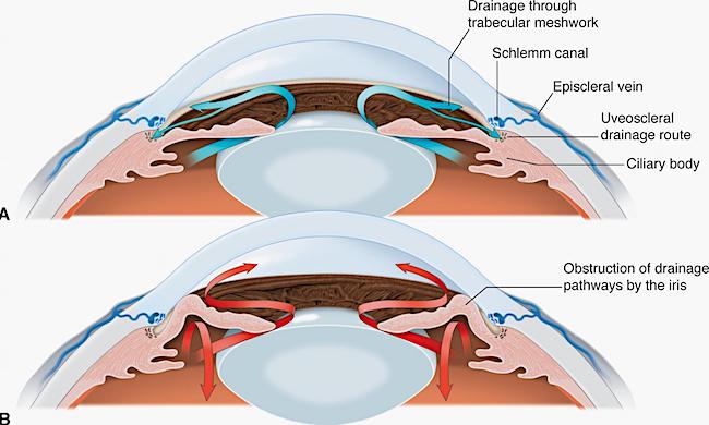
Jocasta: Now, we come to the root of Jocasta’s problem (next image). As intraocular pressure builds because of too much aqueous humor, it presses against the fragile retina (yellow layer) and optic nerve causing irreversible damage.  This means the normal response of the retina to photons of light and subsequent transmission to the brain are diminished or cease altogether. Such changes are typically accompanied by misery and pain.

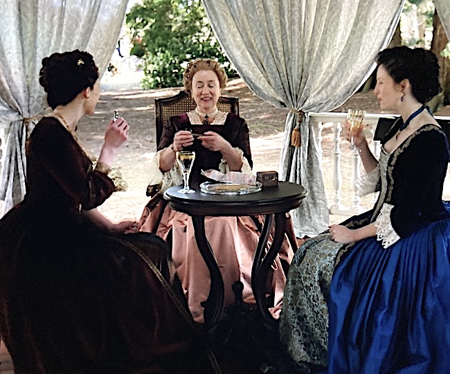
Unfortunately, Claire lacks current methods and medicaments to treat glaucoma. She can provide, however, hemp flowers for a wee pipe!

Now, does this really work?

Yes. Yes. It does.

Smoking hemp decreases intraocular pressure, thus relieving the pain and discomfort of pressure against retina and optic nerve.

Note:  Hemp also reduces blood pressure throughout the entire body, a side effect which might not be beneficial to all individuals, depending on their general health.

But, our Auntie Jo feels better and is even giddy!

(I love those goblets! 😍)

I should clarify that research showing the effects of smoking hemp on glaucoma wasn’t proven until the 1970s, a few years after Claire’s return to the eighteenth century. In her defense, the good doctor may have learned anecdotally about such benefits before she took her long and winding road back to Jamie.

Etiology (cause) of Glaucoma: Although we speak of glaucoma as a single disease, it is not. Glaucoma is a group of eye diseases that cause vision loss and blindness. The two most common types are:

    • Open angle – drainage routes are blocked – progresses slowly
    • Closed angle – drainage routes are blocked –  progresses more rapidly and is usually more severe

Treatments: Treatments include eye drops and surgery. These  may be used separately or together for treatment.

    • Eye drops – reduce intraocular pressure. Glaucoma must be detected early to be effective
    • Laser Eye Surgery – Improves drainage via normal routes
    • Tube Shunt Surgery – Small device is placed in eye to augment fluid drainage
    • Trabeculectomy – Delicate surgery creating new drainage ports 

Claire’s solution of hemp smoking is entirely different than what she does in the book. Read on and you will see why.

Read about Claire’s gutsy and unorthodox treatment of Jocasta’s glaucoma in Diana’s sixth big book, A Breath of Snow and Ashes. Here, she pierces the eyeball with a carpet needle, allowing excess fluid to seep out and relieve the unrelenting pressure and pain.

(Pssst…..  The escaping fluid is aqueous humor, not vitreous 🤫)

Even as I talked, I’d got out the tiny spirit lamp I carried in my case. The fire had been allowed to burn down on the hearth, but there were still live coals; I bent and lit the wick, then opened the needle case I’d taken from the sitting room and abstracted the largest needle in it, a three-inch length of steel, used for mending carpets.

“You aren’t …” Jamie began, then broke off, swallowing.

“I have to,” I said briefly. “There’s nothing else. Hold her hands.”

He was nearly as pale as Jocasta, but he nodded and took hold of the clutching fingers, pulling her hands gently away from her head.

I lifted away the linen bandage. The left eye bulged noticeably beneath its lid, vividly bloodshot. Tears welled up round it and overflowed in a constant stream. I could feel the pressure inside the eyeball, even without touching it, and clenched my teeth in revulsion.

No help for it. With a quick prayer to Saint Clare—who was, after all, patroness of sore eyes, as well as my own patron saint—I ran the needle through the flame of the lamp, poured pure alcohol onto a rag, and wiped the soot from the needle.

Swallowing a sudden excess of saliva, I spread the eyelids of the affected eye apart with one hand, commended my soul to God, and shoved the needle hard into the sclera of the eye, near the edge of the iris.

…  I withdrew the needle carefully, though as fast as I could. Jocasta had stiffened abruptly, frozen stiff, hands clawed over Jamie’s. She didn’t move at all, but made small, shocked panting sounds, as though afraid to move enough even to breathe.

There was a trickle of fluid from the eye, vitreous humor, faintly cloudy, just thick enough to be distinguishable as it flowed sluggishly across the wet surface of the sclera. I was still holding the eyelids apart; I plucked a rag from the goldenseal tea with my free hand, squeezed out the excess liquid, careless of where it went, and touched it gently to her face. Jocasta gasped at the touch of the warmth on her skin, pulled her hands free, and grasped at it.

I let go then, and allowed her to seize the warm rag, pressing it against her closed left eye, the heat of it some relief.

Now, you might think this extreme, but the only other remedy Claire could offer would be to remove one or both of Jo’s eyes!

Well done, Claire! 👏🏻👏🏻👏🏻

See Claire treat Auntie’s glaucoma in Outlander episode 605, Give Me Liberty!

I think Aunt Jo might welcome the hemp just a wee bit more than that needle jab, eye? 😉

The deeply grateful,

Outlander Anatomist

Follow me on:

Photo Credits: Starz, www.aao.org, www.allaboutvision.com, www.majordifferences.com, www.ohioeyesurgeons.com, www.2020pittsburgh.com

Fun Fact: Anatomy of Ian’s Uniform!

Hallo, Outlander fans. Welcome to today’s Fun Fact: Anatomy of Ian’s Uniform! 

This Fun Fact is especially appropriate for Americans as May 16th is  also our national Armed Forces Day!  Pretty timely, no?

If you are like me, you are fascinated with Ian’s uniform in Outlander episode 512, Never My Love, the splendid finale of Season five! In Claire’s dissociative dreamscape, Ian arrives in full dress uniform for Thanksgiving dinner at the Fraser home! 🦃

I wanted to know more about Ian’s uniform, so I turned to Edward Maloney, Lieutenant Colonel, US Army (retired) for assistance. Know that LTC Maloney is also a faithful, long-time fan of Outlander books and show and was more than willing to share his decades-long expertise in this matter.

Just so you know, Colonel Maloney’s former unit is the 101st Airborne Division of the US Army. Below is its beautiful and dramatic Distinctive Unit Insignia which reads “Rendezvous With Destiny!”

Addendum: I just learned “Hang Tough and Currahee!” is the Battle Cry of the 101st Airborne!  Hang tough means “in your parachute harness” and “Currahee” is from the Cherokee word meaning “We stand alone together.” (A good thought for soldiers trained to fight surrounded.)

Thank you, Lieutenant Colonel Maloney, for your service!


So, grab a cuppa (or your fav beverage) and let’s learn about the anatomy of Ian’s uniform, courtesy of LTC Maloney!

Ian’s uniform indicates he belongs with the Infantry, the oldest branch of the US Army. When was this branch formed? Turns out, very close to the date  depicted in the Outlander S5 finale! 

On 14 June 1775, the Continental Congress authorized ten companies of riflemen, the first infantrymen.  Nine years later, the First American Regiment was constituted on 3 June 1784 and it was the 3rd Infantry. Currently, well over two hundred years old, clearly the Infantry is a distinguished branch of the US Army!

So, follow the colored arrows in the below images to discern the anatomical features of Ian’s uniform. (psst…The following image appears twice so you don’t have to keep moving up and down to follow the arrows and explanations!)

Let’s get on with the dissection! 😉

Blue Arrow:  A branch insignia on Ian’s L lapel displays two gold crossed muskets, overlying a disk of Saxony Blue. This insignia is unique to infantry and no other branch of the US Army is allowed this distinction.

The crossed muskets are vintage 1795 Springfields, the first official US shoulder arm made in a government arsenal:

    • caliber .69
    • flint lock
    • smooth bore
    • muzzle loader


Yellow Arrow: Above his L breast, Ian wears a Combat Infantryman’s Badge, unique to those who have served in combat. It is a single flint lock musket on a blue background. 

Green Arrow: Just above Ian’s L breast is a line of Decoration & Award Ribbons, also known as “fruit salad” or “Travel Ribbons.” These are worn in lieu of larger full-size or miniature medals which are awarded for service.  Here, the Ribbon is a line of Ian’s Mohawk beads! 

Gold Arrow:  Over Ian’s L breast is The Maltese Cross with a Bull’s-eye surrounded by a wreath, known as the Expert Marksman Badge. This badge is unique to Army and Marine Corps, although the designs differ. The suspended bars underneath the badge are added for each weapon the soldier qualifies as an expert, such as pistol, rifle, etc. 

White Arrow:  Worn on Ian’s L sleeve, the chevrons signify a soldier’s rank. Two chevrons indicate Corporal, the lowest Noncommissioned Officer Rank (NCO) who leads an infantry fire team. 

Violet Arrow: Ian’s service cap bears the US Coat of Arms. It is backed on a brass disk and in the case of infantrymen, backed by a Saxony Blue disk. 

Khaki Arrow: Ian’s Regimental Coat of Arms is worn above his R breast.  This insignia will indicate his permanent regiment not necessarily the one to which he is currently assigned. 

Tan Arrow: Also worn above his R breast, this insignia indicates a Unit award such as Presidential Unit Citation, Distinguished Unit Citation, etc. 

Next image,  we see Ian’s left shoulder!

Aqua arrow:  Indicates the Distinctive Unit Insignia which is usually a variation of the regiment’s coats of arms but unique to each regiment. This one looks like the 2nd Infantry Regiment but the colors are altered. 

Red Arrow:  L shoulder – Shoulder Sleeve Insignia  indicates the current unit of assignment – Ian serves with a Native American unit, the Mohawk. this insignia was created by Outlander.

And finally, Ian’s infantry blue cord or fourragere (below) is a military decoration worn over the right shoulder of all infantry-qualified US Army soldiers. 

Ian’s fourragere from afar.

And, a closeup. This is Ian’s fourragere in light blue, (dubbed “Infantry Blue” by the US Army), worn under the right shoulder and under the right epaulette of a US Army infantry soldier’s dress uniform jacket.

The cord is composed of a series of alternating left and right half knots that are tied around a leader cord to form a “Solomon bar”.

Fitting that Ian should wear his dress uniform for Thanksgiving, even if that bird is just an illusion!

In summary, Ian’s uniform tells us he is:

    • Infantryman
    • Decorated soldier
    • Served in combat
    • Expert marksman
    • Corporal of an infantry fire team
    • Bears US Coat of Arms for infantrymen on his service cap
    • Wears his permanent regiment’s Coat of Arms
    • Decorated infantryman
    • Member of a distinct infantry unit
    • Member of Native American unit
    • Qualified infantryman

Whew! I don’t know about you, but I am thoroughly impressed with warrior Ian!

And, I am deeply grateful for the men and women who serve in the US Armed Forces.

I could not have done this fun Fact without the aid of LTC Edward Maloney, US Army. Thanks to his expertise for this brief lesson exploring the anatomy of Ian’s US Army insignia!

I hope all Outlander fans, worldwide, will express their gratitude for the warriors who daily protect them, their families, and their homelands. Please take a quiet moment to honor them!

Disclaimer:  If there are any glitches in the insignia descriptions or attributions in this Fun Fact, the fault is entirely my own for not expressing the information with precision.

The deeply grateful,

Outlander Anatomist

Follow me on:

Photo and Video Credits: Sony/Starz; Lt. Col. Edward Maloney; www.wikipedia.com; www.amazon.com

Fun Fact: Conjunctiva

Anatomy Def: Conjunctiva is a clear membrane covering part of the eyeball and lining inside of eyelids.

Outlander Def: Roger’s bloodied and abused tissues as he gazes at the world through his burlap hood!!! 😥

Learn about the conjunctiva in Anatomy Lesson #30, Aye, Eye – The Eyes, Part 2. 

Conjunctiva is a clear, thin mucous (adj.) membrane of the eye. It is a mucous membrane because, indeed, it contains glands and cells which secrete mucus (n.). This sets it apart from serous membranes that do not secrete mucus.

The conjunctiva has two parts:

    • Bulbar conjunctiva: Covers front part of sclera (“white” of the eye)
    • Palpebral conjunctiva:  Lines inner surfaces of eyelids 

Is the conjunctiva useful? Oh, indeed it is!  👏🏻👏🏻👏🏻 It performs two important functions: 

      • Produces mucus and tears to help lubricate the surface of eye
      • Protect against microbes entering the eye which is vulnerable to the outer world

The conjunctiva contains many small blood vessels that are not especially noticeable unless stimulated to dilate. Dilation allows for increased tear and mucus release. What might cause such a response?

    • Allergens, such as pollen
    • Irritants, such as air pollution, makeup, cosmetics
    • infections
    • Contact lenses
    • Fatigue
    • Hypoxia (reduced oxygen supply)

But, Roger’s conjunctivae are bloody! Why??? This occurs because strangling increases pressure in the conjunctival blood vessels and they swell. The vessels then burst from straining for air and against the rope!

Slowly rewatch the color version of the hanging; you will see blood blooming during his conjunctival bleed. Or, more precisely, subconjunctival bleed, as blood collects between conjunctiva and sclera! 

The special effects are excellent, btw! In Rik’s recent Q and A, he explains he was fitted with special contact lenses and thought CGI was also added. I agree, as I see an increase in conjunctival blood during the footage.

Ergo: If Roger every gets his hands on William Buccleigh MacKenzie!!! 💪🏻💪🏻💪🏻

Fun Fact: Some people worry that a contact lens could wander towards the back of the eyeball and reach the brain. For several anatomical reasons, this doesn’t happen. One major reason is the conjunctiva folds on itself forming a closed pouch all the way around the eyeball. The figure explains 👇🏻.

The L panel shows the bulbar and palpebral conjunctiva from a frontal view.  Note: the bulbar conjunctiva stops at the edge (limbus) of the cornea. It does NOT cover the cornea!

The R panel shows a vertical section through the eyeball. The bulbar conjunctiva is shown as blue and the palpebral conjunctiva is red. A red arrow marks the pocket where the two layers meet. This blind pocket encircles the entire eyeball. Hence, a lost contact lens can only travel as far as the blind pocket. Want to know what to do about a “lost” contact lens? This is a pretty good website!

Try This: Gaze into a mirror.  Locate the clear membrane overlying your sclera. Can you see its tiny blood vessels? This is your bulbar conjunctiva. Gently pull down on the lower eyelid. See the glistening surface? This is your palpebral conjunctiva. Got it? Good job!

Herself explains Roger’s horrific ordeal in The Fiery Cross. Accurate details and explanations accompany her descriptions coupled with fantastic story-telling.  Read the books, folks! 🙏🏻

The others had strangled, slowly. The bodies were twisted, held by their bonds in the final postures of their struggle. One man—one body—was cut down as I rode up, and carried past me in the arms of his brother. There was not much to choose between their faces, each contorted, each darkened in its separate agony. They had used what rope was to hand; it was new, unstretched. Roger’s toes trailed in the dust; he had been taller than the rest. His hands had come free; he had managed to hook the fingers of one hand beneath the rope. The fingers were nearly black, all circulation cut off. I couldn’t look at his face at once. I looked at Brianna’s, instead; white and utterly still, each bone and tendon set like death.

See Roger’s bloody conjunctivae in Outlander, episode 508, Famous Last Words!  ❤️ Roger!

The deeply grateful,

Outlander Anatomist

Follow me on:

Photo credits: Sony/Starz; www.aafp.org