
Hello, anatomy students! Today’s Anatomy Lesson #37 covers the ginormous topic of mars and scars, better known as wound healing. A mess of wounds and scars appear in the Outlander books and the Starz series so let’s learn from this perfect anatomical smorgasbord.
You will recall from Anatomy Lesson #35: Outlander Owies! Part One and Anatomy Lesson #36: Outlander Owies! – Part Deux! that pathology is the study of abnormal anatomy. Well, it turns out that wound healing also belongs in the realm of pathology. Our lesson will examine healing of closed and open wounds.
Surprise! Jamie is our model because he has more mars and scars than anyone else of Outlander fame! Let’s also enjoy a drinking game because Jamie’s wounds inspire one to drink more than any others, I ken! Down a wee dram each time you read “Och! Jamie’s puir…”
Drams in hand? Let’s begin…
Blood: Because blood is essential for healing and scar formation, it requires a brief brief mini-lesson of its own. Blood is a connective tissue (yes, it is!) composed of fluid plasma, in which two classes of blood cells are suspended: red blood cells (RBCs or erythrocytes) and white blood cells (WBCs or leukocytes). Blood also contains cell fragments known as platelets (thrombocytes) along with many other suspended or dissolved substances.
RBCs arise in bone marrow as nucleated cells but lose their nuclei just before entering the blood stream. Image A shows a 3-D SEM (Scanning Electron micrograph, Anatomy Lesson #34) image of a blood vessel containing blood cells. Understand that the colors in Image A were computer generated. RBCs are flat, biconcave discs (red in Image A), whose shape and lack of a nucleus allots them maximum oxygen-carrying capacity; think of them as tiny flat bags filled with hemoglobin (Hgb). As RBCs pass through lung capillaries, oxygen binds to Hgb molecules, turning RBCs bright red. As RBCs reach capillaries of other body regions, Hgb molecules release their oxygen burden into the tissues and the RBCs turn a dark, deep red color. Such color changes are important in wound formation and healing.
WBCs are the round, gray fluffy balls in Image A. They arise in bone marrow but retain their nuclei. There are five classes and several subclasses of WBC. All WBCs serve various defensive functions; more about these later in the lesson.
Platelets, which do not appear in Image A, play a crucial role in blood clotting.
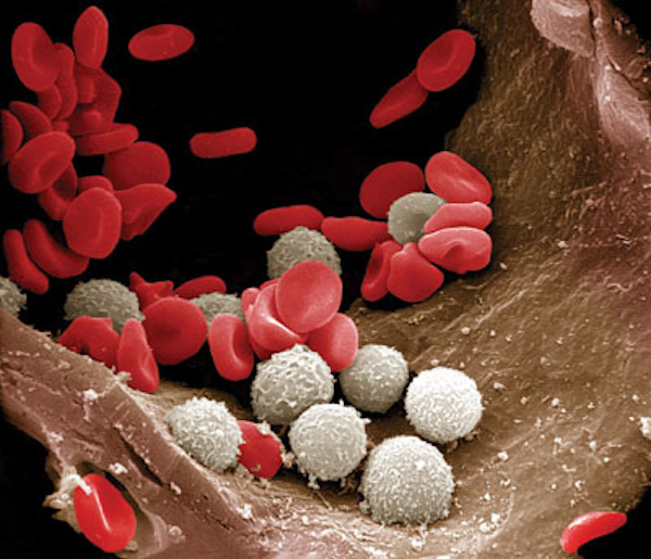
Image A
That was a “quickie” blood lesson. Now, on to healing processes!
The outline for today’s Lesson is:
- Healing Closed Wounds
- Trauma
- Contusion Development
- Contusion Resolution
- Healing Open Wounds
- Trauma
- Hemostasis
- Inflammation
- Proliferation
- Remodeling
- Scars
Healing Closed Wounds: Closed wounds are injuries wherein the skin remans unbroken. The contusion will serve as today’s example of a closed wound, although simple bone fractures are another. Anatomy Lesson #35 explained that a contusion is the medical term for a bruise. Bruises form in subcutaneous tissues (Anatomy Lesson #5: “Claire’s Skin” – “Ivory, Opal and White Velvet.”) where they are often visible through the skin, but they can also form in deep organs such as muscle. Symptoms may include a bump, a hard knot and tenderness. And, bruises do not blanch under pressure. A number of steps occur to form and heal a bruise including:
- trauma
- contusion development
- contusion resolution
Let’s look at each of these steps.
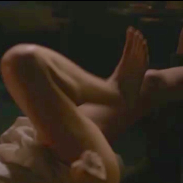
Trauma: Contusions are caused by blunt force trauma wherein the skin is not broken. Claire’s foot presents an formidable blunt force directed at Jamie’s face (Starz episode 109, The Reckoning)! Weil, now, she did warn him not to belt her. This lass is not going down without a stramash!
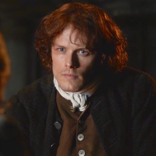
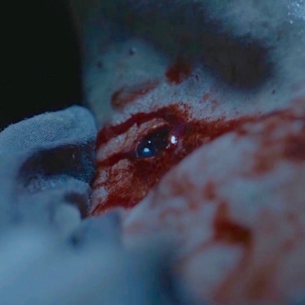
Contusion Development: Contusions pass through an impressive array of colors during development and healing. The first color is redness which occurs because blunt trauma bursts local capillaries allowing oxygen-ladened RBCs (bright red) to spill into nearby tissues. Although time has passed since Claire’s kick, the skin overlying Jamie’s right bony orbit (Anatomy Lesson #30: Aye, Eye – The Eyes Part 2!) shows the redness typical of a fresh contusion (Starz episode 110, By the Pricking of My Thumbs). Or, mayhap he is just blushing because the Duke of Sandringham has a certain fondness for his hindquarters? <G> Och! Jamie’s puir right eyelid! Have a wee dram.
Redness is followed by purple and blue discoloration as Hgb molecules inside the spilled RBCs release their oxygen; the RBCs turn dark red-purple as do the tissues they occupy. Next image is a good example of purple/blue discoloration (Starz episode 116, To Ransom a Man’s Soul). Och! Jamie’s puir left shoulder! (And, another dram). A quote from Outlander book reminds us of the vicious swipe that produced such discoloration!
The spot on his left side where the mallet had struck was an ugly contused swelling…I bit my lip, feeling gingerly down the swell of his biceps. He had one of the worst bruises I had ever seen; a huge mottled splotch of purple-blue—but I was fairly sure the arm wasn’t broken.
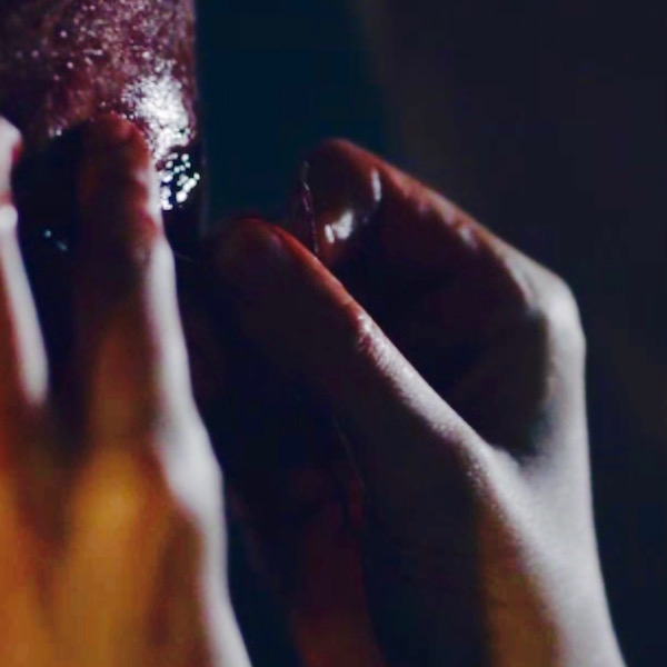
Next, a contusion exhibits the classic black and blue discoloration as spilled RBCs rupture and release their iron-containing Hgb into the soft tissues of the injured area. Och! Jamie’s puir left fingers; they argued with that damnable mallet and lost (Starz episode 116, To Ransom a Man’s soul)! Quick! Take another sip!
Contusion Resolution: Yay! It rhymes! Now, the contusion begins to heal assuming first, green and then, yellow hues. These color changes occur because cells known as macrophages (Greek makros meaning large + phagein meaning to eat) devour freed Hgb, Macrophages break the Hgb into the green compound, biliverdin, (Latin meaning green) which turns the tissues an icky green color. This is followed by yellow hues as the biliverdin is metabolized into the golden chemical, bilirubin. As macrophages clear the last of the debris from the bleed, the yellow fades and if the contusion isn’t too profound, normal coloration and function are restored.
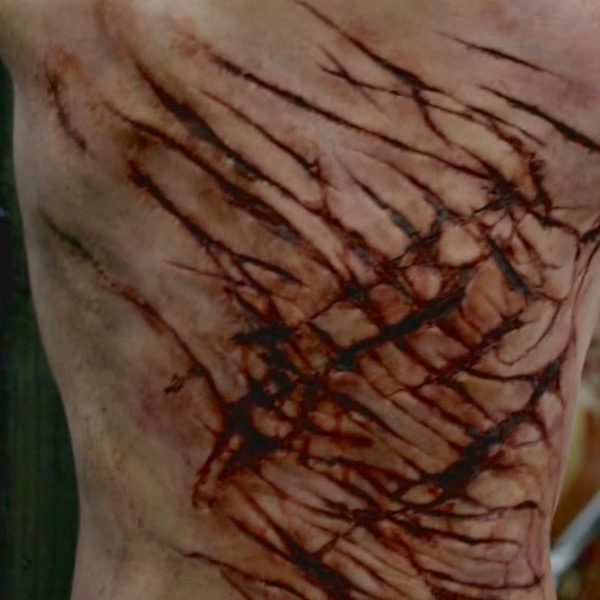
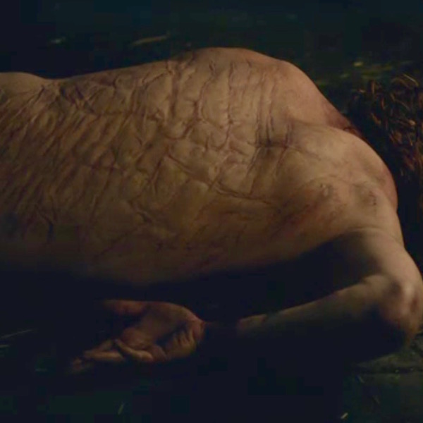
Ergo, changes oin Hgb molecules form the basis for color changes of a healing contusion. A single bruise can simultaneously display all of the above colors because the amount of spilled blood varies in different areas and the stages of healing overlap. Here is a gut-wrenching example (Starz episode 106, The Garrison Commander)! Och! Jamie’s puir back! (Another swallow!) Gah! Skin near the lash marks is red, purple, blue, black, green, and yellow! Herself writes in Outlander book:
Dougal grimaced. “A pitiful sight, it was, too—still raw, no more than half-healed, wi’ the weals turned black and the rest yellow wi’ bruises. The thought of a whip comin’ down on that soreness was enough to make me blench, along wi’ most of those watching.”
Now that we understand the healing of a closed wound, let’s consider open wounds.
Open Wound Healing: Open wounds entail skin breaks; abrasions, lacerations, and incisions (Anatomy Lesson #35) are good examples of open wounds. Such injuries undergo a cascade of events during wound healing.
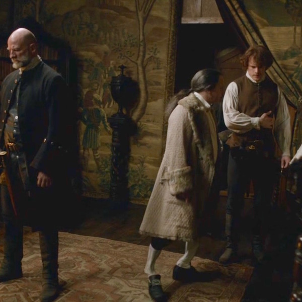
Trauma: First, open wound healing requires trauma; finding good examples of open wounds in Outlander is a no-brainer – as they are pretty much everywhere! This is a fine one: Starz episode 110, By the Pricking of My Thumbs, shows the type of trauma-drama awaiting highlanders that mess with Jamie! The MacDonald lads hurl nasty insults and then attack Jamie. Thalla gu h-Iort – “to St Kilda with ye!” bellows Jamie. With a quick dirk-jerk, Jamie gifts one lad with an incision of his right hamstring tendons! Young MacDonald came to harm, E-I-E-I-Oooooh; that stings!
Open wound healing: Following trauma, the body immediately springs into action and initiates the following four steps of open wound healing (Image B):
- Hemostasis
- Inflammation
- Proliferation
- Remodeling
Wake up, students! Let’s examine each of these four steps.
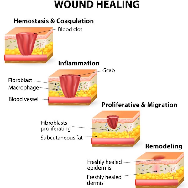
Image B
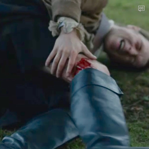
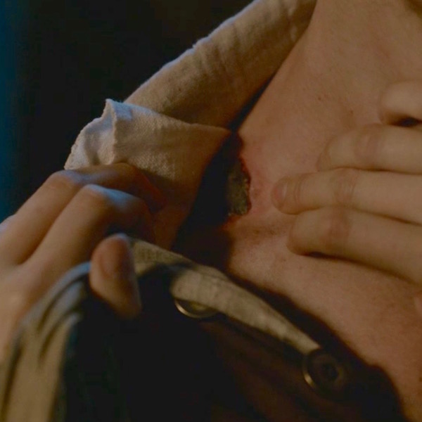
Hemostasis: A fancy word for blood clot, hemostatsis comes from the Latin for blood + Greek for stop. Hemostasis is typically the first step in open wound healing. If trauma ruptures blood vessels, they bleed (duh!) and then undergo spasm to reduce bleeding. Next, platelets stick to the injured site forming a temporary platelet plug, followed by a complex, ten-step!!! cascade of events leading to blood clot formation. If all steps work normally, a clot stoppers each damaged blood vessel to halt blood loss. The dusky red plug of Jamie’s gunshot wound is a massive blood clot formed by the process of hemostasis (Starz episode 101, Sassenach). Och! Jamie’s puir right trapezius muscle! Slàinte!
And, just to keep us honest and abreast of Outlander time line, a common 17-century word for blood clot is grume!
A blood clot with its seeping fluids usually hardens into a scab, the body’s version of a Band-Aid! This firm shell protects the wound from infection and desiccation (drying). Tissues under a scab are repairing so best to leave it alone. If you pull it off, the healing process will be prolonged. Jamie kens better than to tear that scab off his gunshot wound! Nurse Claire will skelp his arse if she catches him doing that (Starz episode 103, The Way Out) From Outlander book:
“That’s good,” I said, clearing my throat of some obstruction that seemed to have lodged there. “It is healing well; it’s scabbed over nicely, and there’s no drainage at all. Just keep it clean, and don’t use the arm more than you must for another two or three days.” I patted the undamaged shoulder, signifying dismissal.
Inflammation: Hemostasis is quickly followed by acute inflammation, a rapid but brief response that is not the same as infection, although both may occur simultaneously. Just so you know, the body also experiences chronic inflammation (e.g. RA), a prolonged response that lies beyond today’s lesson content. So, let’s investigate acute inflammation.
About 2,000 years ago, the Roman, Celsus, described four signs of acute inflammation (rubor, tumor, calor, dolor). The fifth sign (functio laesa), was described centuries later by Rudolf Virchow, father of modern pathology. The signs, in Latin followed by their English equivalents, are:
- Rubor (redness)
- Tumor (swelling)
- Calor (heat)
- Dolor (pain)
- Functio laesa (loss of function)
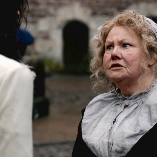
Acute inflammation is the topic of a terse tête-à-tête between Mrs. Fitz and newly arrived Claire (Starz episode 102, Castle Leoch). Claire declares she doesn’t want Jamie’s gunshot wound to become infected. Oops, she means inflamed! Why did Claire change her words? Because, she kens that the terms infected/infection, meaning invasion by micro-organisms, will not be accepted for another 140 years or so! Herself enlightens us from Outlander book:
“But he’s hurt. He was shot yesterday and stabbed last night. I bandaged the wound for riding, but I didn’t have time to clean or dress it properly. I must care for it now, before it gets infected.” “Infected?” “Yes, that is, I mean, inflamed, you know, with pus and swelling and fever.” “Oh, aye, I know what ye mean. But do ye mean to say as ye know what to do for that? Are ye a charmer then? A Beaton?” “Something like that.”
Hee, hee – more like a WW II combat nurse. If only Mrs. F. knew!
The five signs of acute inflammation are caused by dramatic changes in small blood vessels, WBC distribution, and chemical mediators. These changes are very complex so suffice it to say that intact blood vessels near the injury become leaky such that plasma and some types of WBCs pour into the injured tissues. The increased blood flow causes redness. Leaked plasma causes swelling. Freed WBCs release chemical mediators that initiate a host of tissue changes including more redness, swelling, pain, and heat. These responses are designed to eliminate the cause of cell injury, to remove damaged cells, and to initiate tissue repair. Because the injured area hurts, it is hard to use, so it undergoes loss of function. Got it? Grand!
Back to the present (or past): here’s a great example of an injury that leads to acute inflammation. Just before Jamie’s dirk-jerk, he is stabbed by one the MacDonald lads during the big Mac-Attack (Starz episode 110, By the Pricking of My Thumbs). Och! Jamie’s puir left side! Time for another swallow!
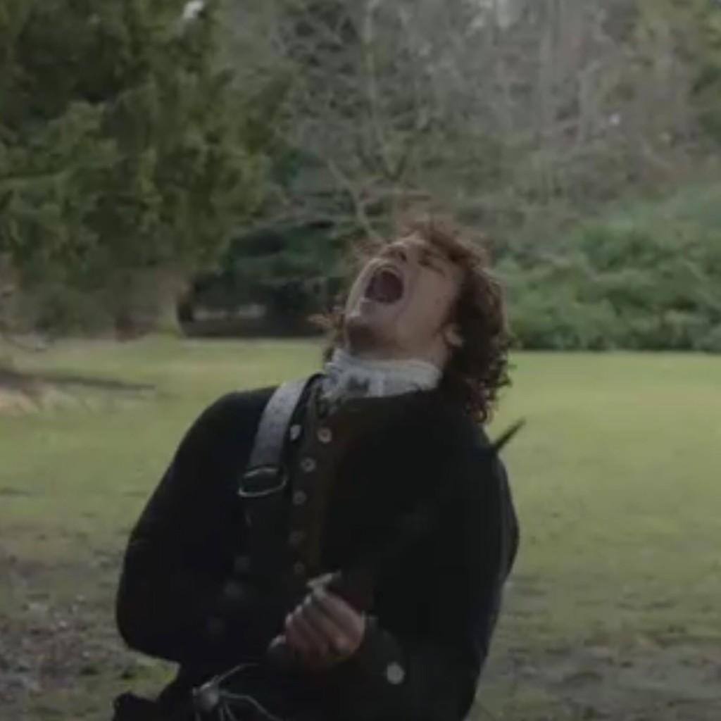
Turning to Claire for a wee bit of TLC (she isna very accommodating) and suturing…well, at least he gets stitched <G>, we see signs of acute inflammation in Jamie’s wound (Starz episode 110, By the Pricking of My Thumbs)? See the redness? See the puffy, swollen wound margins? It hurts even before not-a-closed-mouth-woman, Claire, jabs him with her needle! And, the wound will feel warm to the touch. Och, Jamie’s puir inflamed incision wound! Getting dizzy yet?
Four of the five cardinal signs of acute inflammation accounted for! Where’s the fifth?
Here’s the fifth sign of acute inflammation: Jamie grips his newly-stitched left side to protect and support it as Colum gives the Highlanders Holy Hell (Starz episode 110, By the Pricking of My Thumbs)! This is loss of function: the wound hurts too much to use and the pain is intended to stop the victim from using the wounded area while it heals. Och! Jamie’s puir left side! Another gulp! All five signs of acute inflammation present and accounted for! Now back to the steps of wound healing.
Proliferation: So, the wound has bled and is inflamed, what’s next? The wounded tissues undergo proliferation, a complex process (yes, another one) involving three parts: angiogenesis, fibroplasia, and re-epithelialization (Image C). Whew – more terms! What do these words mean?
Angiogenesis means that new blood vessels grow into the wounded site to supply the healing tissues with oxygen and nutrients and remove waste products (red arcs and loops in Image C).
Fibroplasia means that fibroblasts (connective tissue cells) make gobs of new collagen and other structural proteins to fill the gap left by an open wound. Although laid down in a haphazard fashion, the new collagen fibers provide a structural framework for the repairing tissues (represented by ellipsoid cells and brown and yellow stacks in Image C).
Re-epithelialization means that new epidermis (Anatomy Lesson #5: Claire’s Skin – Ivory, Opal and White Velvet and Anatomy Lesson #6: Claire’s Hair – Jamies Mane or Jesus H. Roosevelt Christ!) regrows to cover the gap caused by the wound (green bricks in Image C). This fascinating step requires old epidermal cells to divide and new epidermal cells to crawl over and cover the breach (blue bumps in Image C). Then, fibroblasts (acting like muscle cells), grip the wound edges and contract, pulling the rim inward and puckering it (not shown in Image C).
If the wound is minor, the three steps of proliferation occur rapidly and the wound closes with little trace. If the wound is deep and/or wide, then proliferation results in a scar (see below).
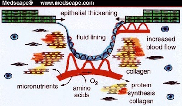
Image C
Remodeling: Finally, we arrive at remodeling, the fourth step of wound healing! Here, the randomly-arranged proliferated collagen is replaced with new collagen fibers that add strength by orienting along stress lines. Near the end of remodeling, scars contract and become smaller and paler. Image D shows that remodeling takes between 21 days for a minor wound and up to 2+ years for significant injuries! NOTE: This graph skips bleeding as the first step in open wound healing.
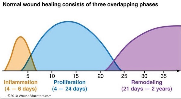 Image D
Necrosis: Before we move to scars, please know this…..If some process interferes with establishment or maintenance of normal blood flow following open wound injury, then injured tissues may undergo cell death, a process known as necrosis.
Scars or Cicatrix: We’ll end this lesson with a discussion about scars, the remnant of a healed wound. The Latin (medical) term for a scar is cicatrix or cicatrice, and has been around since the 17th century. Scars (pl.) are known as cicatrices. With the exception of minor injuries, all wounds (e.g. accident, disease, surgery) result in some degree of scarring. We are familiar with scars of the skin but did you know that our internal organs also scar and the nature of such scars is often organ-specific (e.g. fibrosis of liver).
Let’s address some common wound and scar questions.
Why are wounds stitched? Nowadays, wounds are stitched, stapled, glued, and taped. These processes align and hold together the edges of a wound to minimize bleeding, exclude infectious organisms, and reduce scar size. First recorded in 3,000 B.C., suturing is the oldest of these procedures. Approximating the edges of a wound by any the above methods permits healing by first intention.
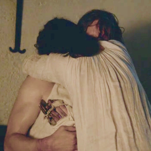
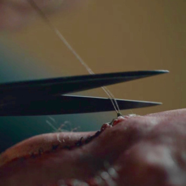
Claire’s careful needlework on Jamie’s smashed fingers approximates his lacerated skin and restores it’s continuity, thus promoting healing by first intention and minimizing scar formation (Starz episode 116, To Ransom A Man’s Soul). She also uses interrupted stitches so if one knot releases, the entire line of stitching does not unravel. Och! Jamie’s puir left fingers! Gulp! Gasp!
Claire stitches Jamie up so many times, she might have welcomed help from this amazing little fellow!
What is the red, grainy “stuff” that fills gaping wounds? If the edges of a wound remain agape or the epidermis is gone (think floor burn), then the gap fills with granulation tissue; this red, grainy “stuff” is a combination of new blood vessels and new collagen. Known as healing by second intention, this type of healing is slower than healing by first intention and usually forms larger scars (Image E).
 Image E
Have you heard the term, “proud flesh” (caro luxurians)? Noooo, I dinna mean the Kardashians, snort! Proud flesh is an oldish term for excess granulation tissue. Image F shows proud flesh, an overgrowth of granulation tissue that developed from a small finger cut. Such overgrowths are atypical but the image is useful because it clearly shows the red blood vessels and grainy appearance of granulation tissue.
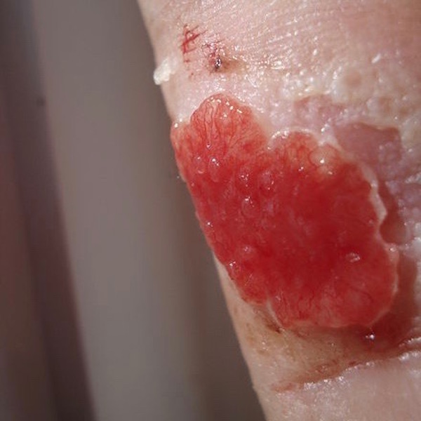
Image F
What is a scar? Scar tissue is remodeled collagen fibers that aligned in one direction for added strength. Unfortunately, scar tissue is not as functional as the tissue it replaces. For example, scar tissue of skin lacks hair follicles and sweat glands (Anatomy Lesson #6: Claire’s Hair – Jamies Mane or Jesus H. Roosevelt Christ!) and is less pliable than normal skin. Scars of heart muscle lead to loss of muscular power during cardiac contractions. Scars of liver are due to death of liver cells and formation of excess collagen – less tissue to preform the ten life-giving functions carried out by the liver. Then, there’s amazing bone that often heals without any structural or functional loss.
Why do scars turn white? Scars usually turn white within a year or two after injury. They do so after collagen remodeling and scar contraction is complete and the need for increased blood flow has diminished.
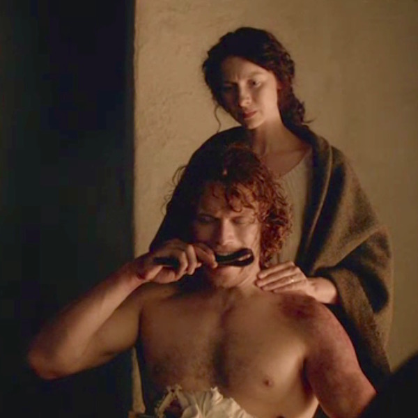
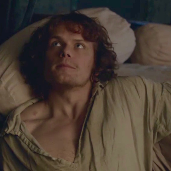
Why do some scars remain red or purple for a long time? Scars remain red or purple as long as WBCs of the area produce chemical messengers to promote increased blood flow. Thus, it may be months or even years before redness completely fades. A good example is the persistent redness of Jamie’s gunshot scar which is apparent months after the event. Oh, no! More sass from Claire who gives him a thorough Sassynach scolding for being a lousy laird (Starz episode 112, Lallybroch). Have ye ever seen a sweeter, more innocent face? Ha, ha! He enjoys a good tongue lashing now and then (hee hee!).
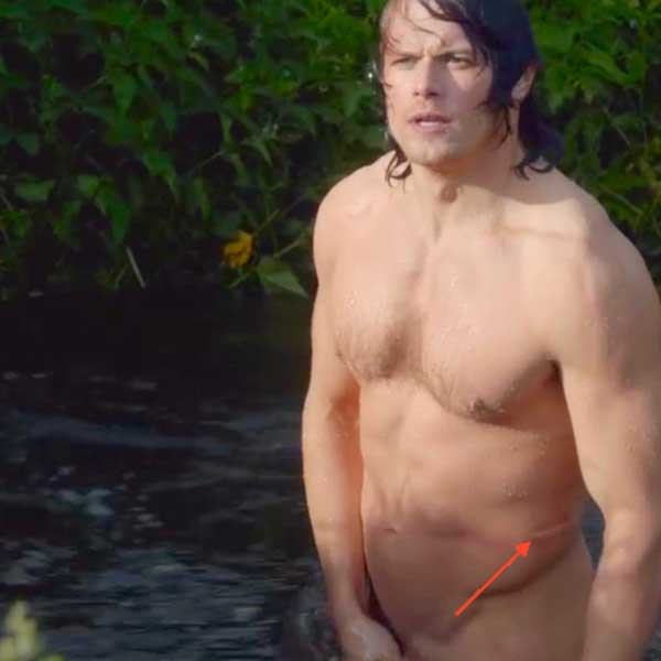
Here is a great … ah, erm, … I forgot what I was writing about! Gah! Oh, I remember! The scar from the sword swipe is white at the millpond even though Jamie’s earlier gunshot scar is red in the same episode (Starz episode 112, Lallybroch). Ahem, students!! The red arrow helps focus your attention on the white scar! Now this can occur because tissues in different regions of the skin may heal at different rates. More likely, it is because that bloody freezing burn contracted Jamie’s skin capillaries: reduced blood flow = paler skin! Och, Jamie’s puir left side! Are we woozy yet?
Why are Jamie’s lash scars so prominent? Back skin is thick and when lacerate, it should be sutured or stapled; unfortunately, neither of these procedures were available to Himself at the time of injury. His deep, wide lash wounds filled with granulation tissue and healed by second intention so the resultant scars are wider and more significant. BTW, his lash scars are also shiny and raised, features of hypertrophic scars, an abnormal healing process due to overproduction of collagen. His lash scars were probably designed for dramatic effect, as Jamie’s skin is an unlikely candidate for hypertrophic scarring. Och! Jamie’s puir back! Glass refill STAT!
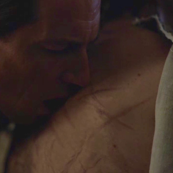
Jamie appears over and over in this lesson because the lad has suffered more than his fair share of mars and scars. Who is to blame for most of these owies? Why, that dark, dastardly devil, BJR, of course. Claire is constantly cleaning up that SOB’s messes! Sadist that he is, Jack-Jerk finds his handwork compelling and erotic (Starz episode 116, To Ransom a Man’s Soul)! Writer Ira Steve Behr explains:
To me, the line that was truest to Black Jack’s character was when he ripped open Jamie’s shirt and said with wonder, “How does it feel to be alive and wear so much dead flesh?”
I go on record as stating, au contraire, repugnant, reptilian Randall! Scars are NOT dead flesh, they are living tissue, the end game of the body’s ability to heal wounds. Scars are molded by our own private first responders; tiny “robots” that dart into action on our behalves. Let us be grateful that our bodies possess such marvelous repair mechanisms!
Shakespeare once wrote, “There’s nothing good or bad, but thinking makes it so” (Hamlet; Act two, Scene two, p. 11). Apparently Will never met BJR – as some handiwork is just plain E-V-I-L!
If you have yet to witness this riveting compilation of Black Jack’s dark and dirty deeds, watch this admirable video – created by Julia LaBlanc and posted by E. Jamie via YouTube. Just keep in mind it grimly showcases the depths of BJR’s depravity.
https://m.youtube.com/watch?v=6GRAsvTFH4s
Closing with my own wee Ode to BJR:
BJ is a fiendish auld cur; a fiendish auld cur is he,
He called for his whip and he called for his brand,
To abuse our darling Jamie!
(Git yer foul tongue off him!)
Hope you are still standing after all those drams! As for me: Hic! Thud!
A deeply grateful,
Outlander Anatomist
Photo creds: Starz, www.bioloby-igcse.weebly.com (Image A – SEM of blood cells), www.medscape.org (Image C – proliferation), www.surpassinc.com (Image B – four steps of wound healing), www.en.wikipedia.org (Image F – proud flesh), www.woundeducators.com (Image D – phases of wound healing), www.wisegeek.com (Image E – granulation tissue)
Love this and as physician myself, I watch the anatomy and its accuracy carefully in the show. I am glad to know I am not the only stickler for accuracy!
Hi fellow traveler! I too watch show and read books carefully! Overall everything is well done IMO!
I have read that animals that can regrow limbs or other parts do not scab or scar.
Indeed Caroline, some animals can do that. Is subject of research!
I said it before. I will say it again, you are too cool. Wish I had thought to put my knowledge to such fun use!
TY Cindy! Appreciate you reading the lessons!
Aside from a few typos (less than the average academic text book these days) an amazing accurate, engaging, and entertaining account of what is usually covered over several dry boring courses.
As a clinician I had no problem with either the gore of the historically accurate brutality of violence described in the books or the realism of the filmed scenes. I know folks who just dissociate or skim over it. But for those scholarly readers, your contribution adds to the phenomenon of the greater Outlander “experience.”
BRAVO! and 5 Starz!-lol ? to your awesome contribution.
Thank you for your kind comments! You’re right! There were typos and I think I’ve caught them. It’s nice to meet a fellow “traveler.”?
Spot on, Anatomist ! I am a Clinical Lab Scientist and thoroughly enjoyed your lesson on mars and scars. Besides being informative, it was HILARIOUS ! Thanks for a bright spot on my day !
Hi May! Thank you so much for your kind comments. So glad you liked the lesson. As a clinical lab scientist you realize there is so much more to wound healing than the lesson detailed. I aimed for the general public to understand. So, it really is pleasing when so eone in the field comments. TY!
I was a Trauma RN for 22 years and i would have run and sat in the first row. You make this learning fun my exams would have been easy. Any you add some levity to such a “crappy” situation. I read all your posts and bookmark them. Thank you
Hi Johanne! Wonderful profession! Good for you. I wish you had been in my classroom. Love having students thar love to learn! You are most welcome!
Learning and laughing, as per usual! Also, stumbling and fumbling thanks to too many drams!!?
Oh, Randi! Careful with thise tots! ?
I learn so very much from you, Prof. And you also have me LAUGHING SO HARD. hic!
Hi Karen! TY! So happy you like my smarty pants comments?