Hemaphrodite – each has one female organ and 9 pairs of testes. 😳
Appear in Proverbs 30:15 as an archetype of insatiable greed!
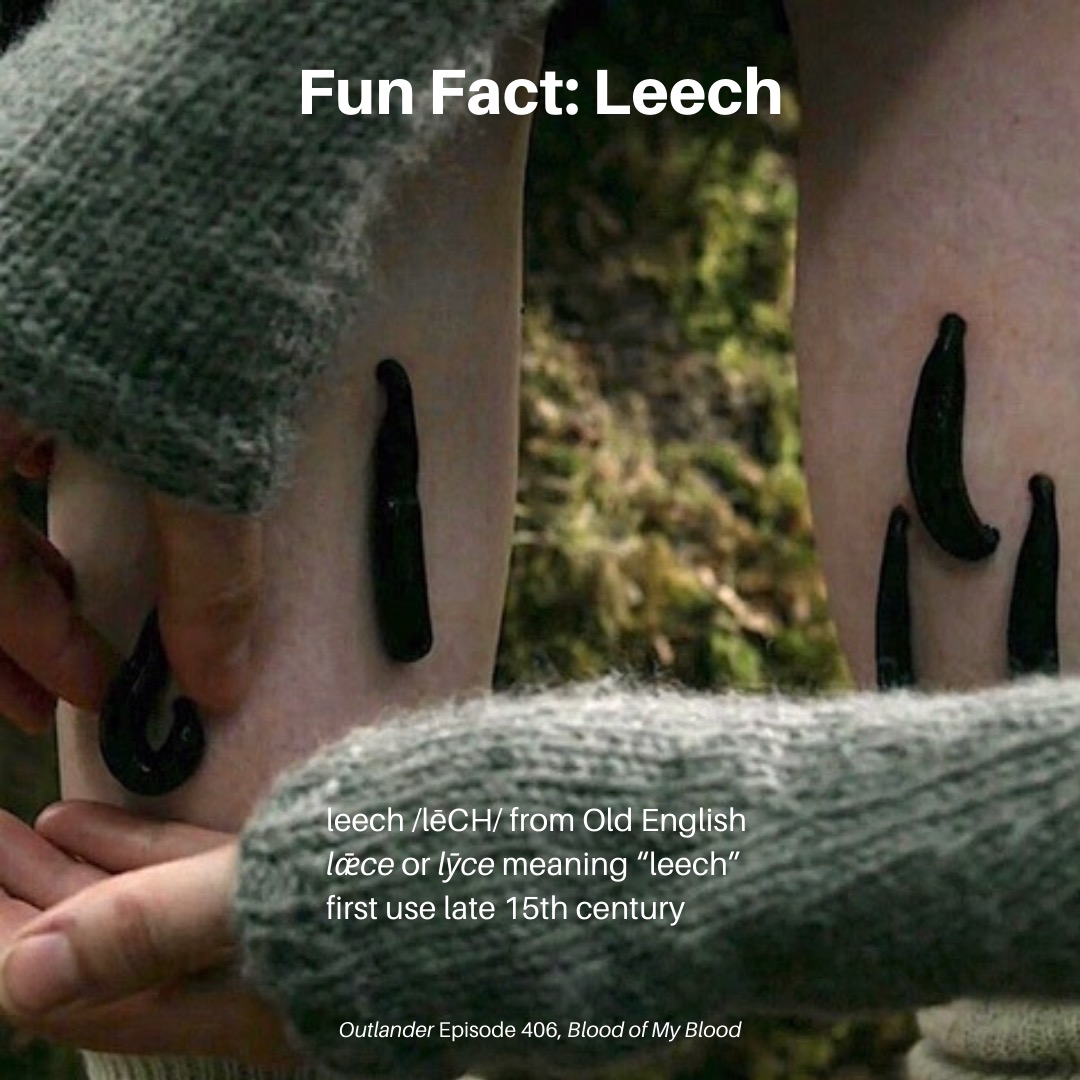
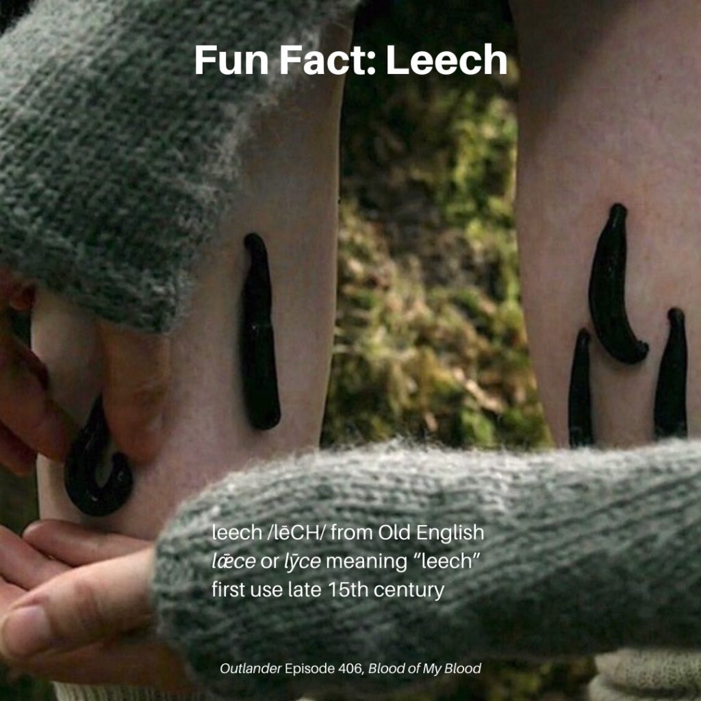
Bloodletting via leech is a time-honored practice dating to ancient Greece and India. Throughout Europe, the medicinal leech, Hirudo medicinalis, was used on ailing patients to rebalance body humors.
Yet more reasons this episode was titled “Blood of My Blood!!!” 😱
In 18th and 19th century Britain, leech-gatherers travelled the marshes gathering leeches from the wild. The practice became so widespread, the population was decimated in many areas. Bloody little buggers! 😈
In Old English, lǣce, was not only the name of the animal but also referred to a physician, and lǣcecraft or leechcraft described the art of healing. Hum….
Beginning in about 1980, leeches enjoyed a resurgence in modern medicine. Today, many hospitals stock them to treat:
Joint disease such as epicondylitis and osteoarthritis
Vein diseases of the extremities
Microsurgery
Blood-clotting disorders using hirudin
WARNING! If you sport a strong wame, watch this PBS video of how a leech attaches, how it feeds and its use in modern medicine. Watch at your own discretion. There will be blood!!! 😷
Important! A leech can be removed by breaking the seal of front and back suckers with a fingernail or other flat, blunt object, flicking the leech away. Irritating the leech with a cigarette, vinegar, salt or soap may cause it to regurgitate stomach contents into the wound and transmit pathogens to its victim. Not common, but it has been reported.
Read about leeches in The Drums of Autumn as Lord John Grey and his “son,” William Ransom, arrive unannounced at Fraser’s Ridge. Make no mistake, Claire recognizes Jamie’s get! 😉
Blundering out of the stream, I shoved my way through the tangled branches, and burst through into the clear space beyond. A boy was dancing on the bank above me, slapping madly at his legs and howling as he hopped and fro.
“What—?” I began, and he glanced up at me, blue eyes wide with startlement at my sudden appearance.
…“Leeches,” I said, professional calm descending by habit over personal tumult. It couldn’t be, I was telling myself, at the same time that I knew it damn well was.“It’s only leeches. They won’t hurt you.”
“I know what they are!” he said. “Get them off me!” He swatted at his calf, shuddering with dislike. “They’re vile!”
“Oh, not so terribly vile,” I said, beginning to get a grip on myself. “They have their uses.”
“I don’t care what use they are!” he bellowed, stamping in frustration. “I hate them, get them off me!”
“Well, stop whacking at them,” I said sharply. “Sit you down and I’ll take care of it.”
He hesitated, glaring at me suspiciously, but reluctantly sat down on a rock, thrusting his leech-spattered legs out in front of him.
“Get them off now!” he demanded.
See leeches feasting on Willie’s blood in Outlander episode 406, Blood of My Blood. 🚫 Leeches for this Lord!
Greetings anatomy students! Hanging is a ghastly topic, but it has a substantial presence in Outlander and is pertinent to anatomy. So, here we go!
The Oxford Dictionary defines hanging as “specifically to put to death by suspension by the neck.” But, in the past, hanging also meant death by crucifixion or impalement!
An interesting fact…. The appropriate terminology is the person was hanged, not the person was hung.
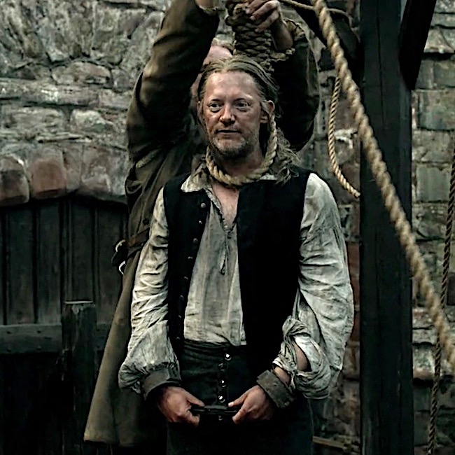
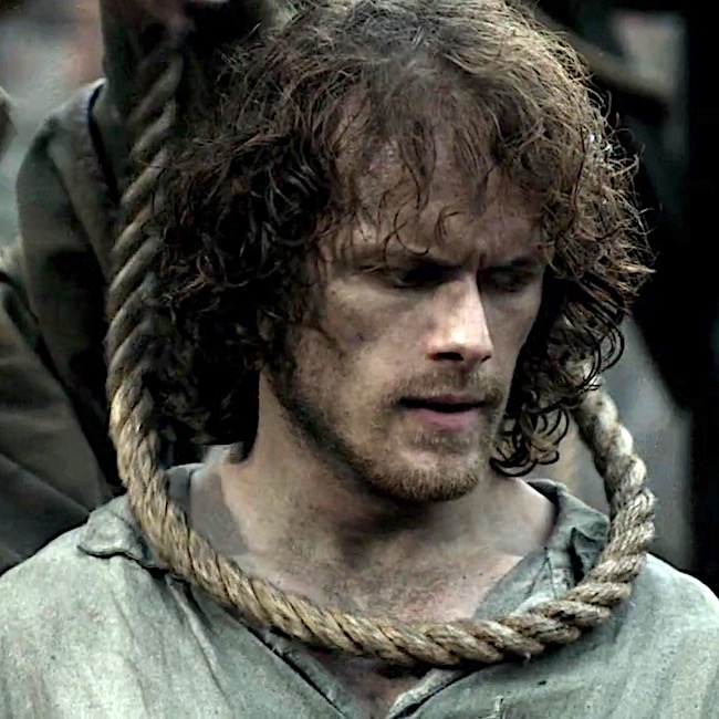
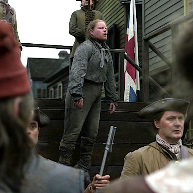
Outlander’s first traditional hanging appeared in episode 115, Wentworth Prison; crusty Taran MacQuarrie of the Watch and Jamie Fraser face their ends with stirring gallows’ humor: Jamie is afraid “My wife will never forgive me for getting myself foolishly hung!” Hanged, Jamie…. the word is hanged!
But, Taran aways knew he was fated to die at the end of a rope – his name is read next.
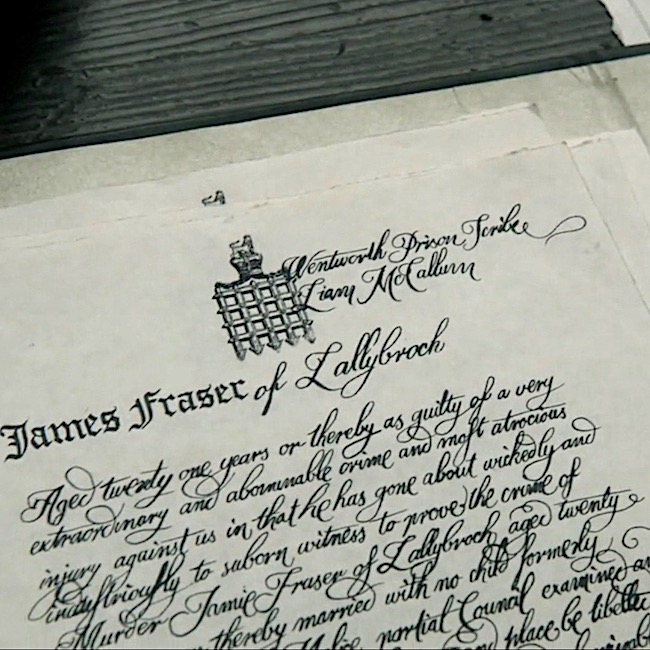
Jamie’s turn! Scribe, Liam McCallum, recorded Jamie as guilty of “a very extraordinary and abominable crime and most atrocious injury against us!” Snort!
Jamie struggles, but is subdued and resigns himself to this ignoble fate. Such an unworthy end for Laird Broch Tuarach!
Then one of Lucifer’s avenging angels swoops in to spare Jamie for a different fate! But, this outcome awaits another lesson.
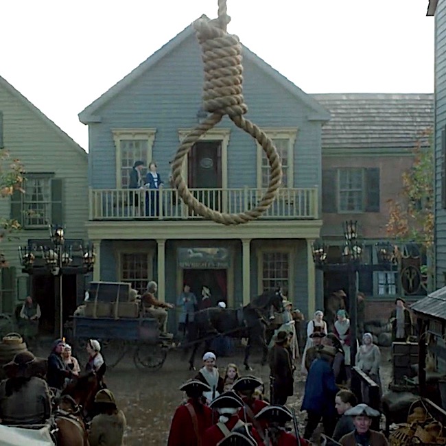
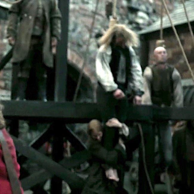
No further traditional hangings, until season four. Episode 401, America the Beautiful, starts off with a bang… Well, with a hangman’s noose, truth be told.
In a drunken state, Gavin Hayes lay with a marrit woman (psst…Jamie warned him!). When the outraged hubby came at him with a pitchfork, Gavin kicked him down the stairs and now, the hangman’s noose awaits. He should have listened to Mac Dubh! (In Drums of Autumn book, Gavin was guilty of stealing six pounds, ten shillings).
Hayes asks Jamie for two favors:
#1. Whisky!
Herself described in Drums of Autumn:
“It was what he asked of me,” he said. “And the best I could manage for him.”
“Brandy or whisky?” asked Fergus, evaluating Hayes’ appearance with a practiced eye.
“The man’s a Scot, wee Fergus.” Jamie’s voice was as calm as his face, but I heard the small note of strain in it. “Whisky’s what he wanted.”
“A wise choice. With luck, he won’t even notice when they hang him,” Fergus muttered
#2. May his last sight be the face of a friend.
Again, from Drums of Autumn:
If Hayes was still sober enough to see anything, the last thing he saw on earth would be the face of a friend.
He could see; Hayes glared to and fro as they lifted him into the cart, twisting his neck, desperately looking.
“Gabhainn! A charaid!” Jamie shouted suddenly. Hayes’ eyes found him at once, and he ceased struggling.
Jamie musters a wan smile for his auld friend and fellow Ardsmuir prisoner.
Drums of Autumn quote continues:
The captain of the guard stood poised, saber raised.
Suddenly, the condemned man drew himself up straight. Eyes on Jamie, he opened his mouth, as though to speak.
The saber flashed in the morning sun, and the drums stopped, with a final thunk! I looked at Jamie; he was white to the lips, eyes fixed wide. From the corner of my eye, I could see the twitching rope, and the faint, reflexive jerk of the dangling sack of clothes. A sharp stink of urine and feces struck…
The rope drops. Hayes death appears mercifully quick! Again from Drums of Autumn:
The hangman had known his business; there had been no undignified struggle, no staring eyes, no protruding tongue; Gavin’s small round head tilted sharply to the side, neck grotesquely stretched but cleanly broken.
OK, but, what about non-traditional hangings? Any of these in Outlander. But, of course!
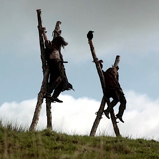
Two Jacobite traitors hang from crossed stakes courtesy of Red Coats in Outlander episode 105, Rent. Dougal and his party find the dead men with “T” for traitor carved into their flesh.Although we don’t know how they died one can reasonably consider this a variation of a crucifixion hanging.
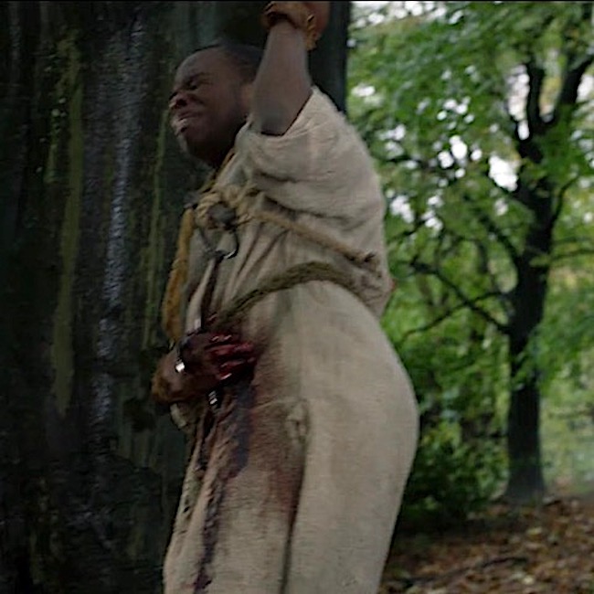
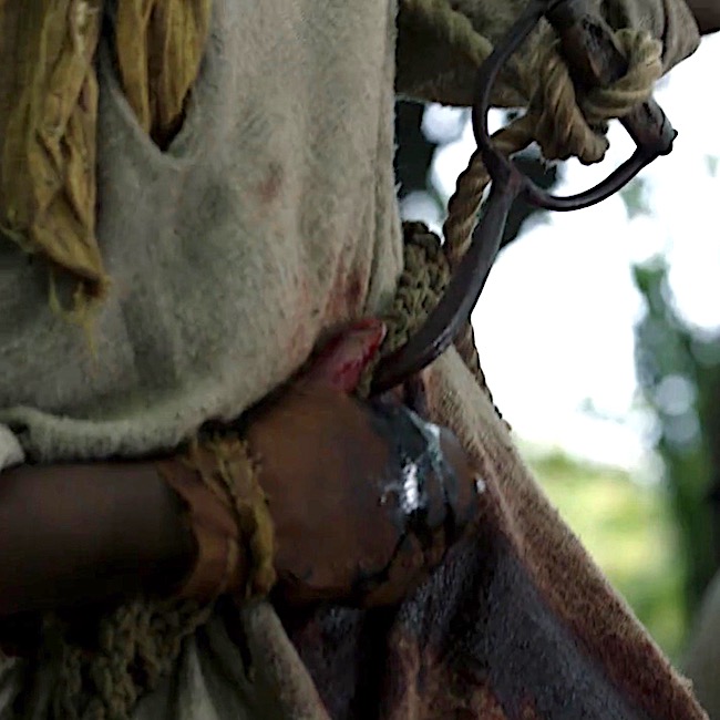
And, finally! In ep 402, Do No Harm, Jocasta’s slave, Rufus, dangles from a hook thrust under his ribs. This type of hanging is known as impalement.
From Drums of Autumn:
From everything I could sense and feel, I thought that the curve of the hook had gone upward through the liver. Likely the right kidney was damaged, and the jejunum or gallbladder might be nicked—but none of those would kill him immediately.
So, one may reasonably conclude that Outlander has dutifully presented both traditional and non-traditional hangings!
So, what does hanging have to do with human anatomy? Quite a lot actually, because hanging destroys normal anatomy. Let’s see how.
Understanding traumatic events from hanging requires a bit of neck anatomy.So….
Neck Anatomy: The neck is complex because it contains a mess of structures. These include:
pharynx for food and air passage
larynx for air passage and vocalization
trachea for air passage
esophagus for food passage
common carotid arteries supply blood to head
jugular veins drain blood from head
cervical vertebrae for support
hyoid bone for speech, swallowing, tongue movements
cervical spinal nerves, for neck and upper limb innervation
muscles for movement of head, neck, scapula and clavicle
Hangings typically traumatize one or more of these neck structures:
cervical vertebrae
hyoid bone
carotid arteries
jugular veins
trachea and larynx
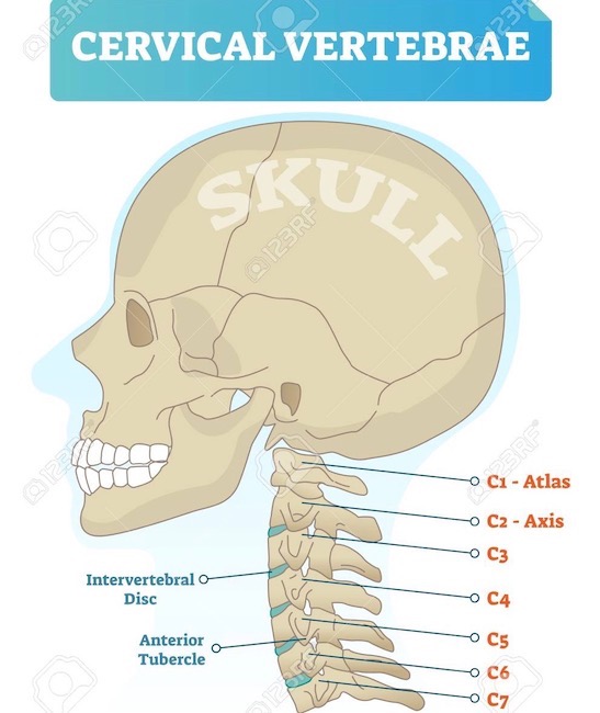
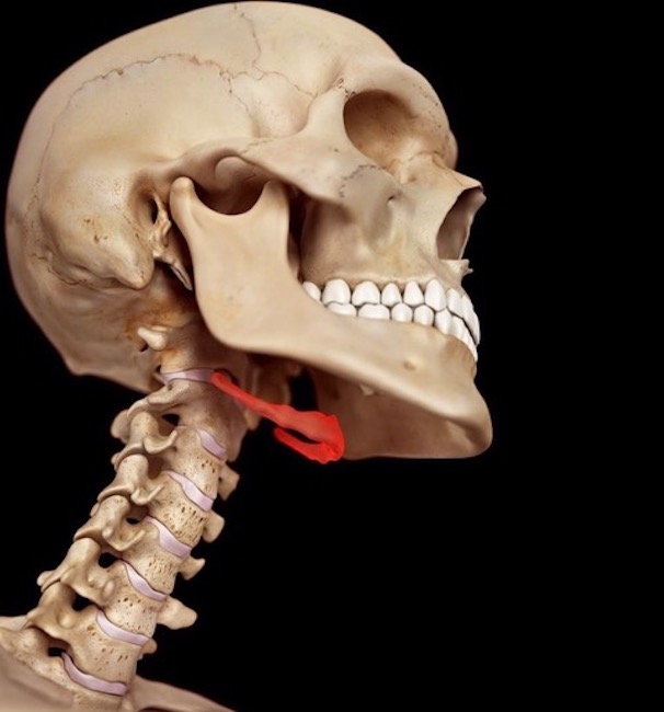
Cervical Vertebrae: The neck contains seven cervical vertebrae numbered from skull downward as C1-C7 (Image A). C1 is also known as the atlas; C2 is the axis.
Cervical vertebrae are smaller and more delicate than thoracic and lumbar vertebrae; this is especially true of C1 and C2. Collectively, all seven cervical vertebrae form the cervical skeleton and are bound together by strong ligaments.
Vector illustration of cervical vertebrae. Medical scheme with close-up skull and isolated C1 atlas, C2 axis, C3, C4, C5, C6 and C7 vertebra. Diagram of Intervertebral disc and anterior tubercle.
Image A
Hyoid Bone: The hyoid bone and cervical vertebrae are the only bones in the neck . The U-shaped hyoid (Image B, red) is unusual because it is the only bone of the body that does NOT articulate with other bones. Rather, it is suspended in the neck via muscles and ligaments connecting to neighboring bones and cartilages. More detail on hyoid and cervical vertebrae in Anatomy Lesson #12, Claire’s Neck – The Ivory tower.
Try This: You can palpate your own hyoid bone. Hold chin parallel to floor. Place thumb and forefinger of one hand on either side of your larynx (voice box). Move fingers upward until just under mandible. Squeeze gently, then swallow. You will feel a thin hard structure on each side, the R and L arms of the hyoid.
And, for book readers, Diana wrote about the hyoid bone in Written in My Own Heart’s Blood.
Image B
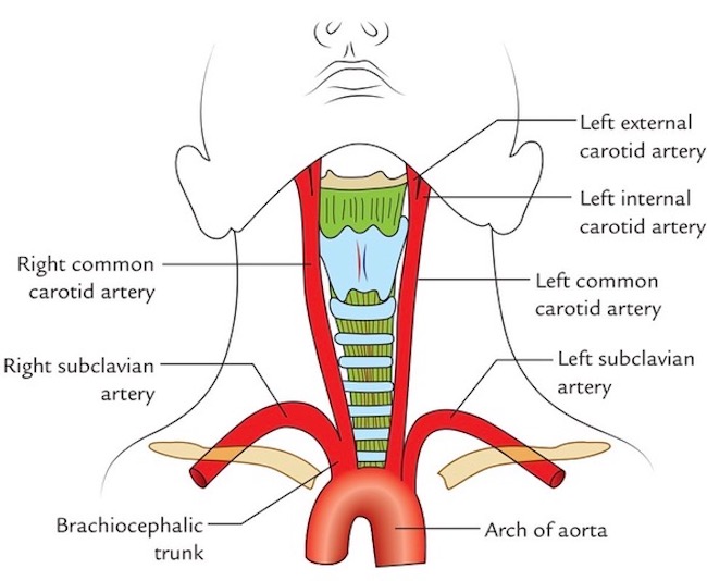
Common Carotid Arteries: The neck has a R and a L common carotid artery; each is a primary or secondary branch of the aorta arch (Image C).Common carotids straddle trachea and larynx (Image C -blue/green structures) before splitting into internal and external carotid arteries which supply blood to neck and head.
Image C
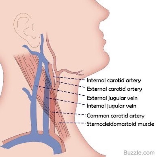
Jugular Veins: The neck has two jugular veins on each side; external and internal jugulars. These drain blood from head and neck structures toward the heart Image D).
Image D
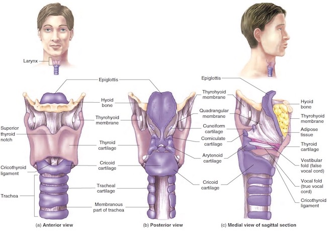
Trachea and Larynx: All of larynx plus upper trachea lie in the neck. Both are conduits for air to reach the lungs during inhalation and to leave the lungs during exhalation. In addition, the larynx produces vocalizations. Trachea and larynx provide the only normal route by which air can move to and from the lungs. Of course, a tracheotomy is an exception, but it is also not normal. (Psst…No S5 spoilers frombook readers, please!)
Image E shows trachea and larynx. Tracheal wall is stiff due to cartilage plates. The complex larynx consists of nine cartilages of various shapes and sizes. Learn more about the larynx in Anatomy Lesson #42, “The Voice – No, not that One!”
Image E
Now, for some info about hanging.
Hanging: One might think hanging is a rather simple act, but it is not. There are many types of hanging although death is typically from either strangulation or neck fracture. Here are three common types of hanging.
*Short Drop: A condemned person is placed on a stool, ladder, cart, horse, etc. with noose around the neck. The object is removed leaving the person dangling from the rope. Although the person is unconscious within 15 seconds, death usually takes 10-20 minutes as the noose tightens causing slow strangulation.
*Standard Drop: A condemned person drops 4-6 feet (1.2-1.8m). This is considered more “humane” than short drop because it typically fractures the neck which traumatizes spinal cord and causes immediate unconsciousness and rapid brain death.
*Long Drop: Drop varies. Height and weight of the condemned were used to calculate the drop needed to fracture the neck, preferably at C2, the axis. This is known as a hangman’s fracture.
Yes, I know! Horrifying to think about, but charts were developed to augment a swift death, the preferred outcome.
Submental Knot: Submental means “under the chin.” Careful placement of the eye or knot of the noose under the chin jerks the head backward, contributing to neck fracture. Side and back knots tend to strangle rather than fracture the neck.
Back to Outlander: So, with knowledge about vulnerable neck structures and types of hangings plus their effect on anatomy, let’s review Outlander hangings.
What about Taran from ep 115, Wentworth Prison? Taran was pushed off a not-very-tall scaffold – so in effect a short drop. Sadly, it didn’t break his neck and the Watchman kicked and struggled at the end of the noose! A member of the execution squad pulled down on his legs to break his neck, an act that was often required to end the misery. Of course, Jamie and other condemned prisoners witnessed this horror!
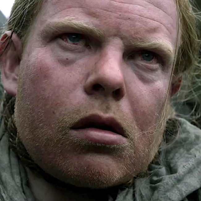
And, then there’s Hayes. Although his end was swift in Drums of Autumn book, not so in the episode. He did experience either a standard or long drop but afterward, his face is red and bloated and his eyes bloodshot. These findings are more consistent with strangulation than neck fracture, meaning his jugular veins were obstructed leading to death by brain edema and ischemia, wherein:
Bleeding in conjunctiva overlying whites of eyeball (subconjunctival hemorrhage)
So the FX were very good but not well-matched for this type of hanging.
Just so we know, if the deeper lying common carotid arteries are occluded by strangulation, the face appears pale or white.
And, sphincters do relax spontaneously so urine and feces are evacuated. Thankfully, these weren’t part of the FX scheme! 😱
And poor Rufus! Impalement by hook is a very old punishment occurring in Babylonia! Later, it was used in 18th-century Ottoman-controlled Bosnia and by Dutch overlords in Suriname (South America) who impaled troublesome slaves under the ribs.
An 18th century observer recorded that an impaled person could hang as long as three days before expiring. The primary source of pain was extreme thirst.Thankfully, this type of hanging has been pretty much abandoned.
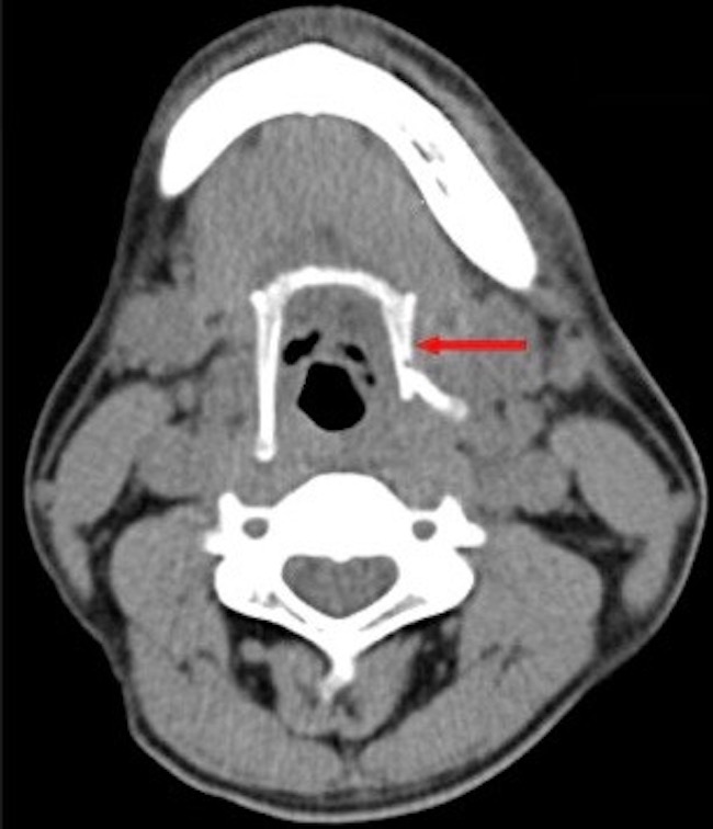
As for the hyoid bone…. It fractures in about a quarter of hangings but is not the cause of death. Forensic scientists look for a fractured hyoid in suspected homicides because about 50% of hyoids fracture with manual choking.
Fracturing the hyoid bone depends on applied force, age of victim, nature of instrument (ligature vs. hands) so even if the bone is not fractured, a murder is still possible (Image F –red arrow).
Image F
Today, we can be grateful that many countries have abandoned hanging as a means of punishment. An excellent film is available on this topic: Pierrepoint: The Last Hangman. Bonus! Tobias Menzies plays Lieutenant Llewelyn!
In Jamie’s time, hanging was an ignoble death. Why, we might ask? Here is one reasonable thought:
“The sword connoted an honourable way of dying, and an honourable return to the earth, but the rope left the body hanging between heaven and earth and was therefore an unseemly death.”
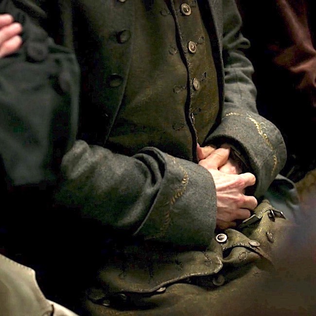
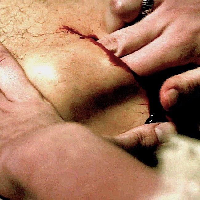
Hey, anatomy students! Are you interested in hernias? If yes, you came to the right place! Today’s lesson, Inguinal Hernia, is prompted by Dr. Claire performing a hernial repair in Outlander episode 408, Wilmington. Let’s pass through the stones and review the scene as it unfolds at a local 18th century theater!
Warning: Two images in this lesson show the groin area. One is of Jamie at Lallybroch millpond, the second is a clinical image. I think all readers are adults and will be OK with these. But, the warning is for those who might find such content objectionable.
After meeting Governor Tryon and his associate, Edmund Fanning, Claire observes Fanning in distress. Turns out, he suffers paroxysms of pain from a strange protrusion, incurred after standing against a mob in Hillsborough. His boots stayed in the mud as his body turned after delivering rum to appease rioters. Oh, my!
Talk about theater! Barely watching that dreadful play, Jamie learns his Godfather is in peril and devises a plan. Hum….mayhap a poke in the puir fellow’s aching belly will buy valuable time? A quick jab to the left and Fanning needs a surgeon!
Talk about belly aching! Call Surgeon Sasseynach….. STAT!
Claire to the rescue! She speedily diagnoses an inguinal hernia – confirming her earlier suspicions:
“The intestines have moved and the blood flow may be cut off!”
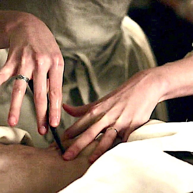
Fanning is quickly laid on a table as Claire marshals helpers, knife, needle, thread, linens and rum. Lots of linens and lots and lots of rum!
That is quite the lump, Edmund! A left inguinal hernia but a bit too high on the abdominal wall!
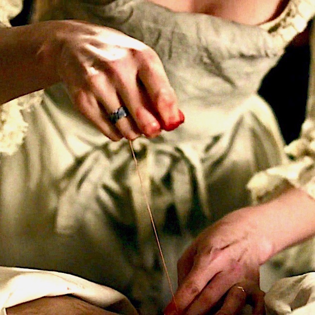
She drapes and swabs the surgical field, sterilizes a knife in alcohol and flame, threads a needle, grinds the wheat and bakes the bread! <G>
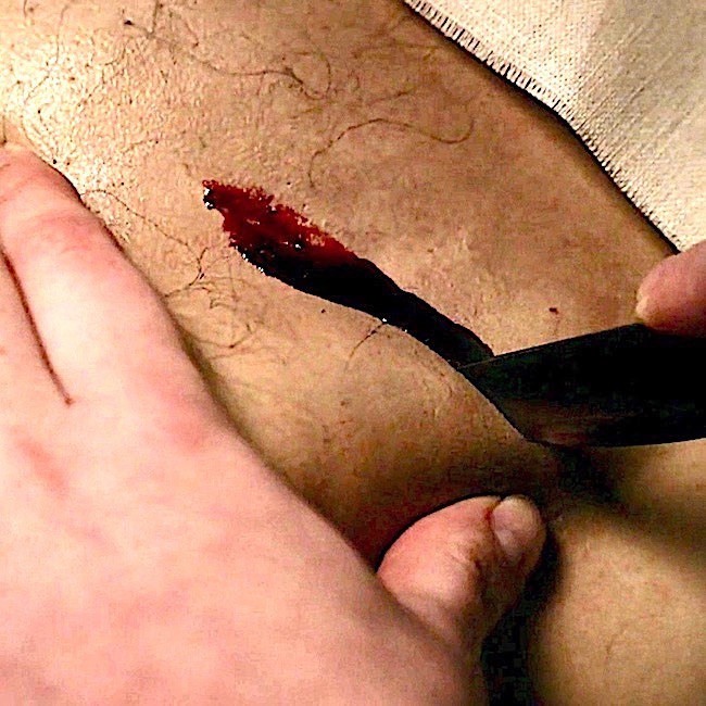
Claire begins surgery with the knife positioned near the hernia, poised to cut above the bulge and parallel to it. Good choice, Claire!
Then, inexplicably, she switches direction and cuts across the hernial bulge!!! Bad choice, Claire! 😱
She cuts very deep and there’s a lot of blood!
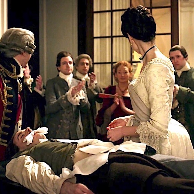
Then, with considerable effort, she shoves the hernia (see below) towards the midline of the body (linea alba). Pushing bowel the wrong direction, Claire!!! 😱
She skillfully sutures the wound with very what appears to be carpet thread. Not surprising, as it was likely salvaged from the costume department.
And, unlike the actors of that dreary, lugubrious play, Claire receives a standing ovation for a job well done!
Now for the science. Yay!
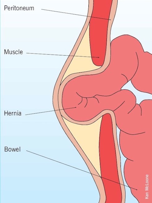
Hernia Defined: Simply put, a hernia is a protrusion or bulge caused by an organ or tissue pushing through the wall enclosing it (Image A).
Image A
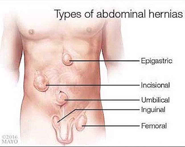
Types of Herniae: Hernae (pl) occur in different body areas, but the most common site is the abdominal wall (Image B). These include:
Understand that groin herniae are the most common type of abdominal herniae; these include both inguinal and femoral types. As Claire diagnosed an inguinal hernia, the lesson will cover only this type.
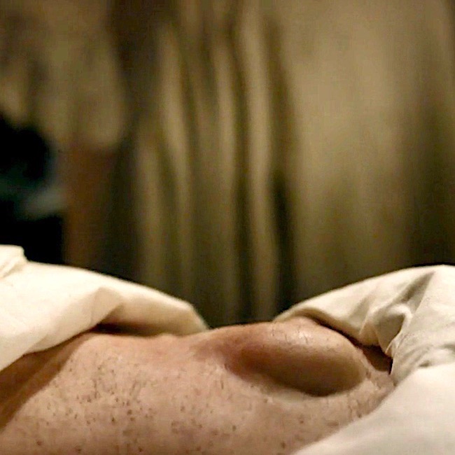
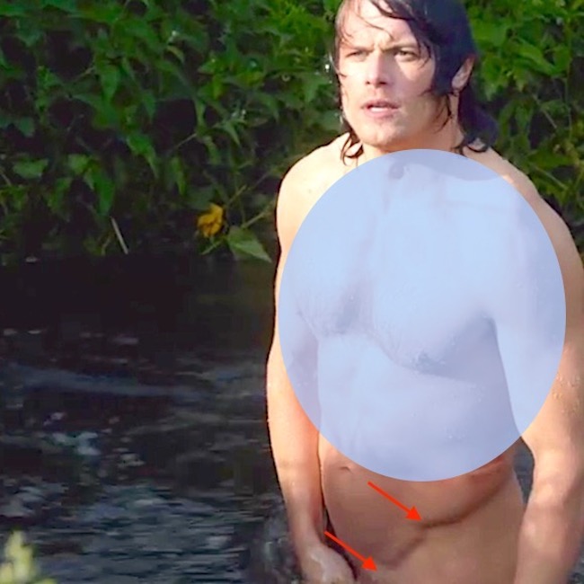
Inguinal Region: Inguinal herniae occur in the inguinal region. But wait! Where, exactly, is the inguinal region? Our fav anatomical model volunteers to demo! Yay, Jamie! Here, from the sky-blue waters of the freezing mill pond (Starz ep 112, Lallybroch), Jamie kindly lends a sneak-peak!
Both ASIS and pubic tubercle are easily palpable landmarks of pelvic bones, especially in the lean and physically fit.
The very strong inguinal ligament spans these two bony points. The ligament is overlaid by a skin crease, the inguinal groove, the site where torso meets thigh. Also, female inguinal grooves are more horizontal; male inguinal grooves are more vertical. This is because female hips are wider and the paired ASIS are further apart.
(Psst…..please forgive the blue mask overlying Jamie’s upper torso.This is to discourage bots from tagging this image as sexually explicit and landing OA in FB jail!!!)
Try This: Lay on your back and feel the prominent point of one hip bone (ilium), this is the ASIS. Now, move finger to pubic bone and feel a bump at the upper-outer margin, this is the pubic tubercle. The inguinal ligament spans these bony landmarks.
Inguinal Hernia: The inguinal hernia is a bulge in the abdominal wall above the inguinal groove. There are direct and indirect inguinal herniae. Both types are strictly defined based on their relationship to an abdominal artery and vein (inferior epigastric vessels, IEV)
Indirect inguinal hernia produces a bulge above the inguinal ligament that is lateral to the IEV.
Direct inguinal hernia produces a bulge above the inguinal ligament that is medial to the IEV.
Why is it important to diagnose the type? Because this may help determine how the hernia will be treated.
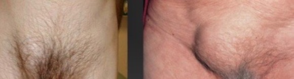
Image C shows right-sided indirect inguinal herniae of a male (L panel) and a female (R panel); both herniae lie above the inguinal groove and developed lateral to the IEV. This image also shows how the female inguinal groove is more horizontal than the male.
Inguinal Herniae Statistics:
can develop at any age
direct inguinal hernia 10x more common in men than in women
indirect inguinal hernia 25x more common in men than in women
more common in men above age 40
more common on R than L side
more common in people with a family history
Image C
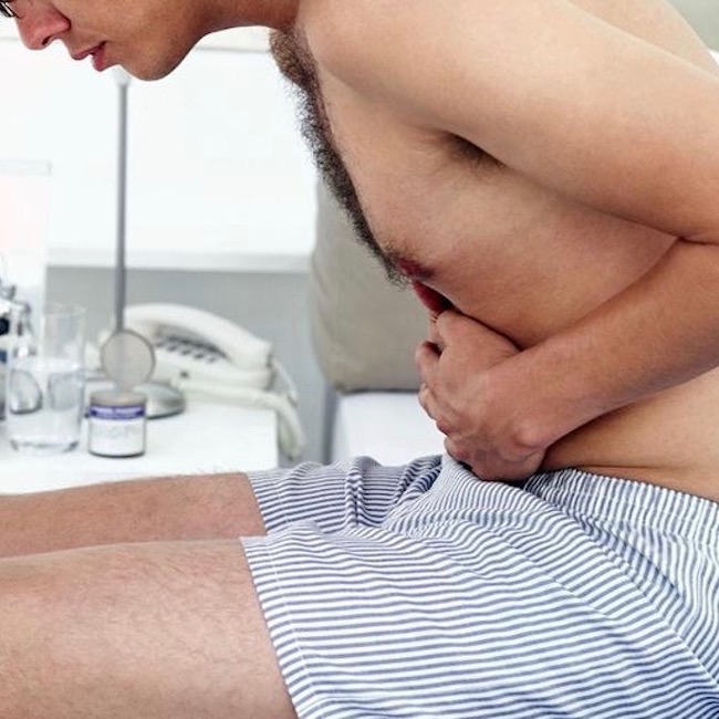
Symptoms: Symptoms of an inguinal hernia include (Image D):
bulge of inguinal region which may extend into scrotum or labia
pain/discomfort with coughing, exercise or defecation
pain increases during the day and lessens when lying down
bearing down enlarges the bulge
heartburn, chest pain, pain with eating
redness or other discoloration of the bulge
Importantly, some inguinal herniae may be asymptomatic! Regular physical exam and complete history should consider this possibility.
Image D
Descent of Testes: There are two very important reason why inguinal herniae are more common in males than in females:
Males tend to do more manual labor requiring heaving lifting thereby straining the abdominal wall. Usually accounts for direct herniae.
Testes descend through the inguinal area during intrauterine life. Usually accounts for indirect herniae. Wait! What???
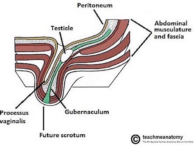
Yes. Ovaries and testes develop in the abdominal cavity.Over time, ovaries descend as far as the pelvis but testes continue to descend into the scrotum, a process that typically completes about week 28 of pregnancy.
Testicular descent is complicated but Image E offers a simplified visual. Descent through the inguinal region involves passing through layers of abdominal muscle and connective tissue (fascia), layers which follow the testes all the way into the scrotum. In addition, a finger of peritoneum, the membranous lining of the abdominal cavity, is dragged along with the descent. This finger of peritoneum is the processus vaginalis.
The channel created by passage through the abdominal wall is dubbed the inguinal canal. Now, this is not a canal in the usual sense, but rather a slit-like passageway. The canal also has internal (deep) and external (superficial) inguinal rings, but these are difficult to explain and not particularly useful in today’s lesson.
If all works as nature intends, each processus vaginalis closes after descent is complete. However, these may fail to close or reopen later in life, leading to an indirect inguinal hernia.
Females also develop an inguinal canal and processes vaginalis but these are smaller and usually close off more readily because no testicular descent is involved.
Image E
Indirect Inguinal Hernia: For your viewing pleasure, this simple cartoon illustrates testicular descent. As you view the video, notice the cream-colored “finger” that accompanies the testis into the scrotum. This finger is an extension of the peritoneum, the membrane that shrink-wraps all surfaces of the abdominal cavity and its organs.
Image E correctly labels this finger-like extension of peritoneum (tan in Image E) as the processus vaginalis. To reiterate, normally, after testicular descent, the processes vaginalis closes off.
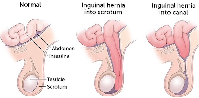
If the processus vaginalis does not close off, or reopens later in life, then fluid, fat or loops of bowel may slither and slide down into the patent processus vaginalis forming a hernia. Not good!
Image F demos such unruly outcomes:
Left panel shows a testis in normal position in the scrotum – no remnant of the processus vaginalis is present (patient facing to your R)
Right panel shows a partially open processus vaginalis containing a loop of inflamed bowel.
Middle panel shows a more extreme situation where the processus vaginalis is open all the way and bowel has slipped down into the scrotum.
If bowel becomes trapped in the processus vaginalis, its blood supply may be diminished, a condition known as incarceration or strangulation. This is a medical emergency because if the bowel dies due to insufficient blood supply, its wall breaks down allowing bacteria to seed sterile body spaces. Untreated, this leads to septicemia and death, especially in the 18th century! So, Claire is correct about surgery being necessary to save Fanning’s life.
A direct hernia works much the same way except the cause is a weaken lower abdominal wall usually from age, pregnancy, heavy lifting, etc. Here, a sac of peritoneum balloons out through the lower abdominal wall wherein bowel may become strangulated with similar fearsome outcomes. Here, intestine cannot enter the scrotum or labia because no processus vaginalis is involved.
Image F
Claire’s Repair: Today, various techniques are used to repair inguinal hernias. Mr. Fanning’s hernia required pushing the bowel back into place followed by suturing the muscle and fascia layers and then the skin. No mesh in those days!
Fanning’s special FX were pretty good. However, I must make the following observations:
Fanning’s hernia lies too high on the abdominal wall for an inguinal hernia. It should be nearer the inguinal groove or pubic bone. Perhaps the site was chosen to avoid TMI?
No surgeon worth their salt would dare cut across a hernial bulge for risk of cutting into the bowel itself! Claire’s initial knife position was correct, why she switched position was puzzling. Perhaps, to make FX more buzz-worthy?
Too much blood oozed from the skin cut which is also too deep – inguinal skin doesn’t bleed that much and is thin. Again, this may have been designed to produce a collective viewer’s gasp.
The FX that really caused me to cringe is the force Claire employs to push the bowel toward the body midline! Nope. That direction, the bowel has no place to go. No wonder Fanning screams! If his is an indirect inguinal hernia, Claire should push the bowel toward his upper left (toward ASIS) following the inguinal groove. If his is a direct inguinal hernia, Claire should push the bowel directly downward so it re-enters the abdominal cavity.
That is one honking thread Claire uses to close the wound! It will likely cause a foreign body reaction accompanied by chronic discomfort but infinitely preferable to dying from an incarcerated bowel!
As Edmund’s bowel was incarcerated, the overlying skin should have appeared inflamed. It didn’t.
Quotes from Outlander books always enrich any anatomy lesson and this is no exception. The inguinal hernia makes its debut In Drums of Autumn book, wherein Claire repairs one on mountain man, John Quincy Myers – atop Auntie Jo’s dining room table – in front of dinner guests!Based on the description, Myers has an indirect inguinal hernia (see Image F, middle panel).
I checked that my supplies and suture needles were ready, took a deep breath, and nodded to my troops.
“Let’s go.”
Myers’s penis, embarrassed by the attention, had already retreated, peeping shyly out of the bushes…Ulysses himself delicately cupping the baggy scrotum away, the hernia was clearly revealed, a smooth swelling the size of a hen’s egg, its curve a deep purple where it pressed against the taut inguinal skin.
I swabbed the perineum thoroughly with pure alcohol, dipped my scalpel in the liquid, passed the blade back and forth through the flame of a candle by way of final sterilization, and made a swift cut.
Not large, not deep. Just enough to open the skin, and see the loop of gleaming pinkish-gray intestine bulging down through the tear in the muscle layer. Blood welled, a thin, dark line, then dribbled down staining the blanket.
I extended the incision, swished my fingers thoroughly in the disinfecting bowl, then put two fingers on the loop and pushed it gently upward.
…I could feel the movement of his intestines as he breathed, the dark wet warmth of his body surrounding my gloveless fingers in that strange one-sided intimacy that is the surgeon’s realm. I closed my eyes and let all sense of urgency, all consciousness of the watching crowd drop away.
…Time stopped. I was acutely aware of each movement, each breath, the tug and pull of the catgut sutures as I tightened the inguinal ring, but my hands did not belong to me.
…Then it was done, and time began again.
“Done,” I said, and the hum from the spectators erupted into loud applause. Still feeling intoxicated—had I caught drunkenness by osmosis from Myers?—I turned on one heel and sank into an extravagant low curtsy, facing the dinner guests.
My favorite part of Fanning’s surgery comes when the 18th century physician bustles in declaring “What hath hell wrought?” Yeah, women didn’t do surgery or openly practice medicine in those days.
Then, he accused Claire of butchering the poor man, finishing with: “All he needed was some smoke up the rear.” Bwahahaha! Priceless!
This entertaining 10 minute video by Dr. Carlo Oller does a terrific job of summarizing much of today’s lesson as well as providing additional tips about hernia prevention and care. Hope you watch!
OK, anatomy students. That is it for today’s lesson. Anatomy of the inguinal region and its associate pathology are complex, but it behooves us all to stay vigilant for signs and symptoms of a hernia.
Let’s close with this simple thought: as inguinal hernias occur more frequently in males than females, shouldn’t these be called, himnias? Wink. Wink.
A deeply grateful,
Outlander Anatomist
Photo Credits: Starz ep 112, Lallybroch, ep 408, Wilmington.